SHORT COMMUNICATION
The Altered Vascular Endothelial Control of Facial Cutaneous Blood Flow in Rosacea
C. Ilinca UNGUREANU1, Megan J. KUBE2, Thad E. WILSON3,4  and Kristen METZLER-WILSON1,5*
1Scholarly Concentrations Program, Indiana University School of Medicine, Indianapolis, IN, 2Trinity Health Livonia Hospital, Livonia, MI, 3Department of Biomedical Sciences, Ohio University College of Osteopathic Medicine, Athens, OH, 4Department of Physiology, University of Kentucky College of Medicine, Lexington, KY, and 5Department of Physical Therapy, University of Kentucky, 900 S. Limestone Ave – CTW 204L, Lexington, KY 40536, KY, USA. *E-mail: Kristen.Metzler@uky.edu
Citation: Acta Derm Venereol 2025; 105: adv42800. DOI: https://doi.org/10.2340/actadv.v105.42800.
Copyright: © 2025 The Author(s). Published by MJS Publishing, on behalf of the Society for Publication of Acta Dermato-Venereologica. This is an Open Access article distributed under the terms of the Creative Commons Attribution-NonCommercial 4.0 International License (https://creativecommons.org/licenses/by-nc/4.0/).
Submitted: Dec 24, 2024. Accepted after revision: Apr 10, 2025. Published: Apr 27, 2025.
Competing interests and funding: The authors have no conflicts of interest to declare.
Laboratory funding was provided by National Institute of Arthritis, Musculoskeletal, and Skin Diseases Grant AR069912 (to KM-W and TEW). This project was funded, in part, with support from the Indiana Clinical and Translational Sciences Institute funded, in part by UL1TR002529 from the National Institutes of Health. The content is solely the responsibility of the authors and does not necessarily represent the official views of the National Institutes of Health.
INTRODUCTION
Rosacea is a common (~5% of adults) chronic regional vascular and inflammatory facial disorder with an unclear aetiology (1, 2). Associated vascular hyperreactivity and the underlying skin blood flow regulation are correlated to specific “triggers” (e.g., certain foods, exercise, or extreme temperatures) (3). In response to trigger events, individuals with erythematotelangiectatic (type 1) rosacea (facial erythema without inflammatory lesions such as papules/pustules [2, 4]) responded with exaggerated supraorbital skin sympathetic nerve activity (SSNA) (5). These direct recordings of a head/neck nerve within the focal region of the disease (e.g., cheek, forehead) provide a possible role of sympathoexcitation in the disease process, as occurs in disorders such as congestive heart failure and obstructive sleep apnoea (6). Additionally, chronic exposure to this heightened activity may contribute to other end-organ changes (e.g., endothelial, vascular smooth muscle, and local inflammatory-induced dysfunction).
Several methods have been employed to assess skin blood flow (7). Non-scanning laser-Doppler flowmetry (LDF) can provide continuous erythrocyte flux on a beat-by-beat basis, permitting analysis in both time and frequency domains, the latter potentially providing mechanistic insight into control and regulation. Skin blood flow oscillations have been proposed to be regulated by several factors including both local and sympathetic control (8–10). Analysis of these oscillations has been performed on the cheek and forehead (11), but not in a dermatological or facial flushing population such as rosacea. Additionally, LDF technology can be coupled with provocation tests to acutely increase skin blood flow. One provocation test, slow local heating, is of interest as it does not cause pain sensations and can induce maximal flow without activating the sympathetic nervous system (7). Thus, neurovascular and neuroinflammatory changes associated with rosacea (2) could be avoided. Thus, this study aims to quantify the spectral power erythrocyte flux in regional areas affected by rosacea within individuals and between people with rosacea and healthy control subjects during vasodilation utilizing slow local heating. We hypothesized that compared with controls, individuals with rosacea would demonstrate greater changes in spectral power in the vascular endothelial (VLF) and sympathetic (LF) ranges during heating of typically rosacea-affected (forehead, cheek) but not rosacea-unaffected (forearm, palm) areas. These data should provide insights into how these skin areas are regionally controlled and regulated in rosacea, as well as having the potential to uncover a rosacea signature response that could aid in developing useful functional diagnostic testing for this clinical population.
Materials and methods
Data were collected in temperate zone Spring within a temperature-humidity controlled laboratory (23–25°C, ~50% RH). We analysed data from 9 otherwise healthy subjects with erythematotelangiectatic rosacea (5 males, 4 females; sex determined by self-report; rosacea classified by a dermatologist using National Rosacea Society guidelines, age 22±3 years (mean±SD)) and 9 age/sex matched healthy control subjects, all of whom completed a health screen. All participants were Caucasian but not classified by Fitzpatrick scale. The data presented here result from a unique analysis of previously published data (Protocol 3 of Ref. [5]). Measurements include beat-by-beat erythrocyte flux (P3 probes connected to MoorLab unit, Moor Instruments, Axminster, UK) of typically affected (cheek, forehead) and typically unaffected (forearm, palm) skin sites and beat-by-beat finger arterial blood pressure (Finometer Pro, Finapres Medical Systems, Amsterdam, The Netherlands) during a slow local heating protocol. Devices were calibrated according to manufacturer guidelines using external references (probe flux standard and brachial sphygmomanometer). Erythrocyte flux was measured on the right side of the body (11) and with anatomical landmarks while avoiding superficial and telangiectatic veins, blemishes, and skin curvature guiding placement. See Ref. (5) for further information regarding experimental protocol and measurements.
We collected data in 6-min data segments (baseline and plateau during 42°C heating) at 62 Hz. The heating perturbation consisted of warming the probe housing (VHP2 heater connected to SH02 unit, Moor Instruments) to 32°C for 6 min, then by 0.1°C every 2–3 s to 42°C and followed by 30 min maintenance. We analysed these 6-min segments using a fast-Fourier transform (FFT) with each Hanning-windowed data segment and averaged to obtain the spectrum. We then binned them into very low frequency (VLF; 0.009–0.02 Hz indicative of local vascular endothelial response), low frequency (LF; 0.02–0.06 Hz indicating a sympathetic response), medium frequency (0.06–0.2 Hz indicating a smooth muscle response), and high frequency (0.2–0.35 Hz indicating respiratory responses) (10). We identified statistical differences via 1-tailed (affected sites) or 2-tailed (unaffected sites) t-tests comparing change from baseline to heat between rosacea and control subjects with alpha set to 0.05 to determine statistical significance.
Results
Unflushed rosacea and control subjects had similar increases in skin blood flow at facial and forearm sites, expressed as a percent increase compared to baseline, when exposed to the slow local heating protocols and similar baseline skin blood flow when expressed as a percentage of 42° C (5). However, these previous analyses artificially constrain baseline erythema and skin blood flow response to 42° C.(5). Using spectral power analysis (Fig. 1), we identified that individuals with rosacea had greater changes in cheek (p = 0.03) and forehead (p = 0.04) VLF spectral power from baseline to maximal heating (Fig. 2A–B). However, there was no difference in changes for forearm (p = 0.76) or palm (p = 0.38) VLF skin blood flow spectral power from baseline to maximal temperature during local heating of individuals with rosacea vs control subjects (Fig. 2C–D). Additionally, we observed no differences in the change in the LF skin blood flow spectral power readings from baseline to heating between rosacea or control subjects (all p > 0.29; Fig. 2E–H). Similarly, there were no differences in changes in MF (cheek p = 0.10, forehead p = 0.38, forearm p = 0.92, palm p = 0.69) or HF (cheek p = 0.08, forehead p = 0.44, forearm p = 0.57, palm p = 0.90) skin blood flow spectral power from baseline to maximal temperature during local heating of individuals with rosacea vs control subjects.
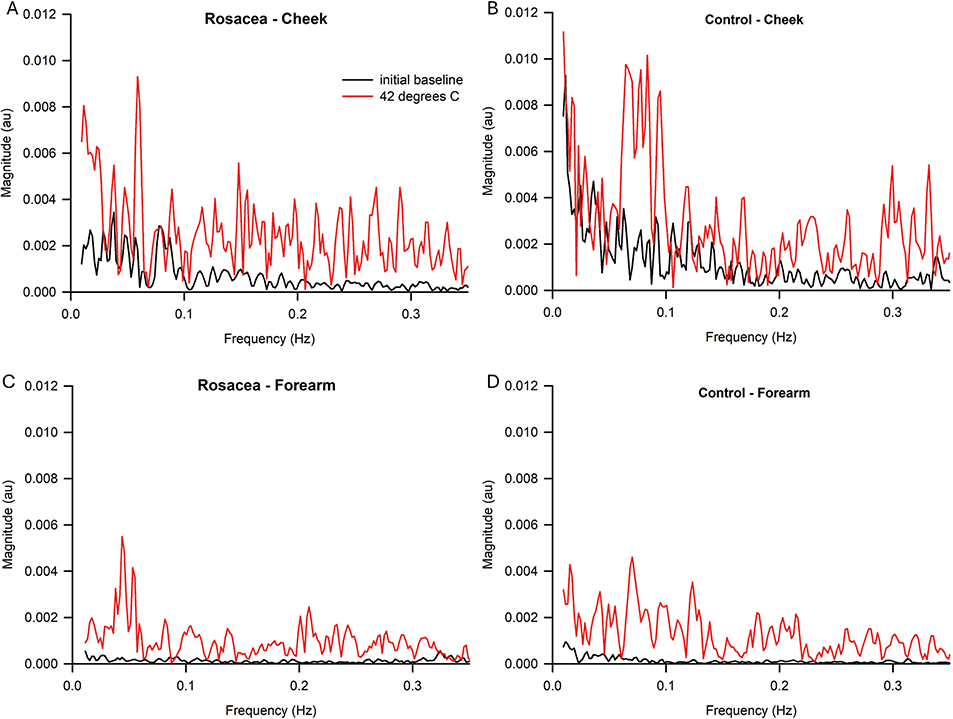
Fig. 1. Representative fast-Fourier transform (FFT) graphs of a typically affected area (cheek) of rosacea (A) and control (B) subjects and a typically unaffected area (forearm) of rosacea (C) and control (D) subjects at baseline vs 42°C.
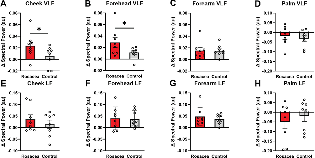
Fig. 2. Change in very low frequency (VLF; 0.009–0.02 Hz; A–D) and low frequency (LF; 0.02–0.06 Hz; E–H) skin blood flow spectral power of the cheek (A & E), forehead (B & F), forearm (C & G), and palm (D & H) in rosacea vs controls (mean±SEM). Significant differences were observed in very low frequency spectral power of the cheek (A) and forehead (B) and are denoted by *.
DISCUSSION
Two previous studies used local heating-induced vasodilation in people with rosacea. Both Guzman-Sanchez et al. (12) and our group (5) identified no change in skin blood flow in affected vs unaffected skin in people with erythematotelangiectatic rosacea (12), although neither analysed the frequency domain of the LDF signal. In the current study, we interrogated the frequency domain to determine whether this approach yielded differences between rosacea and control subjects.
This study investigated the utility of a slow local heating provocation test in characterizing and gaining insights into the underlying mechanism behind erythematotelangiectatic rosacea. Compared with the healthy controls, individuals with rosacea had a greater change in the VLF skin blood flow spectral power readings from baseline to maximal heating in the cheek and forehead, suggesting altered endothelial control in rosacea-affected areas. Neither group had changes in the LF skin blood flow spectral power readings at any location, suggesting that people with rosacea do not have altered basal sympathetic control. Also, a lack of change in the LF readings indicates the slow local heating did not induce a sympathetic response in either healthy controls or people with rosacea.
Our observed vascular endothelial differences in people with rosacea may be linked to either a chronic neuroinflammatory process or an abnormal innate immune pattern. Yamasaki et al. found that people with rosacea had greater Toll-like receptor 2 (TLR2) density when compared with healthy controls (13). Increased TLR2 results in downstream effects of calcium-dependent release of kallikrein 5, which facilitates the inflammatory response (13). Moura et al. similarly found increased expression of not only TLR2, but also Toll-like receptor 4 (TLR4) and inducible oxide nitric synthase (iNOS) in people with rosacea (14). They argue that these abnormal receptors cause faulty innate immunity in rosacea (14). iNOS has been linked to altered cutaneous vasodilation in local heating protocols (15); thus it is possible that these endothelial changes are causing the differences we observed in the VLF domain.
In conclusion, in rosacea-prone regions such as the forehead and cheek, there appear to be altered vascular endothelial control and regulation of skin blood flow. It is possible that these differences are due to the chronic neuroinflammatory processes reported to occur with the disease. By utilizing slow local heating, we were able to uncover these local changes without inducing increased sympathetic nervous system activity. Thus, slow local heating coupled with continuous beat-to-beat measurement of erythrocyte flux spectral power may provide a new rosacea signature that could be applied to clinical and diagnostic testing and characterization. Future large-scale studies on this possibility are encouraged.
ACKNOWLEDGEMENTS
IRB approval status: This protocol was approved by the Ohio University and Indiana University Institutional Review Boards. All subjects provided written and verbal informed consent prior to participating in this study.
REFERENCES
- Gether L, Overgaard LK, Egeberg A, Thyssen JP. Incidence and prevalence of rosacea: a systematic review and meta-analysis. Br J Dermatol 2018; 179: 282–289. https://doi.org/10.1111/bjd.16481
- van Zuuren EJ, Arents BWM, van der Linden MMD, Vermeulen S, Fedorowicz Z, Tan J. Rosacea: new concepts in classification and treatment. Am J Clin Dermatol 2021; 22: 457–465. https://doi.org/10.1007/s40257-021-00595-7
- Gray B, Metzler-Wilson K, Dawes KW, Wilson TE. A neural link to understanding rosacea: focusing on flushing triggers. J AmOsteop College oDermatol 2015; 33: 11–16.
- Gallo RL, Granstein RD, Kang S, Mannis M, Steinhoff M, Tan J, et al. Standard classification and pathophysiology of rosacea: the 2017 update by the National Rosacea Society Expert Committee. J Am Acad Dermatol 2018; 78: 148–155. https://doi.org/10.1016/j.jaad.2017.08.037
- Metzler-Wilson K, Toma K, Sammons DL, Mann S, Jurovcik AJ, Demidova O, et al. Augmented supraorbital skin sympathetic nerve activity responses to symptom trigger events in rosacea patients. J Neurophysiol 2015; 114: 1530–1537. https://doi.org/10.1152/jn.00458.2015
- Elam M, Sverrisdottir YB, Rundqvist B, McKenzie D, Wallin BG, Macefield VG. Pathological sympathoexcitation: how is it achieved? Acta Physiol Scand 2003; 177: 405–411. https://doi.org/10.1046/j.1365-201X.2003.01080.x
- Low DA, Jones H, Cable NT, Alexander LM, Kenney WL. Historical reviews of the assessment of human cardiovascular function: interrogation and understanding of the control of skin blood flow. Eur J Appl Physiol 2020; 120: 1–16. https://doi.org/10.1007/s00421-019-04246-y
- Sasano H, Hayano J, Tsuda T, Katsuya H. Effects of sympathetic nerve blockades on low-frequency oscillations of human earlobe skin blood flow. J Auton Nerv Syst 1999; 77: 60–67. https://doi.org/10.1016/S0165-1838(99)00029-6
- Soderstrom T, Stefanovska A, Veber M, Svensson H. Involvement of sympathetic nerve activity in skin blood flow oscillations in humans. Am J Physiol Heart Circ Physiol 2003; 284: H1638–1646. https://doi.org/10.1152/ajpheart.00826.2000
- Wilson TE, Zhang R, Levine BD, Crandall CG. Dynamic autoregulation of cutaneous circulation: differential control in glabrous versus nonglabrous skin. Am J Physiol Heart Circ Physiol 2005; 289: H385–391. https://doi.org/10.1152/ajpheart.00622.2004
- Benedicic M, Bernjak A, Stefanovska A, Bosnjak R. Continuous wavelet transform of laser-Doppler signals from facial microcirculation reveals vasomotion asymmetry. Microvasc Res 2007; 74: 45–50. https://doi.org/10.1016/j.mvr.2007.02.007
- Guzman-Sanchez DA, Ishiuji Y, Patel T, Fountain J, Chan YH, Yosipovitch G. Enhanced skin blood flow and sensitivity to noxious heat stimuli in papulopustular rosacea. J Am Acad Dermatol 2007; 57: 800–805. https://doi.org/10.1016/j.jaad.2007.06.009
- Yamasaki K, Kanada K, Macleod DT, Borkowski AW, Morizane S, Nakatsuji T, et al. TLR2 expression is increased in rosacea and stimulates enhanced serine protease production by keratinocytes. J Invest Dermatol 2011; 131: 688–697. https://doi.org/10.1038/jid.2010.351
- Moura AKA, Guedes F, Rivitti-Machado MC, Sotto MN. Innate immunity in rosacea. Langerhans cells, plasmacytoid dentritic cells, Toll-like receptors and inducible oxide nitric synthase (iNOS) expression in skin specimens: case-control study. Arch Dermatol Res 2018; 310: 139–146. https://doi.org/10.1007/s00403-018-1806-z
- Miller JT, Turner CG, Otis JS, Sebeh Y, Hayat MJ, Quyyumi AA, et al. Inhibition of iNOS augments cutaneous endothelial NO-dependent vasodilation in prehypertensive non-Hispanic Whites and in non-Hispanic Blacks. Am J Physiol Heart Circ Physiol 2021; 320: H190–H199. https://doi.org/10.1152/ajpheart.00644.2020