QUIZ SECTION
An 81-year-old Woman with a 1-year-old Blue Pigmented Patch on the Right Upper Back: A Quiz
Jin Young LEE and Jun Young KIM*
Department of Dermatology, School of Medicine, Kyungpook National University, 130 Dongdeok-ro, Jung-gu, Daegu 41944, South Korea.
E-mail: 198kjy@hanmail.net
Citation: Acta Derm Venereol 2025; 105: adv42915. DOI: https://doi.org/10.2340/actadv.v105.42915.
Copyright: 2025 © The Author(s). Published by MJS Publishing, on behalf of the Society for Publication of Acta Dermato-Venereologica. This is an Open Access article distributed under the terms of the Creative Commons Attribution-NonCommercial 4.0 International License (https://creativecommons.org/licenses/by-nc/4.0/).
Published: Apr 8, 2025.
Competing interests and funding: The authors have no conflicts of interest to declare.
An 81-year-old woman presented with a solitary, bean-sized, blue-pigmented patch on her right upper back. The lesion had been present for 1 year, with no history of trauma to the area (Fig. 1). The pigmented patch measured 1.0 cm × 0.8 cm and had a texture similar to the surrounding skin. No symptoms and signs, such as itching, oozing, pain, or tenderness, were noted. Dermoscopy revealed a structureless area with a central blue region and a bluish-brownish-grey periphery. The patch was covered by a blue-whitish area and lacked a pigment network (Fig. 2). A 3 mm punch biopsy of the skin lesion was performed for histopathological examination and immunohistochemical analysis. Her medical history included hypertension, hyperlipidaemia, and mild cognitive impairment.
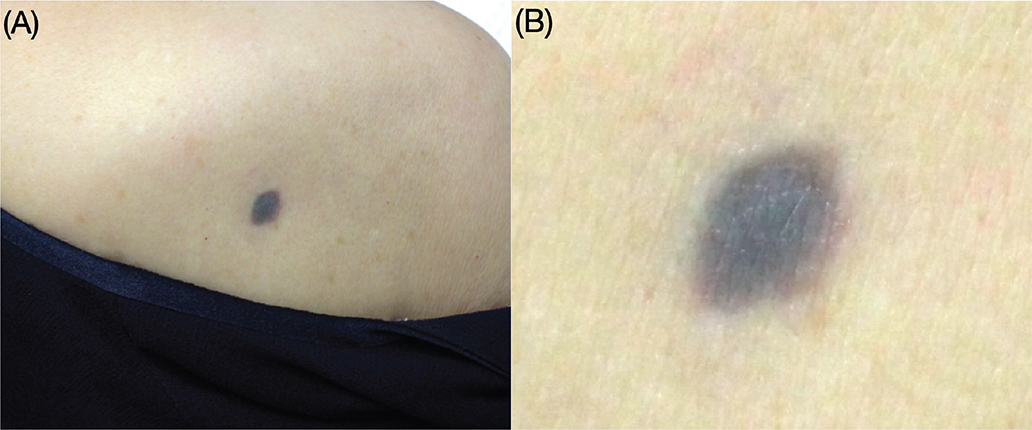
Fig. 1. Skin lesion image. (A) A solitary, 1.0 cm × 0.8 cm asymptomatic blue-grey pigmented patch is present on the right upper back. (B) A magnified view of the pigmented patch shows a well-defined border and a texture similar to the surrounding skin.

Fig. 2. Dermoscopic image. Dermoscopy in polarizing mode shows a structureless area with a blue central region and a bluish-brownish-grey peripheral region. The lesion is covered by a blue-whitish area and lacks a pigment network.
What is your diagnosis?
1: Blue nevus
2: Melanoma
3: Venous haemangioma
4: Pigmented dermatofibrosarcoma protuberans (Bednar tumour)
See next page for answer.
ANSWERS TO QUIZ
An 81-year-old Woman with a 1-year-old Blue Pigmented Patch on the Right Upper Back: A Commentary
Diagnosis: Pigmented dermatofibrosarcoma protuberans (Bednar tumour)
Histopathological examination of the biopsied tissue, stained with haematoxylin and eosin, demonstrated a proliferation of spindle-shaped cells in the dermis and subcutaneous fat (Fig. 3A). A storiform arrangement of these cells was observed in the dermal layers. Fine, scattered melanin granules were also present (Fig. 3B). Immunohistochemical staining revealed that the spindle-shaped cells were positive for CD34 (Fig. 3C). Based on these findings, the patient was diagnosed with pigmented dermatofibrosarcoma protuberans (DFSP), also known as Bednar tumour.
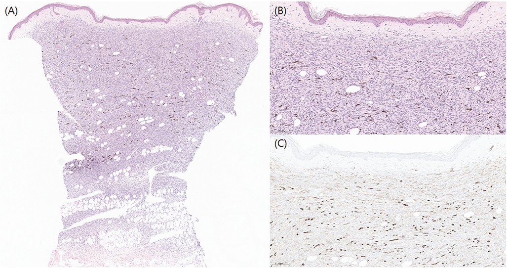
Fig. 3. Histopathological image. (A) Thin epidermis with flattened rete ridges is present above the grenz zone. A proliferation of spindle-shaped cells is observed in the dermis and subcutaneous fat. Pigmented areas are partially observed within the tumour (H&E, 20×). (B) The pigmented areas consist of melanin granules and a storiform arrangement of the cells is observed within the dermal layer, which is consistent with the histological findings of pigmented DFSP (H&E, 150×). (C) Immunohistochemical analysis shows that the spindle cells are positive for CD34 (150×).
When evaluating a patient with a blue pigmented lesion on the skin, it is essential to differentiate between dermal melanocytic and non-melanocytic lesions (1). Dermal melanocytic lesions commonly include blue nevus (or Ito nevus), melanoma, and pigmented basal cell carcinoma, whereas non-melanocytic lesions may include venous haemangioma and post-traumatic haematoma. On dermoscopic evaluation, a blue nevus typically reveals a steel-blue area with ill-defined borders, along with absence of a pigment network and branched streaks (1). Melanoma is characterized by asymmetry, irregular borders, structureless areas, a blue-whitish veil, colour variegation, and an irregular pigment network (2, 3). Pigmented basal cell carcinoma is distinguished by the absence of a pigment network, along with branch-like telangiectasia and tree-like blood vessels (2). Venous haemangioma is marked by bluish to bluish-red lacunae and a structureless area, whereas post-traumatic haematoma presents as homogeneous regions of reddish-black pigmentation, often with red-brown globules at the periphery.
In this case, the patient’s dermoscopic features resembled those of a blue nevus. The lesion was symmetric, lacked branch-like telangiectasia and a pigment network, and displayed a regular border, distinguishing it from those of melanoma and basal cell carcinoma. While previous studies have suggested that the coexistence of a “blue-whitish veil” without a peripheral pigment network and “colour variegation” are characteristic dermoscopic features of pigmented DFSP (4, 5), these features were not prominent in this case. Although the dermoscopic features did not exhibit the typical characteristics of pigmented DFSP, when a pigmented lesion presents with a blue-whitish area on dermoscopy, pigmented DFSP should be considered in the differential diagnosis. In suspicious cases, diagnostic excision is necessary for a definitive diagnosis.
Pigmented DFSP is a rare variant of DFSP, accounting for 1–5% of cases (6). It can develop anywhere on the body, with the trunk being the most commonly affected area, followed by the extremities, head, and neck (6). DFSP most commonly occurs in young adults, typically between 20 and 50 years of age, with peak prevalence occurring in the 30- to 39-year-old age group (6). Although no definitive sex predominance is established, DFSP is found to be slightly more common in males than in females (7). A similar trend has been observed in pigmented DFSP (6). However, the current case is notable because the patient is an 81-year-old woman, which deviates from the typical epidemiological pattern of pigmented DFSP. Although rare, DFSP, including pigmented DFSP, has been reported in an 80-year-old woman with a lesion on the right thumb (8). Therefore, when DFSP is suspected, patient age should be considered only as a reference, not a significant diagnostic criterion.
Surgical excision is the primary treatment for pigmented DFSP (9). A 2- to 3-cm surgical margin is generally recommended for DFSP surgical excision. However, in this case, the lesion was relatively well defined due to its pigmented character, allowing for complete excision with a 1-cm margin. After excision, imaging studies, including MRI and chest-abdominal CT, were performed to check for regional spread to nearby organs, tissues, and lymph nodes, as well as for clinical follow-up. No metastasis was observed.
In conclusion, when an elderly patient presents with a recently developed blue pigmented lesion on the trunk exhibiting dermoscopic features like those of a blue nevus, pigmented DFSP should also be considered in the differential diagnoses. Furthermore, a biopsy is essential to confirm the diagnosis.
The patient’s son provided written consent for the use of the patient’s photographs and medical records.
REFERENCES
- Braun RP, Rabinovitz HS, Oliviero M, Kopf AW, Saurat JH. Dermoscopy of pigmented skin lesions. J Am Acad Dermatol 2005; 52: 109–121. https://doi.org/10.1016/j.jaad.2001.11.001
- Campos-do-Carmo G, Ramos-e-Silva M. Dermoscopy: basic concepts. Int J Dermatol 2008; 47: 712-719. https://doi.org/10.1111/j.1365-4632.2008.03556.x
- Brunsgaard EK, Wu YP, Grossman D. Melanoma in skin of color: Part I. Epidemiology and clinical presentation. J Am Acad Dermatol 2023; 89: 445–456. https://doi.org/10.1016/j.jaad.2022.04.056
- Maeda T, Watabe Y, Yanagi T, Imafuku K, Kitamura S, Hata H, et al. Dermoscopic features of Bednar tumor: report of a case. J Dermatol 2018; 45: e179–e180. https://doi.org/10.1111/1346-8138.14232
- Ehara Y, Yoshida Y, Shiomi T, Yamamoto O. Pigmented dermatofibrosarcoma protuberans and blue naevi with similar dermoscopy: a case report. Acta Derm Venereol 2016; 96: 272–273. https://doi.org/10.2340/00015555-2204
- Liszewski W, Blanchette D, Cunningham AM, Miller DD. Epidemiology of Bednar tumors in the United States. J Am Acad Dermatol 2016; 75: 1064–1066. https://doi.org/10.1016/j.jaad.2016.06.018
- Maghfour J, Genelin X, Olson J, Wang A, Schultz L, Blalock TW. The epidemiology of dermatofibrosarcoma protuberans incidence, metastasis, and death among various population groups: a surveillance, epidemiology, and end results database analysis. J Am Acad Dermatol 2024; 91: 826–833. https://doi.org/10.1016/j.jaad.2024.05.088
- Georgiev GP, Slavchev SA, Ananiev J. A rare case of dermatofibrosarcoma protuberans of the thumb in an 80-year-old woman. Cureus 2018; 10: e2016. https://doi.org/10.7759/cureus.2016
- Chen S, Xiong L, Zhao L, Li Y, Li L. Survival outcomes and prognostic factors of dermatofibrosarcoma protuberans: a population-based retrospective cohort analysis. Dermatol Surg 2023; 49: 825–831. https://doi.org/10.1097/DSS.0000000000003853