QUIZ SECTION
A Solitary Pedunculated Lesion on the Scrotal Skin of a 79-year-old Patient: A Quiz
Anna-Maria FORSTER, Veronika SASSON-SCHMIDT and Beda MUEHLEISEN
Department of Dermatology, University Hospital of Basel, Schoenbeinstrasse 40, CH-4056 Basel, Switzerland
Citation: Acta Derm Venereol 2025; 105: adv42926. DOI: https://doi.org/10.2340/actadv.v105.42926.
Copyright: 2025 © The Author(s). Published by MJS Publishing, on behalf of the Society for Publication of Acta Dermato-Venereologica. This is an Open Access article distributed under the terms of the Creative Commons Attribution-NonCommercial 4.0 International License (https://creativecommons.org/licenses/by-nc/4.0/).
Published: Apr 14, 2025.
A 79-year-old patient presented in our department with an approximately 1 cm x 1.5 cm, polypoid, skin-coloured plaque on the scrotum of unknown duration. There was no obvious enlargement of the lesion throughout the patient’s life. No itching, pain, traumatic events, or local symptoms were reported. The skin overlying the papule was smooth and showed no tenderness on palpation. Clinical history of the patient included latent tuberculosis, seminoma, chronic recurrent urticaria with angioedema, and recurrent polymyalgia rheumatica. The current medications were atorvastatin, oral prednisone, tocilizumab (Actemra®), and an antihistaminic if necessary (Fig. 1).

Fig. 1. Scrotal verrucous papule approximately 1.5 cm x 1.5 cm, with a mild reddish erythema.
What is your diagnosis?
1: Verruciform xanthoma
2: Condyloma acuminatum
3: Verrucous carcinoma
4: Solitary neurofibroma
See next page for answer.
ANSWERS TO QUIZ
A Solitary Pedunculated Lesion on the Scrotal Skin of a 79-year-old Patient: A Commentary
Diagnosis: Verruciform xanthoma
Superficial shave-excision of the scrotal lesion was performed, and histopathologic examination revealed aggregates of lipid-containing foam cells in the dermal stroma of papillary dermis in association with verrucous epidermal hyperplasia without atypia. The dermis contained lymphocytes and plasma cells, and there was prominent neutrophilic exocytosis (Fig. 2). Periodic acid-Schiff (PAS) staining did not show fungal elements. CD68 immunohistochemical staining showed positivity in the foamy macrophages, confirming the diagnosis of verruciform xanthoma. We also performed P16, OCT-2, S100, PU.1, and FXIIIa staining, which showed a nuclear positivity of PU.1 and a slight positivity of FXIIIa while OCT-2 and S100 remained negative. P16 immunostaining, a surrogate marker for human papilloma virus (HPV) infection, was negative (Fig. 3). Due to the benignity of the lesion and as recurrences are rarely reported in the literature no further treatment was initiated. The wound after shave-excision healed within 1 year.

Fig. 2. Verrucous lesion with acanthosis, papillomatosis, and parakeratotic cells in crateriform invagination. (A–B) Elongated rete ridges with broad ends reaching a uniform level and papillary dermis packed with foam cells surrounding dilated capillaries without cytologic atypia in the lesions (haematoxylin and eosin [HE]) (50x magnification, 400x magnification).
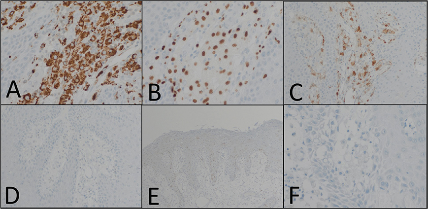
Fig. 3. Foamy macrophages stained strongly positive for CD68 (marker for identifying histiocytes). (A) 200x magnification and PU.1 (nuclear marker for identifying histiocytes and dendritic cells); (B) 400x magnification as well as slightly positive for FXIIIa (marker to identifying a variety of fibrohistiocytic cells); (C) magnification 200x. OCT2 (marker to identifying immature and mature B cells); (D) magnification 200x, P16 (surrogate marker for oncogenic HPV infection); (E) (100x magnification and S100 (marker of Schwann cells and melanocytes); (F) 400x magnification remained negative.
Verruciform xanthoma (VX) is a rare clinicopathologic entity of uncertain aetiology that occurs primarily in the oral mucosa and was first described in 1971 by Shafer. Involvement of an extraoral site was first reported in 1979 by Santa Cruz, with most cases occurring in the anogenital skin (1). Histopathologic features are characterized by epidermal acanthosis with uniform elongation of the rete ridges, papillomatosis, parakeratosis, and filling of the papillary dermis by foamy macrophages (2). The presence of brightly eosinophilic focal dissecting parakeratin is a helpful histologic feature. Considering this, Rush and Bennet coined the term a “wart of fire” (3). Clinically verrucous xanthomas show a verrucous, papillomatous, polypoid, or sessile growth and are often mistaken for viral warts or cutaneous malignancies. Cutaneous verrucous xanthoma should be clinically distinguished from CHILD naevus. The CHILD naevus in the context of inherited X-linked dominant, male-lethal trait often involves the genital mucosa and the inguinal region (4). The predilection of these regions is referred to as ptychotropism (5) and could be excluded in our 79-year-old male patient. Due to the proclivity of verrucous xanthoma for oral and mucosal sites, a viral aetiology, similar to HPV, has been considered but a causal association has not been established (1). Initial damage to keratinocytes by an inciting agent, followed by degeneration and foam cell response, was proposed as a possible pathogenic mechanism in verruciform xanthoma by Mohsin et al. (1). Development of local dystrophic xanthoma can be seen, for instance, at the site of trauma or in other lesions that are common in patients with hyperlipoproteinemia, but can also be seen in normolipidemic patients (6). In our case, immunohistochemically, the biopsy specimen was negative for p16 and an initial trauma or hyperlipoproteinemia could be ruled out. Expression of PU.1 has also been recently described in histiocytoses and in histiocytic lesions. PU.1 is expressed in macrophages, histiocytes, and dendritic cells (7). As a transcription factor that is similarly expressed in multipotent myeloid-lymphoid progenitor cells, PU.1 is required for monocyte as well as B-cell differentiation, and is used as an important marker to distinguish histiocytosis from histiocyte-rich tumours (8). Interestingly PU.1 emerged as a robust marker with sharp nuclear staining in VX histiocytes.
This rare case of cutaneous verrucous xanthoma presenting on the scrotum of a 79-year-old patient highlights the need for integration of clinical data with histopathologic examination to enable a correct diagnosis. We have also reviewed theories behind the aetiology of cutaneous verrucous xanthoma and discuss PU.1 as an interesting immunohistochemical marker that may help to identifying challenging histiocytic entities.
ACKNOWLEDGEMENT
The authors would like to thank the patient for granting permission to publish this information.
Self-assessment credit
This article is eligible for journal-based self-assessment (1 credit) for Maintenance of Certification (MOC) from the American Board of Dermatology (ABD). After completion of an activity, please log on to the ABD website at www.abderm.org to register your credits. This may be done after each exercise or after accumulating many credits.
REFERENCES
- Mohsin S, Lee MW, Amin MB, Stoler, MH, Eyzaguirre, E, Ma CK, et al. Cutaneous verruciform xanthoma: a report of five cases investigating the etiology and nature of xanthomatous cells. Am J Surg Pathol 1998; 22: 479–487. https://doi.org/10.1097/00000478-199804000-00014
- Sinnya S, Wheller L, Carroll M, Williamson R, De’ Ambrosis B. Verruciform xanthoma of the penis: a rare Australian case. Australas J Dermatol 2015; 56: e99–101. https://doi.org/10.1111/ajd.12161
- Roy SF, Prokopetz R, Ayroud Y, Pickett L, Litvinov IV. Wart on fire: a rare entity of verruciform xanthoma arising on a lower leg in a setting of chronic lymphedema. JAAD Case Rep 2017; 3: 36–38. https://doi.org/10.1016/j.jdcr.2016.12.002
- Gantner S, Rütten A, Requena L, Gassenmaier G, Landthaler M, Hafner C. CHILD syndrome with mild skin lesions: histopathologic clues for the diagnosis. J Cutan Pathol. 2014; 41: 787–790. https://doi.org/10.1111/cup.12377
- Happle R. Ptychotropism as a cutaneous feature of the CHILD syndrome. J Am Acad Dermatol 1990; 23: 763–766. https://doi.org/10.1016/0190-9622(90)70285-P
- Rosen T. Dystrophic xanthomatosis in mycosis fungoides. Arch Dermatol 1978; 114: 102–103. https://doi.org/10.1001/archderm.1978.01640130066020
- Kiruthiga KG, Younes S, Natkunam Y. Strong coexpression of transcription factors PU.1 and Oct-2 in Rosai–Dorfman disease. Am J Clin Pathol 2022; 158: 672–677. https://doi.org/10.1093/ajcp/aqac119
- Ungureanu IA, Cohen-Aubart F, Héritier S, Haroche J, Donadieu J, Emile JF. PU.1 is a useful nuclear marker to distinguish between histiocytosis and histiocyte-rich tumours. Histopathology 2023; 83: 320–325. https://doi.org/10.1111/his.14909