QUIZ SECTION
Recurrent Vesiculonecrotic Lesions on the Lower Extremities in a Patient with Dermatomyositis: A Quiz
Zhen-Dong WEI1# , Yi-Ping ZHAO2,3#, Chao YU1, Xin-Yue DING1 and Ao-Xue WANG1*
1Department of Dermatology, The Second Hospital of Dalian Medical University, No.467 Zhongshan Road, Dalian 116027, China, 2Key Laboratory of Immunodermatology, Ministry of Education, Department of Dermatology, The First Affiliated Hospital of China Medical University, Shenyang, China, and 3Institute of School of Basic Medicine, China Medical University, Shenyang, China. *E-mail: wangaxdl@163.com
#These authors contributed equally to this work and should be considered as first authors.
Citation: Acta Derm Venereol 2025; 105: adv42967. DOI: https://doi.org/10.2340/actadv.v105.42967.
Copyright: © 2025 The Author(s). Published by MJS Publishing, on behalf of the Society for Publication of Acta Dermato-Venereologica. This is an Open Access article distributed under the terms of the Creative Commons Attribution-NonCommercial 4.0 International License (https://creativecommons.org/licenses/by-nc/4.0/).
Published: May 5, 2025
Competing interests and funding: The authors have no conflicts of interest to declare.
A 68-year-old male presented with a 2-month history of painful vesicles and necrosis, predominantly on the lower extremities. These lesions subsided naturally within 1 month; however, new rashes reappeared continuously. He had no systemic symptoms, such as fever or fatigue. He had a 6-month history of dermatomyositis (DM) and secondary interstitial pneumonia. Over the past 4 months, his daily prescriptions included prednisone 20 mg, tofacitinib 20 mg, and tacrolimus 2 mg. Physical examination revealed multiple scattered bean-to-nail-sized vesicles and individual pustules surrounded by a purple halo, some with central necrosis and scabs, distributed symmetrically over the lower extremities (Fig. 1). Very few lesions were observed on the trunk and upper extremities. Complete blood cell counts, kidney function, and hepatic function were unremarkable.

Fig. 1. Clinical image. (A) Multiple vesicles and individual pustules distributed on the lower extremities, some with central necrosis. (B) Lesions distributed on the flexor thighs.
What is your diagnosis?
1: Erythema multiforme
2: Cutaneous small vessel vasculitis
3: Bullous impetigo
4: Atypical generalized zoster
See next page for answer.
ANSWERS TO QUIZ
Recurrent Vesiculonecrotic Lesions on the Lower Extremities in a Patient with Dermatomyositis: A Quiz
Diagnosis: Atypical generalized zoster
A skin biopsy revealed intraepidermal vesicles with ballooning degenerated keratinocytes, multinucleated giant cells with steel-grey nuclei, and margination of chromatin. The underlying superficial dermis showed a perivascular and interstitial infiltrate of neutrophils with nuclear dust, fibrinoid necrosis of small vessel walls with thrombi in the lumen, and erythrocyte extravasation (Fig. 2). The blister fluid and tissue blocks were positive for varicella-zoster virus (VZV) by multiplex polymerase chain reaction analysis and pathogenic microorganism DNA high-throughput genetic sequencing, respectively. The patient was diagnosed with atypical generalized zoster associated with cutaneous vasculitis. Tofacitinib and tacrolimus were discontinued, and oral famciclovir was initiated. The patient experienced a rapid regression of the vesicles within 1week, but it took >1 month for the necrosis to heal (Fig. 3).

Fig. 2. Haematoxylin-eosin staining. (A) Original magnification ×40. (B) Original magnification ×200. Histopathology: intraepidermal vesicles with ballooning degenerated keratinocytes, multinucleated giant cells with steel-grey nuclei and margination of chromatin. The underlying superficial dermis showing leukocytoclasis and fibrinoid necrosis of small-sized vessels.
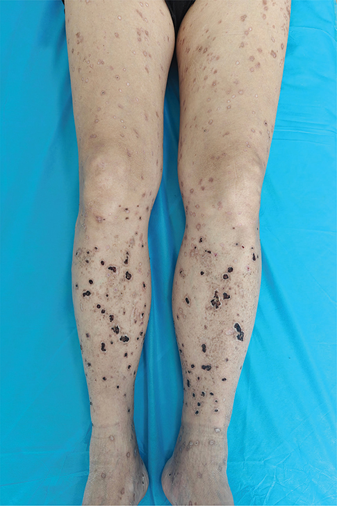
Fig. 3. Follow-up image. Nearly 40 days after the initial treatment, the necrosis has almost healed.
VZV is the aetiological agent of varicella (primary infection) and herpes zoster (HZ, reactivation of a latent infection). The typical clinical presentations of these 2 diseases are distinct and readily recognized by experienced clinicians. However, in the present case, the VZV infection presented with a series of uncommon clinical features, which posed diagnostic challenges: (i) the presence of varicella-like eruptions instead of dermatomal involvement, commonly observed in older adult patients; (ii) dramatic vasculitis-like changes observed clinically and pathologically; (iii) lesions located predominantly on the lower extremities, showing a gravity-dependent distribution; and (iv) a chronic, recurrent course with an atypically prolonged healing period for necrosis post-treatment. Because the patient developed varicella in childhood, the rash that emerged at this time was not considered a primary infection. In 1972, Schimpff et al. (1) first used the term “atypical generalized zoster”, indicating patients with a definite antecedent history of varicella, who developed diffuse lesions resembling disseminated zoster or varicella, but without obvious dermatomal involvement. Unlike the centripetal distribution of varicella, atypical generalized zoster lesions appear primarily in the 4 extremities.
Cutaneous vasculitis is a rare manifestation of VZV infection; however, its underlying mechanism remains unclear. In 2024, Uchiyama et al. (2) reported a vasculitis-like HZ that occurred in a 67-year-old man with ulcerative colitis during the oral administration of tofacitinib. Through RNA in situ hybridization, VZV signals were detected in the epidermis and around the vascular wall of the dermis. It has been hypothesized that VZV can travel to blood vessels either transaxonally or hematogenously, thereby directly causing vasculitis (3). In addition, similar to our case, Álvarez-Salafranca et al. (4) and Zhao et al. (5) have reported 2 other atypical generalized zoster cases with vasculitis-like lesions distributed bilaterally on the lower extremities. Granular IgM and C3 deposits were observed in the superficial vessel walls. These findings indicate that immune complex-mediated damage may be involved in the pathogenesis of vasculitis during VZV infection.
Atypical generalized zoster is most commonly observed in patients who are immunocompromised (1). Patients with DM exhibit significantly lower VZV-specific immunity and an increased incidence of HZ compared with those without DM (6). Janus kinase (JAK) inhibitors are a relatively new class of immunosuppressive drugs that target intracellular signalling pathways involved in the pathogenesis of immune-mediated inflammatory diseases (IMIDs). Tofacitinib, a JAK 1/3 inhibitor, is effective for treating IMIDs, including rheumatoid arthritis, psoriatic arthritis, ulcerative colitis, and DM (7). However, tofacitinib dose-dependently reduces IFN-γ production, proliferation, activation, and CXCR3 expression of VZV-specific CD4+ T cells, thereby increasing the risk of VZV infection (8). Moreover, factors such as a high dose of tofacitinib (20 mg daily), concurrent use of glucocorticoids and immunosuppressants, and advanced age (≥ 65 years) have been shown to increase this risk (9, 10). We hypothesize that these high-risk factors may have worked together to trigger the occurrence of atypical generalized zoster in this case.
The differential diagnosis of atypical generalized zoster should include erythema multiforme (EM), cutaneous small-vessel vasculitis (CSVV), and bullous impetigo (BI). EM typically presents with target lesions, and central bulla and necrosis can be observed in severe cases. Histologically, apoptotic keratinocytes and vacuolar interface dermatitis are key diagnostic indicators. CSVV presents as palpable purpura, haemorrhagic vesicles, and ulcerations that favour the lower extremities. Pathological features of leukocytoclastic vasculitis help confirm the diagnosis. The absence of intraepidermal vesicles and multinucleated giant cells distinguishes CSVV from VZV infection. BI initially presents as small vesicles that rapidly progress to superficial flaccid bullae containing purulent fluid. Subcorneal pustules and gram-positive cocci were observed on histological examination.
In conclusion, diagnosing atypical presentations of VZV infections can be challenging. It is crucial to conduct a thorough assessment of the clinical, pathological, and pathogenic findings. Misdiagnosis may lead to unfavourable prognoses, particularly in diseases that require treatment with glucocorticoids and/or immunosuppressants, which can act as catalysts for VZV infection.
ACKNOWLEDGEMENTS
IRB approval status: Signed informed consent was obtained from the patient for the publishing of his clinical information and images. IRB approval has been obtained from the Ethics Committee of our institution.
REFERENCES
- Schimpff S, Serpick A, Stoler B, Rumack B, Mellin H, Joseph JM, et al. Varicella-zoster infection in patients with cancer. Ann Intern Med 1972; 76: 241–254. https://doi.org/10.7326/0003-4819-76-2-241
- Uchiyama E, Yamaguchi R, Anzawa K, Fujii T, Watanabe D, Shimizu A. Vasculitis-like herpes zoster in the course of treatment with tofacitinib in ulcerative colitis: an assessment of local viral distribution by RNA in situ hybridization. J Dermatol 2024; 51: 1248–1251. https://doi.org/10.1111/1346-8138.17169
- Clark AK, Dhossche J, Korcheva VB, Keller JJ. Herpes zoster presenting as unilateral vasculitis. Dermatol Online J 2018; 24: 10. https://doi.org/10.5070/D32411042007
- Álvarez-Salafranca M, Garces-Horna V, Garcia-Garcia M, Ara-Martin M. Atypical vasculopathic varicella-zoster infection mimicking cutaneous small-vessel vasculitis. Int J Dermatol 2020; 59: e214–e216. https://doi.org/10.1111/ijd.14816
- Zhao R, Nadelman DA, Harms PW, Eshaq M. Disseminated zoster with vasculitis. JAAD Case Rep 2022; 28: 28–30. https://doi.org/10.1016/j.jdcr.2022.08.002
- Lin CF, Hong CT, Lee WH, Wu D, Hu CJ, Chung CC. Disseminated cutaneous herpes zoster and multiple cerebral infarcts in an adult with diabetes mellitus. J Neurovirol 2020; 26: 130–132. https://doi.org/10.1007/s13365-019-00790-7
- Min MS, Alsarheed A, Kassamali B, Mazori DR, Schaefer M, Merola JF, et al. Tofacitinib as treatment for refractory dermatomyositis: a retrospective study from 2 academic medical centers. J Am Acad Dermatol 2022; 86: 423–425. https://doi.org/10.1016/j.jaad.2021.07.003
- Almanzar G, Kienle F, Schmalzing M, Maas A, Tony HP, Prelog M. Tofacitinib modulates the VZV-specific CD4+ T cell immune response in vitro in lymphocytes of patients with rheumatoid arthritis. Rheumatology (Oxford) 2019; 58: 2051–2060. https://doi.org/10.1093/rheumatology/kez175
- Lichtenstein GR, Bressler B, Francisconi C, Vermeire S, Lawendy N, Salese L, et al. Assessment of safety and efficacy of tofacitinib, stratified by age, in patients from the Ulcerative Colitis Clinical Program. Inflamm Bowel Dis 2023; 29: 27–41. https://doi.org/10.1093/ibd/izac084
- Lin QX, Meng HJ, Pang YY, Qu Y. Recurrent herpes zoster in a rheumatoid arthritis patient treated with tofacitinib: a case report and review of the literature. World J Clin Cases 2022; 10: 8703–8708. https://doi.org/10.12998/wjcc.v10.i24.8703