QUIZ SECTION
Multiple Erythematous Nodules on the Trunk: A Quiz
Yoshihito MIMA1,2 and Tsutomu OHTSUKA2
1Department of Dermatology, Tokyo Metropolitan Police Hospital, 4-22-1 Nakano, Nakano-ku, Tokyo, Japan, and 2Department of Dermatology, International University of Health and Welfare Hospital, Tochigi, Japan. E-mail: yoshihito11.mima@gmail.com
Citation: Acta Derm Venereol 2025; 105: adv43091. DOI: https://doi.org/10.2340/actadv.v105.43091.
Copyright: 2025 © The Author(s). Published by MJS Publishing, on behalf of the Society for Publication of Acta Dermato-Venereologica. This is an Open Access article distributed under the terms of the Creative Commons Attribution-NonCommercial 4.0 International License (https://creativecommons.org/licenses/by-nc/4.0/).
Published: Jun 2, 2025.
Competing interests and funding: The authors have no conflicts of interest to declare.
A 75-year-old male patient presented with multiple erythematous skin nodules. The rash began as a solitary abdominal lesion a month before presentation and gradually spread across the trunk, including the back and abdomen. Three years earlier, the patient had been diagnosed with myelodysplastic syndromes (MDS) based on a bone marrow examination after pancytopenia was detected during a routine health checkup. However, as he has mild cytopenia with no systemic symptoms and exhibited a normal karyotype on G-banding analysis, he was classified as having low-risk MDS and remained under observation without any treatment. His medical history included heart failure and diabetes mellitus, both of which he had been managed with stable medication regimens for over 10 years. Physical examination revealed multiple erythematous nodules on the back and abdomen (Fig. 1). The lesions were non-pruritic and non-tender. No mucosal involvement or lymphadenopathy was noted, and no other systemic abnormalities were detected. Laboratory tests revealed anaemia (8.9 g/dL), thrombocytopenia (91 × 10³/μL), and leukopenia (2.7 × 10³/μL); however, no blast cells were detected in the peripheral blood. Inflammatory markers, infection screening, antinuclear antibodies, and eosinophil counts were all within normal limits. Computed tomography revealed no significant abnormalities.

Fig. 1. Clinical photo: multiple erythematous nodules on the trunk.
What is your diagnosis?
1: Leukaemia cutis in myelodysplastic syndromes
2: Sweet syndrome
3: Primary cutaneous lymphoma
4: Lichen planus
See next page for answer.
ANSWERS TO QUIZ
Multiple Erythematous Nodules on the Trunk: A Commentary
Diagnosis: leukaemia cutis in myelodysplastic syndromes
Histopathological examination of the multiple nodules revealed lymphocyte-like cell infiltration in the dermis (Fig. 2). Immunostaining showed positivity for myeloperoxidase (MPO) and CD117, whereas CD3, CD20, CD34, and CD30 were negative (Fig. 3A, B). Bone marrow analysis revealed a hypercellular marrow with a predominance of myeloid precursors and 5% blasts. Ineffective haematopoiesis was observed, consistent with myelodysplastic syndromes (MDS). There was no evidence of transformation to acute leukaemia. Immunohistochemical staining of bone marrow cells showed positivity for MPO and CD117 but negativity for CD3, CD20, and CD34 (Fig. 3C, D). These similar cellular compositions, observed in both bone marrow and skin biopsy specimens through immunohistochemical analysis, confirmed the diagnosis of MDS-associated leukaemia cutis (LC), resulting in the observation without any topical treatment.

Fig. 2. Histopathological findings. Histopathological examination of the multiple nodules reveals significant infiltration of lymphocyte-like cells into the dermis. (A) Haematoxylin and eosin (HE) staining; ×40. (B) HE staining; ×200.
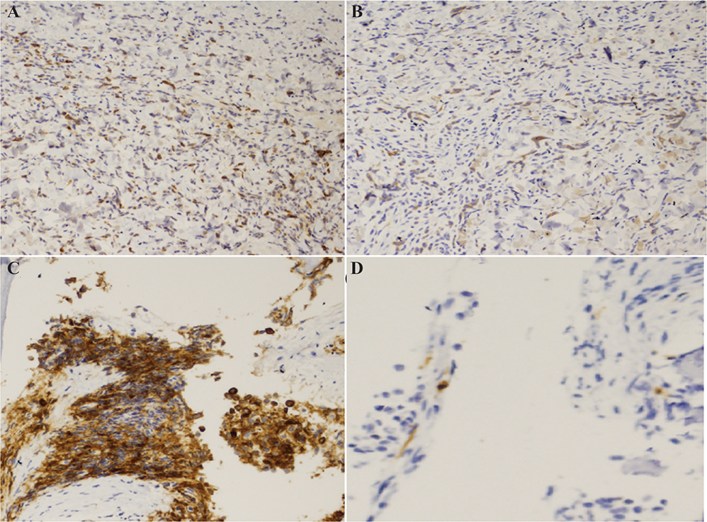
Fig. 3. Histopathological findings. Immunostaining of the multiple nodules shows positivity for (A) myeloperoxidase and (B) CD117 (x100). Immunostaining of the marrow cells was positive for (C) myeloperoxidase (x100) and (D) a little bit positive for CD117 (x400).
MDS was first defined as a distinct disease entity by the French-American-British (FAB) Group in 1982 (1). MDS is a haematologic disorder characterized by cytopenia, particularly anaemia, with typically hypercellular bone marrow (1). Dysplastic haematopoiesis is observed across 1 or more cell lineages (1). MDS can present with various skin manifestations, classified as non-specific or specific lesions (2, 3). Non-specific lesions, including Sweet’s syndrome, erythema nodosum-like eruptions, and pernio-like eruptions, are thought to be induced by excessive inflammatory cytokine production in MDS (2). Conversely, specific lesions, referred to as LC, involve direct infiltration of malignant hematopoietic cells into the skin (3). LC presents with diverse clinical manifestations, including skin-coloured or violaceous papules, plaques, ulcers, and nodules (4). LC is observed in approximately 3% of patients with acute leukaemia, and its incidence is considered to be even lower in chronic leukaemia (5). Although LC is commonly observed in patients with acute leukaemia, its occurrence in MDS is rarely reported. In most reported cases, the transformation of MDS into acute leukaemia coincides with the onset of skin lesions (4–6). Therefore, LC is considered an early indicator of the transformation into acute leukaemia and is associated with a poor prognosis (4–6). Even in the absence of visible skin lesions, leukemic blasts may infiltrate into the skin (7), suggesting that the transformation into acute leukaemia may begin even before the emergence of LC. Thus, when LC develops in patients with MDS, the possibility of the transformation into acute leukaemia must always be considered, necessitating early and proactive management (4–6).
No standardized treatment approach exists for LC in MDS, and a therapeutic consensus has yet to be established (4–6). Given that LC arises as a manifestation of MDS, its treatment focuses primarily on managing the underlying MDS (4–6). Most cases of MDS-associated LC progress to acute leukaemia and are ultimately fatal, with LC lesions persisting without regression. However, Katagiri et al. reported a case of MDS-associated LC in which azacitidine therapy led to the disappearance of bone marrow blasts and the resolution of LC lesions (8). Conversely, Kaiserling et al. reported a case of MDS-related LC that showed regression followed by transformation into acute leukaemia within months, resulting in death within 6 months (9). This suggests that LC regression does not necessarily indicate an improvement in the underlying haematologic disorders and may, in some cases, signal an impending aggressive transformation. In many cases of myeloid neoplasms presenting with cutaneous manifestations, shared clonal mutations have been identified in both skin and bone marrow specimens (10). Therefore, performing mutational analysis, such as next-generation sequencing (NGS), on both bone marrow and skin samples is considered highly valuable for understanding whether the clones at both sites share a common origin. However, in the present case, mutational analysis using a targeted panel with NGS was not performed on either the bone marrow or skin specimens, which represents a limitation in terms of fully investigating and confirming the clonal relationship between the 2 sites.
In our case, the bone marrow blast percentage remained at 5%, and the degree of pancytopenia was consistent with pre-existing levels before the appearance of LC lesions. Therefore, we are currently monitoring the patient closely for potential acute transformation while following a careful disease management strategy without the administration of chemotherapies. Skin manifestations in haematologic disorders may serve as crucial markers of disease progression. Dermatologists must collaborate with haematologists and other specialists to ensure comprehensive patient care. Further research is needed to elucidate the mechanisms underlying the development of MDS-associated LC and establish effective treatment strategies for LC in MDS.
REFERENCES
- Aractingi S, Bachmeyer C, Miclea JM, Vérola O, Rousselot P, Dubertret L, et al. Unusual specific cutaneous lesions in myelodysplastic syndrome. J Am Acad Dermatol 1995; 33: 187–191. https://doi.org/10.1016/0190-9622(95)90232-5
- Namba Y, Koizumi H, Nakamura H, Tarumi T, Sawada K, Ohkawara A. Specific cutaneous lesions of the scalp in myelodysplastic syndrome with deletion of 20q. J Dermatol 1999; 26: 220–224. https://doi.org/10.1111/j.1346-8138.1999.tb03460.x
- Semenic D, Aleksovska N. An atypical chronic ulcer caused by acute myeloid leukemia. Wounds 2019; 31: 25–28.
- Teri AL, Bruce RS. Leukemia cutis: analysis of 50 biopsy-proven cases with an emphasis on occurrence in myelodysplastic syndromes. Am J Clin Pathol 1993; 100: 276–284. https://doi.org/10.1093/ajcp/100.3.276
- Bakst RL, Tallman MS, Douer D, Yahalom. How I treat extramedullary acute myeloid leukemia. Blood 2011; 118: 3785–3793. https://doi.org/10.1182/blood-2011-04-347229
- Findakly D, Amar S. A rare case of leukemia cutis as the first presentation of a myelodysplastic syndrome to acute myeloid leukemia transformation. Cureus 2020; 12: e8698. https://doi.org/10.7759/cureus.8698
- Yavorkovsky LL, Zain J, Wu CD, Trivelli L, Cook P. Monocytic leukemia cutis diagnosed simultaneously with refractory anemia with monocytosis: a case report. Am J Hematol 2001; 66: 120–122. https://doi.org/10.1002/1096-8652(200102)66:2<120::AID-AJH1027>3.0.CO;2-V
- Katagiri T, Ushiki T, Masuko M, Tanaka T, Miyakoshi S, Fuse K, et al. Successful 5-azacytidine treatment of myeloid sarcoma and leukemia cutis associated with myelodysplastic syndrome: a case report and literature review. Medicine 2017; 96: e7975. https://doi.org/10.1097/MD.0000000000007975
- Kaiserling E, Horny HP, Geerts ML, Schmid U. Skin involvement in myelogenous leukemia: morphologic and immunophenotypic heterogeneity of skin infiltrates. Mod Pathol 1994; 7: 771–779.
- Sadigh S, DeAngelo DJ, Garcia JS, Hasserjian RP, Hergott CB, Lane AA, et al. Cutaneous manifestations of myeloid neoplasms exhibit broad and divergent morphologic and immunophenotypic features but share ancestral clonal mutations with bone marrow. Mod Pathol 2024; 37: 100352. https://doi.org/10.1016/j.modpat.2023.100352