QUIZ
Capillaroscopic Changes in a Patient with Undifferentiated Connective Tissue Disease – A Diagnostic Challenge: A Quiz
Angelo NIGRO
Department of Rheumatology of Lucania – UOSD of Rheumatology, “Madonna delle Grazie” Hospital, Matera, Italy. E-mail: angelo.nigro@asmbasilicata.it
Citation: Acta Derm Venereol 2025; 105: adv43121. DOI: https://doi.org/10.2340/actadv.v105.43121.
Copyright: 2025 © The Author(s). Published by Medical Journals Sweden, on behalf of the Society for Publication of Acta Dermato-Venereologica. This is an Open Access article distributed under the terms of the Creative Commons Attribution-NonCommercial 4.0 International License (https://creativecommons.org/licenses/by-nc/4.0/).
Published: June 9, 2025.
A 45-year-old woman with a 3-year history of undifferentiated connective tissue disease (UCTD), characterized by ANA positivity (1:640, speckled), intermittent arthralgias, and episodes of Raynaud’s phenomenon in cold environments, who was on treatment with hydroxychloroquine 200 mg/day and prednisone 5 mg/day, presented with new onset of plantar psoriasis (Fig. 1) and worsening acrocyanosis. She had no history of digital ulcers or systemic sclerosis. Previously, nailfold capillaroscopy revealed a pattern substantially within normal limits, with only isolated apical capillary ectasias (Fig. 2A). At this visit, capillaroscopy revealed a distinct change: more than 30% enlarged capillaries, reduced capillary density, pericapillary oedema, and absence of megacapillaries and microhaemorrhages (Fig. 2B). Physical examination confirmed scaly erythematous plaques on the soles and mild synovitis. Laboratory tests were unchanged. The clinical and capillaroscopic shift raised questions about its underlying cause.
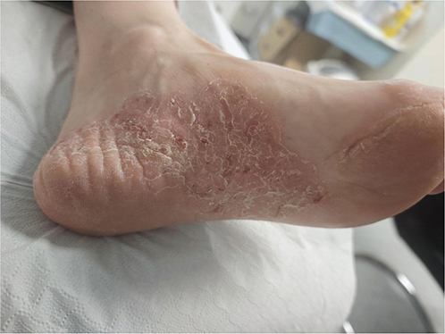
Fig. 1. Image documenting the onset of plantar psoriasis in a patient with undifferentiated connective tissue disease (UCTD). Informed consent was obtained from the patient for the publication of this case report.

Fig. 2. (A) Capillaroscopy image from 2023 shows the presence of capillaries in their normal form (in a patient with undifferentiated connective tissue disease (UCTD) prior to the onset of plantar psoriasis). (B) The capillaroscopic image obtained in November 2024, subsequent to the onset of plantar psoriasis, demonstrates significant morphological alterations. Noteworthy findings include the presence of enlarged capillaries, pericapillary edema, and a reduction in capillary density.
What is your diagnosis:
1: Progression to systemic sclerosis
2: Drug-induced microangiopathy
3: Coexistence of psoriatic microangiopathy with UCTD
4: Non-specific reactive microangiopathy
See next page for answer.
ANSWERS TO QUIZ
Capillaroscopic Changes in a Patient with Undifferentiated Connective Tissue Disease – A Diagnostic Challenge: A Commentary
Diagnosis: Coexistence of psoriatic microangiopathy with UCTD
This case highlights the dynamic nature of nailfold capillaroscopy and its ability to reflect overlapping disease processes. The patient had a stable diagnosis of UCTD (1) with minimal capillaroscopic changes for years. Only after the development of clinically evident plantar psoriasis (2) did her capillaroscopic pattern evolve, with the appearance of multiple enlarged capillaries and reduced capillary density.
Psoriatic arthritis and psoriasis can both induce specific capillaroscopic changes, although these are usually non-specific or mild. Enlarged capillaries have been described in psoriatic patients, especially when inflammatory skin involvement is active (3). However, such changes are generally insufficient to fulfil the criteria for scleroderma-pattern microangiopathy. In this patient, the abrupt appearance of more pronounced capillaroscopic alterations coincided with the development of cutaneous psoriasis, suggesting a contributory effect of psoriatic inflammation on the microvascular bed.
The differential diagnosis includes progression to systemic sclerosis, but the absence of scleroderma symptoms, megacapillaries, microhemorrhages capillary loss, or avascular areas makes this unlikely (4, 5). Drug-induced microangiopathy is also improbable, as hydroxychloroquine, the only immunomodulatory therapy used, rarely causes vascular damage and is typically associated with retinopathy or myopathy rather than nailfold changes. Non-specific reactive microangiopathy might be considered in inflammatory or mechanical conditions, but it typically lacks the reproducible pattern of multiple enlarged capillaries observed here and is often transient or poorly defined.
UCTD is defined by the presence of autoimmune features that do not meet the criteria for a specific connective tissue disease (1). In this patient, the coexistence of UCTD and new-onset psoriasis likely resulted in a composite capillaroscopic picture. Such overlaps are not rare; shared microvascular and inflammatory pathways may modulate the morphology of the nailfold capillaries.
The case underscores how psoriasis, even when arising in a patient with a different autoimmune background, can modify the capillaroscopic profile. Awareness of such overlaps is essential to avoid misclassification or overdiagnosis of systemic sclerosis or other progressive vasculopathies.
REFERENCES
- Mosca M, Tani C, Vagnani S, Carli L, Bombardieri S. The diagnosis and classification of undifferentiated connective tissue diseases. J Autoimmun 2014; 48–49: 50–52. https://doi.org/10.1016/j.jaut.2014.01.019
- Griffiths CEM, Barker JNWN. Pathogenesis and clinical features of psoriasis. Lancet 2007; 370: 263–271. https://doi.org/10.1016/S0140-6736(07)61128-3
- Smith V, Ickinger C, Hysa E, Snow M, Frech T, Sulli A, et al. Nailfold capillaroscopy. Best Pract Res Clin Rheumatol 2023; 37: 101849. https://doi.org/10.1016/j.berh.2023.101849
- Lledó-Ibáñez GM, Sáez Comet L, Freire Dapena M, Mesa Navas M, Martín Cascón M, Guillén Del Castillo A, et al. Capi-score: a quantitative algorithm for identifying disease patterns in nailfold videocapillaroscopy. Rheumatology (Oxford) 2025 Feb 7 [Online ahead of print]
- Lambova SN, Muller-Ladner U. Nailfold capillaroscopy within and beyond the scope of connective tissue diseases. Curr Rheumatol Rev 2018; 14: 12–21. https://doi.org/10.2174/1573397113666170615093600