QUIZ
Extensive Scaly Erythema with Recurring Ulcers and Crusts: A Quiz
Danfeng ZHU1#, Bingjie CHEN2# and Ying ZHOU2* 
1Department of Dermatology, The Fifth People’s Hospital of Hainan Province, China, and 2Institute of Dermatology and Venereal diseases, Department of Dermatology and Venereal diseases, Affiliated Hospital of Guangdong Medical University, No. 57 Renmin South Road, Xiashan District, Zhanjiang, Guangdong Province, 524001 China. *E-mail: zhouy10000@126.com
#These authors contributed equally to this article.
Citation: Acta Derm Venereol 2025; 105: adv43151. DOI: https://doi.org/10.2340/actadv.v105.43151.
Copyright: © 2025 The Author(s). Published by MJS Publishing, on behalf of the Society for Publication of Acta Dermato-Venereologica. This is an Open Access article distributed under the terms of the Creative Commons Attribution-NonCommercial 4.0 International License (https://creativecommons.org/licenses/by-nc/4.0/).
Published: Mar 31, 2025.
Competing interests and funding: The authors have no conflicts of interest to declare.
This project was supported by Hainan Province Clinical Medical Center.
A 29-year-old man presented to the dermatology clinic with a 1-year history of papular abscesses and crusts, alongside a 10-year history of generalized erythema and desquamation. He had previously been diagnosed with “eczema” at an outside hospital and was treated with glucocorticoids and antihistamines. While these treatments provided temporary relief, his symptoms recurred following discontinuation, resulting in a fluctuating disease course. The patient had no history of syphilis or HIV/AIDS, and laboratory evaluations revealed normal liver and kidney function as well as a normal electrocardiogram.
On physical examination, extensive, ill-defined erythema was observed on the patient’s face, neck, and trunk (Fig. 1A). The erythematous regions exhibited scattered atrophic scars, some of which demonstrated heterochromia and significant surface desquamation. Additionally, scattered dark-red papules, dark-brown nodules with areas of surface necrosis, and some black, oyster-like crusts were noted (Fig. 1B). Scattered brown blood blisters were also present on both hands (Fig. 1C). Biopsies were obtained from a blood blister on the right hand and a nodule on the abdomen for histopathological examination and immunohistochemical analysis.
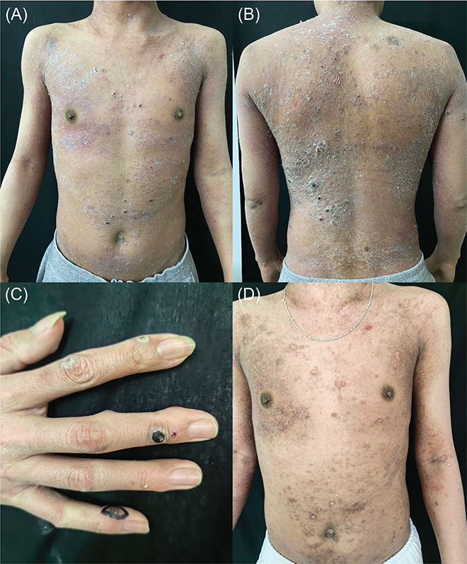
Fig. 1. Clinical image. (A) Clinical image showing extensive, ill-defined erythema on the patient’s face, neck, and trunk. (B) Scattered dark-red papules, dark-brown nodules with areas of surface necrosis, and some black, oyster-like crusts were noted. (C) Scattered brown blood blisters on both hands. (D) The improvement in the patient’s condition after 3 weeks of medication.
What is your diagnosis?
1: Lichenoid pityriasis
2: Primary cutaneous anaplastic large cell lymphoma
3: Lymphoid papulosis type E with mycosis fungoides
4: Extranodal NK/T-cell lymphoma
See next page for answer.
ANSWERS TO QUIZ
Extensive scaly erythema with recurring ulcers and crusts: A Commentary
Diagnosis: Lymphoid papulosis type E with mycosis fungoides
Lymphomatoid papulosis type E (LyPE) is a self-limiting lymphoproliferative disorder characterized by CD30+ and CD8+ lymphoid cells, accompanied by destructive vascular infiltration and necrosis (1). Clinically, LyPE presents with papular nodules that often ulcerate and scar, typically resolving within several weeks. Mycosis fungoides (MF), the most common primary cutaneous T-cell lymphoma, is derived from CD4+ effectors and follows a slow disease course (2). Its clinical manifestation includes pruritic erythematous patches covered by scales, typically distributed in “swimsuit” areas not exposed to sunlight, and often associated with telangiectasia (3). In cases where LyPE and MF co-occur, a combination of clinical benignity and histologic malignancy is observed, while the combination of immunohistochemical (CD30+) T cell cloning helps us differentiate the diagnosis from other dermatitis and lymphoma.
Microscopic examination revealed abnormal keratinization, central necrosis, surface erosion, and dyskeratotic cells. Additional features included epidermal hyperplasia with mild oedema, nuclear hyperchromasia, basal cell degeneration, and dense lymphoid cell infiltration extending into the dermis (Fig. 2A). Pleomorphic T cells exhibit marked aggressiveness and destructive angiotropic growth, leading to vascular occlusion, thrombus formation, haemorrhage, extensive necrosis, and ulceration (Fig. 2B). Pathological sections demonstrated epidermotropism, with atypical lymphocytes infiltrating the epidermis. The infiltrating cells appeared slightly enlarged, showing the characteristic structure of MF – Pautrier microabscesses (Fig. 2C). Immunohistochemistry identified cells positive for CD30 (Fig. 3A), CD3, CD4 (Fig. 3B), CD56, CD7 and CD8 (Fig. 3C), TIA-1, and Ki-67, and negative for EBV-encoded RNA (EBER). T-cell clonality assessment revealed a monoclonal rearrangement.
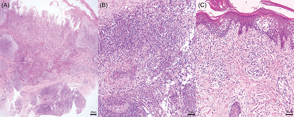
Fig. 2. Histopathological image. (A) Histopathology reveals dyskeratotic cells, epidermal hyperplasia with mild oedema, basal cell degeneration, and dense lymphoid cell infiltration extending into the dermis (H&E 40x). (B) Pleomorphic T-cells exhibit marked aggressiveness and destructive angiotropic growth, leading to vascular occlusion, thrombus formation, haemorrhage, extensive necrosis, and ulceration (H&E 200x). (C) The infiltrating cells appeared slightly enlarged, and Pautrier microabscesses were present (haematoxylin-eosin) (H&E 200x).
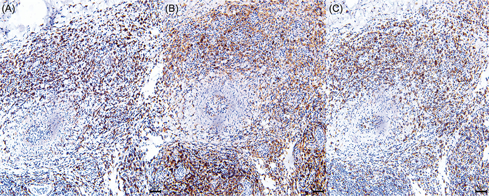
Fig. 3. Results of immunohistochemical analysis. (A) Moderate CD30 positive (original magnification ×200). (B) Moderate CD4 positive (original magnification ×200). (C) Moderate CD8 positive (original magnification ×200).
The differential diagnosis for this presentation includes primary cutaneous anaplastic large-cell lymphoma (PC-ALCL), extranodal NK/T cell lymphoma (ENKTL), and lichenoid pityriasis. Diagnosis relies on a comprehensive assessment of clinical course, histopathology, and immunohistochemical findings. PC-ALCL is a rare subtype of CD30+ T-cell non-Hodgkin lymphoma, manifesting clinically as localized masses on the face, trunk, and limbs, with positive CD30 and negative CD8 expression. Given the overlapping histopathological, phenotypic, and genetic features of PC-ALCL and LyP, careful clinical differentiation is required. PC-ALCL typically presents as ulcerative erythematous nodules, whereas LyP is marked by recurrent episodes of multiple papular nodular lesions that resolve spontaneously within weeks (4, 5). ENKTL is a rare, aggressive subtype of non-Hodgkin’s lymphoma, histologically characterized by vascular destruction, coagulative necrosis, and associated with Epstein–Barr virus infection (6). While LyP also demonstrates vascular destruction, ENKTL’s aggressive clinical course and EBER positivity aid in differentiation. The clinical features of LyPE can resemble those of pityriasis lichenoides, with spontaneous resolution of papules and evidence of lymphocytic vasculitis in the dermis on histopathology. Pityriasis lichenoides is more common in younger patients, has a shorter disease course, and lacks nodular lesions; CD30 is also not expressed histologically. In this case, the distinctive histopathological findings – CD3, CD4, CD8, and CD30 positivity – along with clinical manifestations and T-cell clonality, support a diagnosis of LyPE combined with MF.
LyPE belongs to CD30+ lymphoproliferative diseases; MF does not belong to CD30+ lymphoproliferative diseases, but can express CD30, especially in the case of large-cell transformation. CD30-induced apoptosis may lead to the self-regression seen in LyP lesions (2). Meanwhile, Viral CD30 binds with high affinity to the CD30 ligand and blocks its interaction with CD30. CD30 is co-expressed on CD4+ T cells that express T helper 2 (Th2) but not Th1 cytokines (7), which may explain why it can be seen in the late stages of MF.
LyP and MF share a common monoclonal T-cell receptor gene rearrangement, suggesting a common origin between the clinicopathologies of the 2 diseases (8). That is, the 2 diseases are different clinical manifestations of the same T-cell clone. Based on this, we consider the possibility of clinical disease based on LyPE based on existing MF risk genes.
The current treatment regimen includes methotrexate tablets (15 mg/week), anti-inflammatory immune modulation, and topical application of halometasone cream and allantoin cream. Regular follow-up visits were conducted post-treatment, and the patient’s condition showed signs of improvement (Fig. 1D).
To our knowledge, only 1 prior case of LyPE coexisting with MF has been reported (9). A recent large retrospective study has confirmed a biological link between LyP and MF (10), though type E has been less frequently documented. Our case provides references for future diagnosis and treatment strategies for both diseases.
ACKNOWLEDGEMENTS
IRB approval status: The authors thank the patient for granting permission to publish this information.
REFERENCES
- Kempf W, Kazakov DV, Schärer L, Rütten A, Mentzel T, Paredes BE, et al. Angioinvasive lymphomatoid papulosis: a new variant simulating aggressive lymphomas. Am J Surg Pathol 2013; 37: 1–13. https://doi.org/10.1097/PAS.0b013e3182648596
- Duvic M. CD30+ neoplasms of the skin. Curr Hematol Malig Rep 2011; 6: 245–250. https://doi.org/10.1007/s11899-011-0096-8
- Jawed SI, Myskowski PL, Horwitz S, Moskowitz A, Querfeld C. Primary cutaneous T-cell lymphoma (mycosis fungoides and Sézary syndrome): part I. Diagnosis: clinical and histopathologic features and new molecular and biologic markers. J Am Acad Dermatol Venereol 2014; 70: 205.e1–16; quiz 21–22. https://doi.org/10.1016/j.jaad.2013.07.049
- Atmaja B, Millard T, Wotherspoon A, Attygalle AD, Cunningham D, Sharma B. A rare case of primary cutaneous-anaplastic large cell lymphoma (PC-ALCL) localized to the eyelid. Br J Haematol 2023; 200: 539. https://doi.org/10.1111/bjh.18573
- Yan X, Ren W, Li S, Zhu Z, Gao L, Zhi K. Primary cutaneous anaplastic large-cell lymphoma resembling infratemporal space infection: a case report. BMC Oral Health 2024; 24: 470. https://doi.org/10.1186/s12903-024-04178-w
- Akbar M, Clasen-Linde E, Specht L. Extranodal NK/T-cell lymphoma, nasal type, with extranasal presentation: a case report and a review of the literature. Acta Oncol 2020; 59: 1480–1487. https://doi.org/10.1080/0284186X.2020.1795250
- Del Prete G, De Carli M, Almerigogna F, Daniel CK, D’Elios MM, Zancuoghi G, et al. Preferential expression of CD30 by human CD4+ T cells producing Th2-type cytokines. FASEB J 1995; 9: 81–86. https://doi.org/10.1096/fasebj.9.1.7821763
- de la Garza Bravo MM, Patel KP, Loghavi S, Curry JL, Torres Cabala CA, Cason RC, et al. Shared clonality in distinctive lesions of lymphomatoid papulosis and mycosis fungoides occurring in the same patients suggests a common origin. Human Pathology 2015; 46: 558–569. https://doi.org/10.1016/j.humpath.2014.12.008
- Kiavash K, Abner SM, Malone JC. New variant lymphomatoid papulosis type E preceding and coexisting with mycosis fungoides: a case report and review of the literature. J Cutan Pathol 2015; 42: 1018–1023. https://doi.org/10.1111/cup.12606
- Melchers RC, Willemze R, Bekkenk MW, de Haas ERM, Horvath B, van Rossum MM, et al. Frequency and prognosis of associated malignancies in 504 patients with lymphomatoid papulosis. J Eur Acad Dermatol Venereol 2020; 34: 260–266. https://doi.org/10.1111/jdv.16065