QUIZ SECTION
Congenital Segmental Erosions and Hyperkeratotic Plaques in a Male Infant: A Quiz
Kaan YILMAZ1, Christina EVERS2,3, Oliver BRANDAU2, Marthe-Lisa SCHAARSCHMIDT1 and Cyrill GÉRAUD1,4,5*
1Department of Dermatology, University Medical Center and Medical Faculty Mannheim, Heidelberg University, Mannheim, Germany, 2SYNLAB MVZ Humangenetik Mannheim GmbH, Mannheim, Germany, 3Institute of Human Genetics, Heidelberg University, Heidelberg, Germany, 4Section of Clinical and Molecular Dermatology, Medical Faculty Mannheim, Heidelberg University, Mannheim, Germany, 5European Center for Angioscience, Medical Faculty Mannheim, Heidelberg University, Mannheim, Germany. *Email: cyrill.geraud@umm.de
Citation: Acta Derm Venereol 2025; 105: adv43154. DOI: https://doi.org/10.2340/actadv.v105.43154.
Copyright: © 2025 The Author(s). Published by MJS Publishing, on behalf of the Society for Publication of Acta Dermato-Venereologica. This is an Open Access article distributed under the terms of the Creative Commons Attribution-NonCommercial 4.0 International License (https://creativecommons.org/licenses/by-nc/4.0/).
Published: June 4, 2025
Competing interests and funding: The authors have no conflicts of interest to declare.
A 9-month-old, otherwise healthy male infant presented with disseminated, asymptomatic skin lesions, which were readily visible at birth. Initially, the patient exhibited erythema and erosions, which gradually transitioned into progressive hyperkeratotic plaques with increasing age. Paediatric screening revealed age-appropriate attainment of developmental milestones. Cardiological and orthopaedic examinations were completely unremarkable. There was no history of skin disease in his non-consanguineous parents or his 2 maternal half-sisters. On physical examination, multiple brownish, reticular, hyperkeratotic macules and plaques were observed on the extremities, neck, and trunk. These lesions were distributed partly in narrow bands following Blaschko’s lines and partly in a chequerboard pattern with midline separation (Fig. 1). The palms and soles as well as mucosal surfaces were spared. No abnormalities of hair or teeth were detected. Histopathological assessment of 2 lesional skin biopsies from the knee and abdomen revealed hyperkeratosis, multifocal vacuolar and granular degeneration, as well as cytolysis in the stratum granulosum and spinosum, alternating with unaffected areas (Fig. 2).
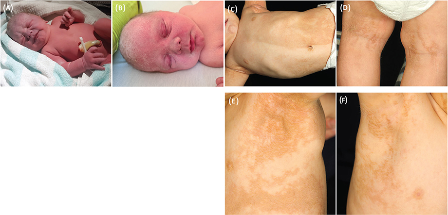
Fig. 1. Clinical manifestations of the patient. (A) Generalized congenital erythroderma observed on the day of birth. (B) Diffuse erythema with fine scaling affecting the scalp and face on postnatal day 2. (C) Multiple brown, partly reticular, hyperkeratotic plaques on the trunk, partly arranged in a chequerboard pattern with midline separation (age 9 months). (D) Reticular, brownish, hyperkeratotic plaques in the popliteal fossa following Blaschko’s lines (age 9 months). (E, F) Blaschkolinear hyperkeratotic macules and plaques on the lateral trunk and the axillary region (at age 18 months).

Fig. 2. Histopathological findings. (A, B) Haematoxylin and eosin stain of a lesional skin biopsy specimen shows hyperkeratosis with multifocal vacuolar and granular degeneration and epidermolysis in the granular and spinous layer, interrupted by uninvolved areas of normal appearing epidermis. Scale bars, 200 μm.
What is your diagnosis?
1: Congenital hemidysplasia with ichthyosiform erythroderma and limb defects (CHILD) syndrome
2: Type 1 segmental Darier disease
3: Epidermolytic ichthyosis
4: McCune–Albright syndrome
See next page for answer.
ANSWERS TO QUIZ
Congenital Segmental Erosions and Hyperkeratotic Plaques in a Male Infant: A Commentary
Diagnosis: Epidermolytic ichthyosis
Due to a high index of suspicion for cutaneous mosaicism, molecular testing was performed using full-thickness skin samples from a lesional (abdominal) and an uninvolved (pectoral) area, as well as EDTA blood. Exome-based next-generation sequencing (NGS) with copy number variation (CNV) analysis revealed a missense pathogenic variant in the keratin 10 gene (KRT10) (c.466C > T, (p.Arg156Cys)) in heterozygous state. Mosaic variants were detected with a variant allele frequency (VAF) of approximately 10% in DNA from lesional skin, at a lower level in blood-derived DNA (approx. 5%) and to an even lesser extent in nonlesional skin (< 5%) (Fig. 3). The combination of clinical signs, histopathological features, and molecular results led to the diagnosis of epidermolytic ichthyosis (EI).

Fig. 3. Analysis for the KRT10 variant by next-generation sequencing (NGS) on full-thickness skin and blood samples. Representative views of DNA read analysis with the respective base-pair exchange from G to A illustrate that the variant is present in 10% of reads in affected skin (left panel). In contrast, the proportion of reads containing the variant is below 5% in unaffected skin (middle panel) and approximately 5% in blood samples (right panel).
EI (MIM 113800), formerly known as epidermolytic hyperkeratosis (EHK) or bullous congenital ichthyosiform erythroderma (Brocq), is a very rare cornification disorder predominantly inherited in an autosomal dominant manner (1). It is caused by mutations in the genes encoding keratin 1 (KRT1) or KRT10 and belongs to the group of keratinopathic ichthyoses (KPI), along with superficial EI and minor KPI variants (1, 2). Notably, de novo mutations of KRT1 or KRT10 have recently been identified in approximately 61% of cases in a large German cohort (3). Epidermolytic nevi, a minor variant of KPI, represent epidermal nevi with histopathological features of EHK and are considered to reflect a postzygotic somatic type 1 mosaicism for KRT1 or KRT10 mutations, which may give rise to generalized EI in an offspring generation (1, 4–6).
A histopathological study conducted by Ross et al. comparing generalized EI, mosaic EI, and superficial EI identified hyperkeratosis, hypergranulosis, and epidermolysis with perinuclear vacuolization as shared features across all 3 entities (7). However, generalized EI was associated with continuous epidermolytic hyperkeratosis along the entire horizontal epidermis and focal parakeratosis, while multifocal involvement with skip areas of normal appearing epidermis was indicative of mosaic EI (7).
Cutaneous manifestations of EI usually follow a chronological sequence, characterized by erythroderma, blisters, and erosions at birth, followed by subsequent resolution and development of hyperkeratosis, especially in areas prone to friction (8). Interestingly, EI patients with KRT10 mutations usually exhibit no involvement of the palms and soles, whereas KRT1 mutations have been associated with epidermolytic palmoplantar keratoderma (1). Based on the modified Ichthyosis Area Severity Index (mIASI) and the Investigator’s Global Assessment (IGA), the disease severity of EI was recently categorized into localized, intermediate, and severe, similar to inherited epidermolysis bullosa (EB) (3).
The therapeutic arsenal for EI includes symptomatic topical treatments to improve hydration (e.g., glycerine, dexpanthenol, urea), antiseptics, balneotherapy, keratolytic agents, and adequate wound management. Systemic treatment with low-dose retinoids may be indicated in patients with marked hyperkeratosis; however, caution is particularly warranted in KRT1-associated EI due to the risk of increased blistering (8). Remarkably, more recent evidence also suggests that treatment with cultured epidermal autografts produced from revertant skin may be a potential therapeutic option in EI and ichthyosis with confetti (9). Characterization of immune fingerprints in ichthyosis, such as Th17/IL-23 polarization, and further advances in gene therapy, as utilized in EB, might allow targeted and/or molecular therapies to emerge as promising frontiers for patients with ichthyosis (8–10).
In conclusion, we present an exceedingly rare case of a molecularly confirmed mosaic EI, highlighting the clinicopathological and genotype–phenotype correlations.
ACKNOWLEDGEMENTS
IRB approval status: Written informed consent was obtained from the parents of the patient to publish his case details and accompanying images.
REFERENCES
- Oji V, Tadini G, Akiyama M, Blanchet Bardon C, Bodemer C, Bourrat E, et al. Revised nomenclature and classification of inherited ichthyoses: results of the First Ichthyosis Consensus Conference in Sorèze 2009. J Am Acad Dermatol 2010; 63: 607–641. https://doi.org/10.1016/j.jaad.2009.11.020
- Oji V, Preil ML, Kleinow B, Wehr G, Fischer J, Hennies HC, et al. S1 guidelines for the diagnosis and treatment of ichthyoses – update. J Dtsch Dermatol Ges 2017; 15: 1053–1065. https://doi.org/10.1111/ddg.13340
- Frommherz L, Giehl K, Hofmann J, Huebner S, Kiekbusch K, Sabkova T, et al. Epidermolytic ichthyosis: clinical spectrum and burden of disease in a large German cohort. J Eur Acad Dermatol Venereol 2024; 10.1111/jdv.20096. https://doi.org/10.1111/jdv.20096
- Paller AS, Syder AJ, Chan YM, Yu QC, Hutton E, Tadini G, et al. Genetic and clinical mosaicism in a type of epidermal nevus. N Engl J Med 1994; 331: 1408–1415. https://doi.org/10.1056/NEJM199411243312103
- Nazzaro V, Ermacora E, Santucci B, Caputo R. Epidermolytic hyperkeratosis: generalized form in children from parents with systematized linear form. Br J Dermatol 1990; 122: 417–422. https://doi.org/10.1111/j.1365-2133.1990.tb08292.x
- Tsubota A, Akiyama M, Sakai K, Goto M, Nomura Y, Ando S, et al. Keratin 1 gene mutation detected in epidermal nevus with epidermolytic hyperkeratosis. J Invest Dermatol 2007; 127: 1371–1374. https://doi.org/10.1038/sj.jid.5700712
- Ross R, DiGiovanna JJ, Capaldi L, Argenyi Z, Fleckman P, Robinson-Bostom L. Histopathologic characterization of epidermolytic hyperkeratosis: a systematic review of histology from the National Registry for Ichthyosis and Related Skin Disorders. J Am Acad Dermatol 2008; 59: 86–90. https://doi.org/10.1016/j.jaad.2008.02.031
- Süßmuth K, Traupe H, Metze D, Oji V. Ichthyoses in everyday practice: management of a rare group of diseases. J Dtsch Dermatol Ges 2020; 18: 225–243. https://doi.org/10.1111/ddg.14049
- Tanahashi K, Kono M, Yoshikawa T, Suzuki Y, Inoie M, Kuwatsuka Y, et al. Treating epidermolytic ichthyosis and ichthyosis with confetti with epidermal autografts cultured from revertant skin. Br J Dermatol 2024; 191: 397–404. https://doi.org/10.1093/bjd/ljae193
- Paller AS, Renert-Yuval Y, Suprun M, Esaki H, Oliva M, Huynh TN, et al. An IL-17-dominant immune profile is shared across the major orphan forms of ichthyosis. J Allergy Clin Immunol 2017; 139: 152–165. https://doi.org/10.1016/j.jaci.2016.07.019