QUIZ SECTION
Multiple Erythematous Oedematous Plaques on the Neck of a 68-year-old Man: A Quiz
Phanitanun PHUSANAWIWAT1, Patcharawadee THONGKUMKOON2, Siri CHIEWCHANVIT1, Chatree CHAI-ADISAKSOPHA3* and Mati CHUAMANOCHAN1*
1Division of Dermatology, Department of Internal Medicine, Faculty of Medicine, Chiang Mai University, Chiang Mai, 2Center of Multidisciplinary Technology for Advanced Medicine (CMUTEAM), Faculty of Medicine, Chiang Mai University, Chiang Mai, and 3Division of Hematology, Department of Internal Medicine, Faculty of Medicine, Chiang Mai University, Chiang Mai, Thailand. E-mails: chatree.chai@cmu.ac.th and mati.c@cmu.ac.th
Citation: Acta Derm Venereol 2025; 105: adv43241. DOI: https://doi.org/10.2340/actadv.v105.43241.
Copyright: © 2025 The Author(s). Published by MJS Publishing, on behalf of the Society for Publication of Acta Dermato-Venereologica. This is an Open Access article distributed under the terms of the Creative Commons Attribution-NonCommercial 4.0 International License (https://creativecommons.org/licenses/by-nc/4.0/).
Published: July 10, 2025
Competing interests and funding: This work was supported by Faculty of Medicine Research Fund, grant No. MED-2567-0052.
A 68-year-old male patient presented to the dermatology clinic with multiple tender skin lesions on his neck, shoulders, and upper extremities that had persisted for 1 month. His medical history included relapsing polychondritis. Upon examination, the patient was found to be afebrile, although anaemia was noted. Dermatological examination revealed multiple erythematous oedematous plaques on the neck (Fig. 1A), as well as on both arms and forearms (Fig. 1B). Skin biopsy demonstrated papillary dermal oedema and dense neutrophilic infiltration with marked leukocytoclasia in the upper dermis (Fig. 1C). Laboratory findings indicated a haemoglobin of 6.0 g/dL, white blood cell count of 5.2 x 10⁹/L (neutrophils 90%, lymphocytes 2%, monocytes 8%), and platelet count of 8,000 × 109/L. Bone marrow aspiration revealed age-related hypercellularity with trilineage dysplastic features in the erythroid, myeloid, and megakaryocytic series along with vacuolisation (Fig. 1D). Further laboratory analyses revealed an elevated C-reactive protein level of 172 mg/L (normal reference range: 0–5 mg/L) and an erythrocyte sedimentation rate of 88 mm/h (normal reference range: 0–10 mm/h). A positive antinuclear antibody test, characterised by a centromere pattern at a titre of 1:1,280, was observed. In contrast, the tests for anti-SSA/Ro and anti-La/SSB autoantibodies were negative.
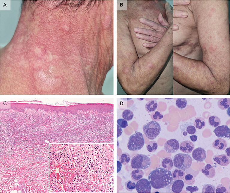
Fig. 1. Clinical images. (A) multiple erythematous oedematous plaques on the neck and (B) both arms and forearms. (C) Histopathology of skin biopsy shows papillary dermal oedema and dense neutrophilic infiltration with marked leukocytoclasia in the upper dermis. (D) Bone marrow reveals age-related hypercellularity with trilineage dysplastic features in the erythroid, myeloid, and megakaryocytic series, along with vacuolization.
What is your diagnosis?
1: VEXAS syndrome
2: Systemic vasculitis
3: Systemic lupus erythematosus
4: Mixed connective tissue disease
See next page for answer.
ANSWERS TO QUIZ
Multiple Erythematous Oedematous Plaques on the Neck of a 68-year-old Man: A Commentary
Diagnosis: VEXAS syndrome
In the present case, an elderly male patient with a history of relapsing polychondritis presented with skin lesions consistent with Sweet syndrome, a diagnosis confirmed by skin biopsy. Bone marrow analysis revealed dysplastic features, including the presence of multiple vacuolations. Taken together, these clinical findings strongly suggested VEXAS syndrome. The diagnosis of VEXAS syndrome was further substantiated by the identification of a UBA1 gene mutation (c.121 A > C; p.Met41Leu) through next-generation sequencing (NGS) in peripheral blood mononuclear cells. The skin lesions were partially controlled using multiple immunomodulatory drugs, including systemic corticosteroids, colchicine, acitretin, dapsone, and mycophenolate mofetil. Despite intensive treatment, the patient succumbed to septicaemia and respiratory failure.
VEXAS syndrome (Vacuoles, E1 enzyme, X-linked, Autoinflammatory, Somatic) is a newly recognized adult-onset autoinflammatory disease caused by somatic mutations in the ubiquitin-like modifier-activating enzyme 1 (UBA1) gene. UBA1 encodes a ubiquitin-activating enzyme (E1), which plays a pivotal role in the ubiquitination pathway and is essential for cellular protein regulation. Pathogenic missense mutations at codon 41 of UBA1 result in diminished ubiquitination, leading to the accumulation of misfolded proteins and aberrant activation of the innate immune system. This dysregulation contributes to the inflammatory processes observed in VEXAS syndrome (1, 2).
The clinical course of VEXAS syndrome is severe, progressive, and predominantly affects individuals later in life. The median age at diagnosis, typically ranging from 65 to 70 years, is a key characteristic that differentiates VEXAS syndrome from other autoinflammatory and autoimmune disorders, which generally present at a younger age. The syndrome involves multiple organ systems and is frequently characterized by recurrent episodes of non-infectious fever accompanied by systemic constitutional symptoms. These are the most frequent manifestations, followed by dermatological and haematological abnormalities (3, 4).
The most prominent cutaneous features are Sweet syndrome-like lesions, characterised by inflammatory, erythematous, and firm oedematous nodules that progressively involve the trunk, back, neck, and limbs (2, 4). These lesions commonly appear red or violaceous, are non-pruritic yet notably tender, and frequently resolve with post-inflammatory desquamation (4). In addition to these features, vasculitis and/or vasculopathy, particularly small-vessel vasculitis, are frequently reported in affected individuals. Other cutaneous findings include septal panniculitis or erythema nodosum, livedo racemosa, lupus tumidus-like eruptions, and polyarteritis nodosa (3, 5). Notably, localised erythematous plaques have been observed at injection sites in patients with VEXAS syndrome undergoing treatment with anakinra, an interleukin-1 receptor antagonist (6).
Histopathologically, skin biopsies typically reveal features of neutrophilic dermatosis characterized by infiltrates composed of mature neutrophils with prominent leukocytoclasia. Perivascular neutrophilic infiltration with interstitial oedema is a hallmark finding, often involving the entire dermis and occasionally extending into the subcutaneous tissue (4).
Haematological abnormalities are a common feature of VEXAS syndrome. Patients typically present with macrocytic anaemia despite normal folic acid and vitamin B12 levels, in addition to thrombocytopenia and lymphopenia. Furthermore, VEXAS syndrome is frequently associated with concurrent haematological conditions, including myelodysplastic syndrome (MDS), multiple myeloma, and monoclonal gammopathy of undetermined significance (3, 7).
In typical cases, the bone marrow morphology exhibits cytoplasmic vacuoles within myeloid and erythroid precursor cells. However, these findings are neither specific nor pathognomonic for VEXAS syndrome, as they can also be observed in a variety of other conditions, including alcoholism, MDS, zinc toxicity, and copper deficiency (3, 7). Other characteristic bone marrow findings in patients with VEXAS syndrome include hypercellularity with granulocytic hyperplasia, typically in the absence of overt dysplasia (7).
Less commonly affected systems in VEXAS syndrome include the cartilage, with manifestations such as nasal or auricular chondritis, and the musculoskeletal system, which may present with arthralgia or arthritis. Pulmonary involvement can present as infiltrates or pleural effusion, whereas ocular involvement may include uveitis, scleritis, or episcleritis. Lymphadenopathy is also observed in some cases. Additionally, cardiovascular manifestations such as pericarditis and myocarditis as well as neurological features such as neuropathy may be present (3).
Enzyme-linked immunosorbent assay has demonstrated elevated levels of inflammatory markers in patients with VEXAS syndrome, including C-reactive protein, interleukin-6, interleukin-8, interferon-inducible protein 10, interferon-γ, and tumour necrosis factor, among others. Additionally, increased levels of antinuclear antibodies, rheumatoid factor, and antiphospholipid antibodies were observed in these patients (1).
For the identification of pathogenic gene mutations, NGS enables precise molecular characterization across a range of tissue types, including bone marrow and cutaneous biopsy specimens – particularly those demonstrating features of neutrophilic dermatoses (8). This approach is particularly valuable when hematologic tissue is not readily available or when cutaneous manifestations are prominent and biopsied early in the disease course. Beyond confirming mutations in the UBA1 gene, whole-exome or whole-genome sequencing via NGS allows for the concurrent detection of additional autoinflammatory or haematological variants. Such comprehensive genomic profiling enhances diagnostic accuracy and supports a more nuanced differential diagnosis in patients with complex multisystem disease.
Effective treatments for VEXAS syndrome remain elusive. This syndrome primarily occurs in patients with persistent autoinflammatory symptoms who fail to respond to conventional therapies. Management strategies primarily focus on addressing UBA1 mutations and regulating the inflammatory response (9).
High-dose systemic corticosteroids are considered the most effective anti-inflammatory therapy for patients with VEXAS syndrome owing to their broad-spectrum suppression of the inflammatory response. Nevertheless, symptom recurrence following steroid tapering is nearly universally observed. Prolonged use of high-dose corticosteroids is associated with significant adverse effects, particularly infectious and cardiovascular complications, which pose substantial risks and may contribute to morbidity and mortality in these patients (2, 3, 9).
Disease-modifying antirheumatic drugs, including methotrexate, mycophenolate mofetil, azathioprine, hydroxychloroquine, cyclophosphamide, dapsone, cyclosporine, and sulfasalazine, have demonstrated limited efficacy in managing VEXAS syndrome. Additionally, there are reports of biologic and targeted therapies being utilized, including anti-TNF agents (adalimumab, infliximab, etanercept, golimumab), anti-IL-1 therapies (anakinra, canakinumab), anti-Th17 agents (secukinumab, ustekinumab), JAK inhibitors (tofacitinib, baricitinib), as well as tocilizumab, rituximab, and abatacept (3, 9). However, the overall effectiveness of these treatments remains variable, and further clinical trials are necessary to establish their role in managing this challenging condition.
In terms of prognosis, the VEXAS mutation with the lowest UBA1b production (c.121A > G, Met41Val) was associated with the poorest outcomes among the 3 mutations studied, suggesting a potential link between UBA1b production and disease severity. Conversely, the UBA1 p.Met41Leu mutation is associated with a more favourable prognosis (7). VEXAS syndrome exhibits significant variability in its clinical presentation and is associated with a high mortality rate, ranging from 20% to 50% in various studies. Factors contributing to increased mortality include gastrointestinal and pulmonary involvement, mediastinal lymphadenopathy, and transfusion dependence. Poor prognosis is frequently attributed to delayed diagnosis, progression of haematological disorders, and lack of effective treatment options (2).
REFERENCES
- Beck DB, Ferrada MA, Sikora KA, Ombrello AK, Collins JC, Pei W, et al. Somatic mutations in UBA1 and severe adult-onset autoinflammatory disease. N Engl J Med 2020; 383: 2628–2638. https://doi.org/10.1056/NEJMoa2026834
- Zhang Y, Dong X, Wang H. VEXAS syndrome. Glob Med Genet 2023; 10: 133–143. https://doi.org/10.1055/s-0043-1770958
- Nicholson LT, Cowen EW, Beck D, Ferrada M, Madigan LM. VEXAS syndrome—diagnostic clues for the dermatologist and gaps in our current understanding: a narrative review. JID Innov 2024; 4: 100242. https://doi.org/10.1016/j.xjidi.2023.100242
- Saad AJ, Patil MK, Cruz N, Lam CS, O’Brien C, Nambudiri VE. VEXAS syndrome: a review of cutaneous findings and treatments in an emerging autoinflammatory disease. Exp Dermatol 2024; 33: e15050. https://doi.org/10.1111/exd.15050
- Hines AS, Mohandesi NA, Lehman JS, Koster MJ, Cantwell HM, Alavi A, et al. Cutaneous involvement in VEXAS syndrome: clinical and histopathologic findings. Int J Dermatol 2023; 62: 938–945. https://doi.org/10.1111/ijd.16635
- Georgin-Lavialle S, Terrier B, Guedon A, Heiblig M, Comont T, Lazaro E, et al. Further characterization of clinical and laboratory features in VEXAS syndrome: large-scale analysis of a multicentre case series of 116 French patients. Br J Dermatol 2022; 186: 564–574. https://doi.org/10.1111/bjd.20805
- Al-Hakim A, Savic S. An update on VEXAS syndrome. Expert Rev Clin Immunol 2023; 19: 203–215. https://doi.org/10.1080/1744666X.2023.2157262
- Zakine E, Schell B, Battistella M, Vignon-Pennamen MD, Chasset F, Mahévas T, et al. UBA1 variations in neutrophilic dermatosis skin lesions of patients with VEXAS syndrome. JAMA Dermatol 2021; 157: 1349–1354. https://doi.org/10.1001/jamadermatol.2021.3344
- Heiblig M, Patel BA, Groarke EM, Bourbon E, Sujobert P, editors. Toward a pathophysiology-inspired treatment of VEXAS syndrome. Semin Hematol 2021; 58: 200–206. https://doi.org/10.1053/j.seminhematol.2021.09.001