QUIZ SECTION
Recurrent Red Papules in a Satellite Pattern Following Laser Treatment: A Quiz
Xilan CHEN1, Jia CHEN2, Bingjie ZHENG1 and Xiao LIN1*
1Department of Dermatology, Shanghai Children’s Hospital, School of Medicine, Shanghai Jiao Tong University, Shanghai, and 2Department of Dermatopathology, Shanghai Skin Disease Hospital, Tongji University School of Medicine, Shanghai, China. *E-mail: linx@shchildren.com.cn
Citation: Acta Derm Venereol 2025; 105: adv43255. DOI: https://doi.org/10.2340/actadv.v105.43255.
Copyright: © 2025 The Author(s). Published by MJS Publishing, on behalf of the Society for Publication of Acta Dermato-Venereologica. This is an Open Access article distributed under the terms of the Creative Commons Attribution-NonCommercial 4.0 International License (https://creativecommons.org/licenses/by-nc/4.0/).
Published: Aug 3, 2025
A healthy 10-year-old boy presented to the dermatology clinic with a 1-month history of a solitary red nodule on his right infrascapular region. It was asymptomatic but bled easily. He denied any history of trauma. The papillary nodule measured 7 mm in diameter (Fig. 1). A diagnosis of pyogenic granuloma was made, and it was treated with Nd: YAG laser. Two weeks later, the nodule sloughed, leaving hyperpigmentation behind. However, two months later, numerous bright red papules appeared around the scar. These papules ranged from 0.5 to 2 mm in diameter and were arranged in a satellite pattern (Fig. 2). Hospital level project of Shanghai Children’s Hospital (2020YLYM01).
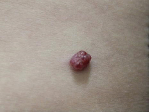
Fig. 1. Primary solitary red papillary nodule with a diameter of 7 mm.

Fig. 2. Recurrent bright red papules in a satellite pattern.
What is your diagnosis?
1: Recurrent pyogenic granuloma
2: Cherry angioma
3: Angiokeratoma
4: Folliculitis
See next page for answer.
ANSWERS TO QUIZ
Recurrent Red Papules in a Satellite Pattern Following Laser Treatment: A Commentary
Diagnosis: Recurrent pyogenic granuloma
A skin biopsy was performed. The histopathological features revealed a benign lobular capillary proliferation in the dermal papillae, consistent with lobular capillary pyogenic granuloma (Fig. 3). The boy’s lesions did not show any spontaneous regression after 2 months of observation. Consequently, the entire patch was excised due to frequent bleeding. At the 1-year follow-up visit, no recurrence was noted.
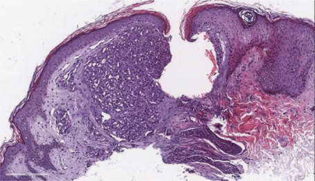
Fig. 3. A benign lobular capillary proliferation in the dermal papillae (haematoxylin and eosin, ×400).
Pyogenic granuloma (PG) is a common benign vascular proliferation that typically presents as a single lesion. It is a rapidly growing, red, and fragile nodule that may bleed easily. It can occur anywhere on the skin or mucosa, but most commonly on the head and neck, and may present at any age (1).
The recurrence of multiple satellite lesions is a rare complication following treatment for the primary lesion. It occurs most often on the back, particularly over the scapular regions of young individuals under the age of 25 years (2, 3). Most satellite lesions appear between 1 week and 4 months after the treatment for the primary lesion (2, 3). The satellite lesions tend to be smooth, bright red, sessile papules measuring 1 to 10 mm in diameter, located around the primary site (2, 3).
The histopathological findings of the recurrent lesions are consistent with pyogenic granuloma, except that some of the small recurrent papules closely resemble simple capillary haemangiomas (4).
The pathogenesis of satellite metastasis is not well known. The study by Groesser et al. revealed BRAF and RAS mutations as major driver mutations in the pathogenesis of PG (5). Whether these findings are relevant to the pathogenesis of satellitosis requires further investigation. Most authors hold the view that after treatment or irritation of the primary lesion, angiogenic factors promote the angiogenesis during the natural repair process. This complication has been reported in cases after surgical excision, electrodesiccation, curettage, and carbon dioxide laser excision (2–4). Therefore, the risk of recurrent lesions seems unrelated to the treatment methods used for the primary lesion. It has also been observed after scratching of the primary lesion (4).
Most untreated recurrent lesions are reported to resolve spontaneously within 2 to 12 months (3), making conservative treatment a suitable option. Depending on the location, size, and patient’s preference, recurrent lesions can also be treated with the same methods as the solitary pyogenic granuloma. Dermatologists should be aware of this complication, inform patients in advance, and select the appropriate treatment.
REFERENCES
- Giblin AV, Clover AJ, Athanassopoulos A, Budny PG. Pyogenic granuloma – the quest for optimum treatment: audit of treatment of 408 cases. J Plast Reconstr Aesthet Surg 2007; 60: 1030–1035. https://doi.org/10.1016/j.bjps.2006.10.018
- Allen RK, Rodman OG. Pyogenic granuloma recurrent with satellite lesions. J Dermatol Surg Oncol 1979; 5: 490–493. https://doi.org/10.1111/j.1524-4725.1979.tb00702.x
- Pitarch G, Pérez-Ferriols A, Millán F. Granuloma piogénico recidivante [Recurrent pyogenic granuloma]. Actas Dermosifiliogr 2012; 103: 536–539. https://doi.org/10.1016/j.ad.2011.04.017
- Zaynoun ST, Juljulian HH, Kurban AK. Pyogenic granuloma with multiple satellites. Arch Dermatol 1974; 109: 689–691. https://doi.org/10.1001/archderm.1974.01630050035007
- Groesser L, Peterhof E, Evert M, Landthaler M, Berneburg M, Hafner C. BRAF and RAS mutations in sporadic and secondary pyogenic granuloma. J Invest Dermatol 2016; 136: 481–486. https://doi.org/10.1038/JID.2015.376