QUIZ SECTION
A 37-year-old Woman with a Solitary Flaccid Bulla on the Left Wrist: A Quiz
Zhe WU1-3#, Chenglong WU1-3#, Kaili ZHOU1-3# and Zhirong YAO1-3*
1Dermatology Center, Xinhua Hospital, Shanghai Jiaotong University School of Medicine, 1665 Kongjiang Road, Yangpu District, Shanghai, 2Department of Dermatology, Xinhua Hospital, Shanghai Jiaotong University School of Medicine, and 3Institute of Dermatology, Shanghai Jiaotong University School of Medicine, Shanghai, China. *E-mail: yaozhirong@xinhuamed.com.cn
#These authors contributed equally to this work.
Citation: Acta Derm Venereol 2025; 105: adv43323. DOI: https://doi.org/10.2340/actadv.v105.43323.
Copyright: © 2025 The Author(s). Published by MJS Publishing, on behalf of the Society for Publication of Acta Dermato-Venereologica. This is an Open Access article distributed under the terms of the Creative Commons Attribution-NonCommercial 4.0 International License (https://creativecommons.org/licenses/by-nc/4.0/).
Published: Mar 26, 2025.
A 37-year-old woman presented with a solitary, asymptomatic, translucent bulla on her left wrist that had developed insidiously over a period of 3 months without any identifiable triggers. Despite the patient’s self-administration of topical antibiotics and povidone-iodine, the lesion persisted and gradually enlarged.
Physical examination revealed a 2-cm dark red, flaccid bulla with intact surrounding skin and a negative Nikolsky sign (Fig. 1). No evidence of mucocutaneous lesions, a history of trauma, or systemic symptoms was observed.
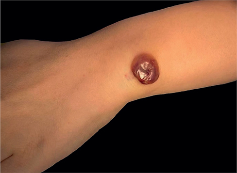
Fig. 1. Clinical findings. A solitary, 2-cm, dark red, flaccid bulla is observed on the left wrist. The Nikolsky sign was negative.
What is your diagnosis?
1: Bullous dermatofibroma
2: bullous pemphigoid
3: acquired epidermolysis bullosa
4: lymphangioma circumscriptum
See next page for answer.
ANSWERS TO QUIZ
A 37-year-old Woman with a Solitary Flaccid Bulla on the Left Wrist: A Commentary
Diagnosis: Bullous dermatofibroma
Histopathological examination revealed a subepidermal bulla lacking inflammatory infiltrates, accompanied by diffuse proliferation of bland dermal spindle cells and marked dilation of superficial lymphatic and blood vessels (Fig. 2). Immunohistochemical analysis demonstrated a low proliferation index (Ki67: 5% positive) with negative staining for p53, AE1/3 (epithelial markers), SMA, DES (muscle markers), SOX10 (neural crest marker), CD34 (vascular marker), and STAT6 (a marker for dermatofibrosarcoma protuberans). Indirect immunofluorescence studies were negative for autoantibodies, thereby effectively excluding autoimmune blistering disorders, including bullous pemphigoid. The absence of anchoring fibril abnormalities and type VII collagen autoantibodies further ruled out acquired epidermolysis bullosa.

Fig. 2. Histopathologic evaluation of the lesion. The specimen reveals a subepidermal bulla devoid of inflammatory infiltrates, associated with diffuse proliferation of dermal spindle cells and marked dilatation of superficial lymphatic and blood vessels. Original magnifications: ×10, with an inset (blacked-boxed area) at ×50.
The diagnosis of bullous dermatofibroma is established through the integration of clinical and histopathological findings. Unlike classic dermatofibromas, which typically present as firm, hyperpigmented papules, this variant is characterized by a cystic morphology resulting from the mechanical compression of dermal lymphatics by proliferating fibroblasts. This mechanism is analogous to that observed in bullous pilomatricoma, in which lymphatic obstruction results in fluid accumulation (1, 2). Moreover, the bland spindle cell proliferation, absence of cytologic atypia, and lack of immunoreactivity for markers associated with aggressive neoplasms (such as CD34 in dermatofibrosarcoma protuberans) distinguish this entity from malignant spindle cell tumours. Furthermore, lymphangioma circumscriptum, another differential consideration, typically exhibits dilated lymphatic channels within the papillary dermis without accompanying spindle cell proliferation.
Complete surgical excision was performed, resulting in both diagnostic confirmation and therapeutic resolution. No recurrence was observed during a 6-month follow-up period. Bullous dermatofibroma represents a rare yet clinically significant variant, characterized by an excellent prognosis and the absence of malignant potential. Recognizing its distinct clinicopathological profile is essential to prevent misdiagnosis as autoimmune bullous diseases or malignancies, thereby avoiding unnecessary systemic therapies.
REFERENCES
- Chen S, Wu F, Qian Y, Zhu L, Tu Y, Huang C. Pilomatricoma with bullous appearance: a case report and review of literature. Int J Dermatol 2011; 50: 615–618. https://doi.org/10.1111/j.1365-4632.2010.04586.x
- Cao H, Chen J, Yan J, Wang J. Pilomatricoma with a bullous appearance. Indian J Dermatol Venereol Leprol 2015; 81: 77–78. https://doi.org/10.4103/0378-6323.148588