QUIZ SECTION
An Ulcerated Nodule on the Toe in an Elderly Patient: A Quiz
Ziqi LIU#, Qiaoan ZHANG#, Lianjun CHEN and Qiong HUANG*
Department of Dermatology, Huashan Hospital, Fudan University, No.12 Wulumuqi Road, Jinan District, Shanghai, China. *E-mail: blueqiong1972@163.com
#These authors contributed equally to this work.
Citation: Acta Derm Venereol 2025; 105: adv43431. DOI: https://doi.org/10.2340/actadv.v105.43431.
Copyright: 2025 © The Author(s). Published by MJS Publishing, on behalf of the Society for Publication of Acta Dermato-Venereologica. This is an Open Access article distributed under the terms of the Creative Commons Attribution-NonCommercial 4.0 International License (https://creativecommons.org/licenses/by-nc/4.0/).
Published: Apr 16, 2025.
A 61-year-old female presented with an ulcerated prominent brown nodule present on her left toe for 3 months (Fig. 1). Physical examination revealed a 1 x 1 cm brown prominent nodule with ulceration on the left big toe and palpable lymph nodes in the left groin. She denied preceding trauma or exposure to the local site, and had no fever, malaise, or weight loss. Her medical and family histories were unremarkable. A biopsy of the toe lesion was performed for histopathological examination.
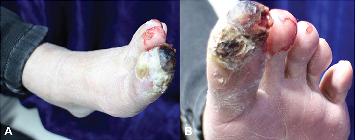
Fig. 1. An ulcerated prominent brown nodule on the toe. (A) Overview, (B) close-up view. Written patient consent for publication was obtained.
Histopathological examination revealed sheets of large, pleomorphic lymphoid cells with abundant amphophilic cytoplasm and horseshoe/kidney-shaped nuclei (Fig. 2A, B). Immunohistochemical staining results were positive for CD30 (Fig. 2C), EMA, anaplastic lymphoma kinase (ALK, Fig. 2D), CD2, LCA, and perforin, while negative for HMB45, A103, SOX10, CD20, CD79a, and EBER. A positron emission tomography (PET)-CT scan found multiple enlarged lymph nodes and nodular lesions in the left neck, clavicular and inguinal regions, posterior peritoneum, left toe, muscle space of left lower limb, and multiple sites in the bones with significant fluro-2-deoxy-D-glucose uptake. Puncture biopsy of the enlarged inguinal lymph node revealed similar histological and immunochemical features. Bone marrow aspiration and biopsy revealed no abnormalities.
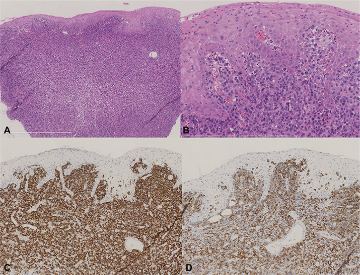
Fig. 2. Histopathological image demonstrates sheets of large, pleomorphic lymphoid cells in the dermis (A, haematoxylin eosin, x50) with abundant amphophilic cytoplasm, some of which have horseshoe/kidney-shaped nuclei (B, haematoxylin eosin, x200). Immunohistochemical staining revealed the lymphoid cells were positive for CD30 (C: CD30 x100) and ALK (D: ALK x100).
What is your diagnosis?
1: Primary cutaneous anaplastic large cell lymphoma
2: ALK-positive anaplastic large cell lymphoma
3: Amelanotic melanoma
4: Lymphomatoid papulosis
See next page for answer.
ANSWERS TO QUIZ
An Ulcerated Nodule on the Toe in an Elderly Patient: A Commentary
Diagnosis: ALK-positive anaplastic large cell lymphoma
ALK-positive anaplastic large cell lymphoma (ALK+ALCL) is a subtype of peripheral T-cell lymphoma (PTCL) histologically characterized by distinct cell morphology and expression of CD30 and ALK. ALK+ALCL usually occurs in young subjects (median age: 34 years) with a male predominance (M:F ratio: 1.5), accounting for 3% of adult non-Hodgkin lymphomas and 10–15% of childhood lymphomas (1). As it is an aggressive CD30-positive T-cell lymphoma, patients with ALK+ALCL frequently present with advanced-stage disease (stage III–IV in 66% of cases) (1). Extranodal involvement has been observed in 60% of cases, most commonly in the soft tissue and bone. According to a retrospective study including 1,306 systemic ALK+ALCL patients with extranodal involvement, 47.5% of patients had primary cutaneous/soft tissue lesions (2). Moreover, patients with primary disease presentations in the head and neck, cutaneous/soft tissue areas, and bone had a better prognosis than those located in the central nervous system and chest/abdomen/pelvis (2).
Although ALK+ALCL shows a broad morphologic spectrum, all cases contain a variable proportion of large anaplastic cells with horseshoe/kidney/embryo-shaped nuclei, referred to as hallmark cells. The common pattern exclusively consists of large-sized cells with hallmark appearance, accounting for 60% of cases (3). Other morpho-logic variants including lymphohistiocytic, small cell, and Hodgkin-like patterns are more difficult to diagnose as the tumour cells are mixed with reactive lymphocytes and histiocytes, and cluster around blood vessels, which might resemble a metastatic tumour. Immunostaining of CD30 and/or ALK could highlight tumour cells and aid in differential diagnosis. The majority of ALK+ALCL are positive for EMA, and expresses 1 or more T-cell antigens and cytotoxic markers (TIA1, granzyme B, perforin). Some 90% of ALK+ALCL cases show clonal rearrangement of TCR genes and harbour t(2;5) (p23;q35)/NPM-ALK translocation, which results in up-regulation of ALK (3). ALK+ALCL is consistently negative for Epstein–Barr virus.
The first clinical differential diagnosis of a progressing ulcerated erythematous nodule on the toe is amelanotic melanoma, which contains little or no pigmentation by naked eye or histologic examination. Pathologically, melanoma cells have various morphologies which could mimic carcinoma, sarcoma, and lymphoma. Notably, rare CD30+ melanoma cases have been reported (4), which highlighted the importance of immunohistochemical panels including S100, SOX10, and HMB45. Skin-involved nodal ALCL needs to be differentiated from primary cutaneous CD30+ T cell proliferative disorders (LPDs). Primary cutaneous CD30+ LPDs are the second most common cutaneous lymphoma after mycosis fungoides and Sezary syndrome, which include primary cutaneous anaplastic large cell lymphoma (pcALCL), lymphomatoid papulosis (LyP), and borderline lesions. While LyP is characterized by disseminated self-regressing papules with a benign but recurrent clinical course, pcALCL usually manifests as a single or grouped reddish-brown plaques and nodules that sometimes ulcerate, which needs to be distinguished from secondary involvement of nodal ALK-negative ALCL based on correlation between clinical, histological, and immunophenotypical aspects.
Traditional therapy of anthracycline-based polychemotherapy CHOP (cyclophosphamide, vincristine, doxorubicin liposome, methylprednisolone) with or without etoposide is associated with an overall response rate of 90% and a 5-year overall survival of 70% in ALCL patients (5). Recently, CD30 antibody-drug conjugates (brentuximab vedotin) plus cyclophosphamide, doxorubicin, and prednisone (BV+CHP) have become the first-line treatment for systemic ALCL based on the ECHELON-2 trial data (6). Our patient, due to economic considerations, completed 6 cycles of CHOEP polychemotherapy with complete remission of the enlarged lymph nodes and skin lesion.
In summary, we present a case of ALK+ALCL manifested as an ulcerated nodule on the toe, which broadens the clinical differential diagnosis of acral tumour. More importantly, our case illustrates the importance of ALK staining in the diagnosis of CD30-positive lymphoma or lymphoproliferative disease and emphasizes the ultimate need for systemic evaluation, especially in ALK-positive cases.
REFERENCES
- Chiattone C, Civallero M, Fischer T, Miranda E, Manni M, Zing NPC, et al. Characteristics and clinical outcomes of patients with ALK-positive anaplastic large cell lymphoma: report from the prospective international T-cell lymphoma project. Hematol Oncol 2022; 40: 953–961. https://doi.org/10.1002/hon.3074
- Nguyen KA, Su C, Bai HX, Zhang Z, Xiao R, Karakousis G, et al. Disease site as a determinant of survival outcome in patients with systemic anaplastic lymphoma kinase positive anaplastic large cell lymphoma with extranodal involvement: an analysis of 1306 cases from the US National Cancer Database. Br J Haematol 2018; 181: 196–204. https://doi.org/10.1111/bjh.15145
- Amador C, Feldman AL. How I diagnose anaplastic large cell lymphoma. Am J Clin Pathol 2021; 155: 479–497. https://doi.org/10.1093/ajcp/aqab012
- Polski JM, Janney CG. Ber-H2 (CD30) immunohistochemical staining in malignant melanoma. Mod Pathol 1999; 12: 903–906.
- Rigaud C, Knörr F, Brugières L, Woessmann W. Diagnosis and management of ALK-positive anaplastic large cell lymphoma in children and adolescents. Best Pract Res Clin Haematol 2023; 36: 101444. https://doi.org/10.1016/j.beha.2023.101444
- Horwitz S, O’Connor OA, Pro B, Trümper L, Iyer S, Advani R, et al, The ECHELON-2 Trial: 5-year results of a randomized, phase III study of brentuximab vedotin with chemotherapy for CD30-positive peripheral T-cell lymphoma. Ann Oncol 2022; 33: 288–298. https://doi.org/10.1016/j.annonc.2021.12.002