RESEARCH LETTER
Papular Clear Cell Hyperplasia of the Eccrine Duct with Histological Pitaya Sign: Not Just “Heat Rash”!
Anna GIORDANO-ROSENBAUM  1, Heidemarie SCHINAGL2, Stefan W. SCHNEIDER 1 and Christian ROSE 3
1Department of Dermatology and Venereology, University Medical Centre Hamburg-Eppendorf, Martinistraße 52, DE-20251, Hamburg, Germany, 2Dermatology in Schenefeld, Hamburg, Germany, and 3Dermatopathology, Lübeck, Germany. E-mail: a.giordano-rosenbaum@uke.de
Citation: Acta Derm Venereol 2025; 105: adv43492. DOI: https://doi.org/10.2340/actadv.v105.43492.
Copyright: © 2025 The Author(s). Published by MJS Publishing, on behalf of the Society for Publication of Acta Dermato-Venereologica. This is an Open Access article distributed under the terms of the Creative Commons Attribution-NonCommercial 4.0 International License (https://creativecommons.org/licenses/by-nc/4.0/).
Submitted: Mar 28, 2025; Accepted after revision: Apr 8, 2025; Published: May 7, 2025
Competing interests and funding: The authors have no conflicts of interest to declare.
To the Editor,
Papular clear cell hyperplasia of the eccrine duct (PCCH) is a very rare disease that has only been described in 4 patients to date.
A 48-year-old woman presented with severely itchy (VAS 8/10), 1- to 3-mm large, grouped, slightly erythematous papules on the extensor sides of both upper extremities that had been present for a year. Occasionally, similar papules were also found on the neck and décolleté. The skin changes had a rough, grater-like consistency without any recognizable connection to the hair follicles (Fig. 1) and increased in summer or after exposure to sunlight and heat, while they completely regressed in winter and in unheated environments. Routine laboratory tests, including HbA1c and fasting blood glucose, were normal. Apart from a mild form of atopic eczema in early childhood, there were no previous illnesses and no long-term medication. A skin biopsy of the right forearm showed clearly widened eccrine ducts consisting of clear cells with optically empty cytoplasm and small chromatophilic nuclei (Fig. 2a, b). The secretory portions themselves were not affected and there was no inflammatory infiltrate. Apart from the focal granular PAS positivity in the clear cytoplasm of the epithelial cells, further staining (alcian blue staining, Berlin blue, and naphthol-AS-D-chloroacetate esterase reaction) did not reveal any inflammatory infiltrate. Due to the very characteristic histological findings, the diagnosis of papular clear cell hyperplasia of the eccrine duct (PCCH) was made.
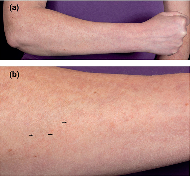
Fig. 1. Clinical manifestations. (a) General view of the miliary-sized, discrete erythematous papules on the extensor side of the right forearm with (b) corresponding detailed view.
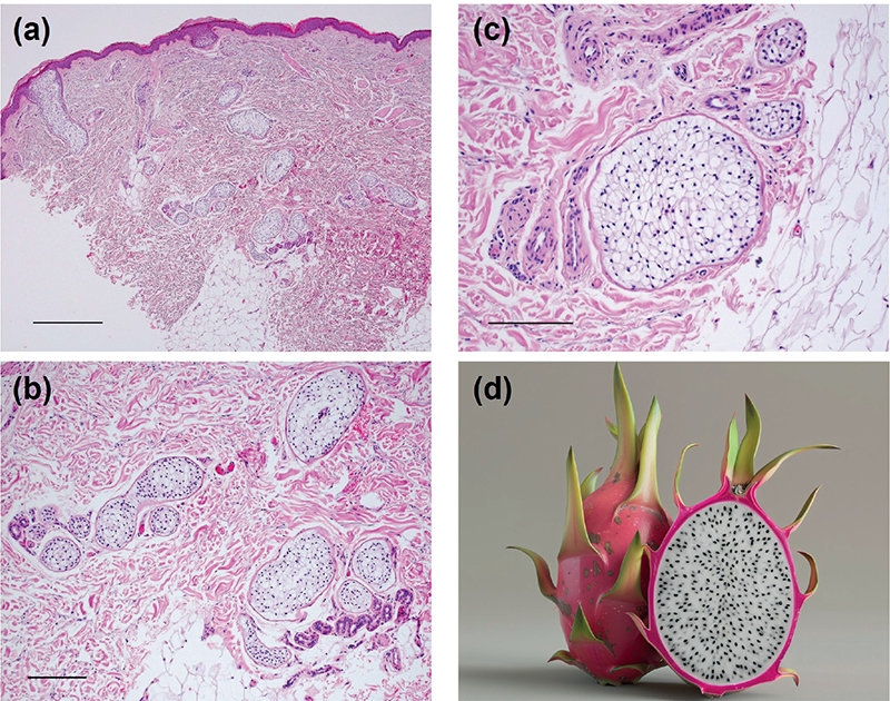
Fig. 2. Histological findings of the biopsy from the forearm. (a) Eccrine secretory ducts, spread by clear cells (haematoxylin-eosin stain, bar: 1000 μm); (b) detailed view of the unaffected secretory portion with dilated ducts without inflammatory infiltrate (haematoxylin-eosin stain, bar: 500 μm); (c) comparison of the histological image with a wide eccrine secretory duct (haematoxylin-eosin stain, 250 μm); and (d) a cross-section of a pitaya (dragon fruit).
To date, only 4 patients with PCCH have been documented in the literature (Table I). The clinical and pathological findings of the case presented show notable parallels to the earlier case reports (1–4). The patients were all women aged between 37 and 77 years. The asymptomatic to pruritic papules occurred in the 3 younger patients on the upper extremities and trunk and predominantly after exposure to heat (1–3).
| Author | Gender | Age, years | Diabetes mellitus | Clinical characteristics |
| Izaki et al. (1) | Female | 41 | No | Skin-coloured, itchy papules on the upper limb |
| Signoretti et al. (2) | Female | 46 | No | Asymptomatic, skin-coloured papules on the torso |
| Tiju et al. (3) | Female | 37 | Yes | Asymptomatic, skin-coloured papules on the upper limb and torso |
| Alonso-Riaño et al. (4) | Female | 77 | Yes | Asymptomatic hyperkeratosis in the scar area on the capillitium |
| Presented case | Female | 48 | No | Skin-coloured, itchy papules on the upper limb and torso |
Histologically, clear cell syringoma (CCS) should be mentioned as a differential diagnosis. However, this is a benign tumour of the eccrine duct. A localized fibrosis is found in the centre of the cell clusters. Clear cell syringomas have been described in association with type 2 diabetes mellitus, although this association was not confirmed in a further study (5, 6). Two of the total of 5 PCCH patients were diabetic (3, 4). Ultimately, the pathogenesis of PCCH remains unclear.
The very distinct histological image of the wide eccrine ducts, which appear to be inflated due to clear epithelia with small cell nuclei, is reminiscent of the cross-section of a pitaya (dragon fruit) (Fig. 2c, d). This visual pattern could therefore serve as a mnemonic for easier recognition of PCCH.
ACKNOWLEDGEMENTS
Ethics statement: The patient in this manuscript has given written informed consent to the publication of her case details.
Data availability statement: The data that support the findings of this study are available from the corresponding author upon reasonable request.
REFERENCES
- Izaki S, Kono E, Hirai A, Kitamura K. Eruptive clear cell hamartoma of sweat duct. J Cutan Pathol 1994; 21: 271–273. https://doi.org/10.1111/j.1600-0560.1994.tb00272.x
- Signoretti S, Annessi G, Occhiuto S, Ruatti P, Faraggiana T. Papular clear cell hyperplasia of eccrine duct in a diabetic. Br J Dermatol 1996; 135: 139–143. https://doi.org/10.1046/j.1365-2133.1996.d01-952.x
- Tjiu JW, Hsiao CH, Wang SH, Wu JJ, Lu CF, Chen HC. Papular clear cell hyperplasia of the eccrine duct. J Eur Acad Dermatol Venereol 2009; 23: 199–200. https://doi.org/10.1111/j.1468-3083.2008.02771.x
- Alonso-Riaño M, Cámara-Jurado M, Garrido MC, Rodríguez-Peralto JL. Papular clear cell hyperplasia of the eccrine duct: a precursor lesion of clear cell syringoma? Am J Dermatopathol 2015; 37: 701–703. https://doi.org/10.1097/DAD.0000000000000339
- Saitoh A, Ohtake N, Fukuda S, Tamaki K. Clear cells of eccrine glands in a patient with clear cell syringoma associated with diabetes mellitus. Am J Dermatopathol 1993; 15: 166–168. https://doi.org/10.1097/00000372-199304000-00012
- Cherian P, Beer TW. Clear cell change in eccrine glands is not associated with diabetes. Am J Dermatopathol 2010; 32: 56–60. https://doi.org/10.1097/DAD.0b013e3181b3ad76