SHORT COMMUNICATION
Real–world Experience with JAK Inhibitors and Dupilumab in Dystrophic Epidermolysis Bullosa: A Case Report and Literature Review
1Department of Dermatology, Peking University Shenzhen Hospital, No. 1120 Lianhua Road, Futian District, Shenzhen, Guangdong Province, 518036, China, and 2Shantou University Medical College, Shantou, China. *E-mail: drdouxia@163.com
Citation: Acta Derm Venereol 2025; 105: adv43588. DOI: https://doi.org/10.2340/actadv.v105.43588.
Copyright: © 2025 The Author(s). Published by MJS Publishing, on behalf of the Society for Publication of Acta Dermato-Venereologica. This is an Open Access article distributed under the terms of the Creative Commons Attribution-NonCommercial 4.0 International License (https://creativecommons.org/licenses/by-nc/4.0/).
Submitted: Apr 10, 2025; Accepted after revision: Apr 25, 2025; Published: May 27, 2025.
Competing interests and funding: This work was supported by Shenzhen High-level Hospital Construction Fund and Peking University Shenzhen Hospital Scientific Research Fund (KYQD2024401) and Shenzhen Key Medical Discipline Construction Fund (No. SZXK040).
The authors have no conflict of interest to declare.
INTRODUCTION
Epidermolysis bullosa (EB) is a heterogeneous group of inherited skin-fragility disorders caused by mutations in structural proteins of the dermal–epidermal junction. It is classified into 4 main types based on the cleavage level: EB simplex, junctional EB, dystrophic EB (DEB), and Kindler syndrome. Pruritus is a common symptom in all subtypes of EB and affects both intact skin and healing wounds, with several internal or external factors contributing (1). Patients with DEB exhibit a prevalence of pruritus comparable to that observed in patients with atopic dermatitis (2). However, conventional antipruritic therapies, such as topical corticosteroids, tacrolimus, thalidomide, and systemic immunosuppressants, are often inadequate to provide effective relief of pruritus in these patients. Recently, biologics and small molecule inhibitors have expanded the therapeutic options for the management of this condition.
CASE PRESENTATION
We report a 20-year-old man with DEB who was successfully treated with a Janus kinase (JAK) inhibitor. The patient presented with recurrent vesicles and bullae on his lower legs, triggered by minor trauma over a 2-year period. Over time, the lesions had spread to his trunk and were accompanied by severe pruritus with a visual analogue scale (VAS) score of 8. On examination, numerous firm papules and nodules were scattered across the trunk and extremities, some with central keratotic crusts and punctate white scarring (Fig. 1A–F). His nails were normal. A skin biopsy from a nodule on the right lower leg confirmed the diagnosis of DEB (Fig. 1G). Due to the severe pruritus, we initiated oral tofacitinib 5 mg twice daily, with topical halometasone/triclosan cream. The patient’s pruritus improved rapidly, with the VAS score decreasing to 4 at the 2-week follow-up visit. At week 20, his VAS score decreased to 0 and the nodules on the extremities and trunk showed significant flattening (Fig. 1H–J). No adverse events were reported during treatment.
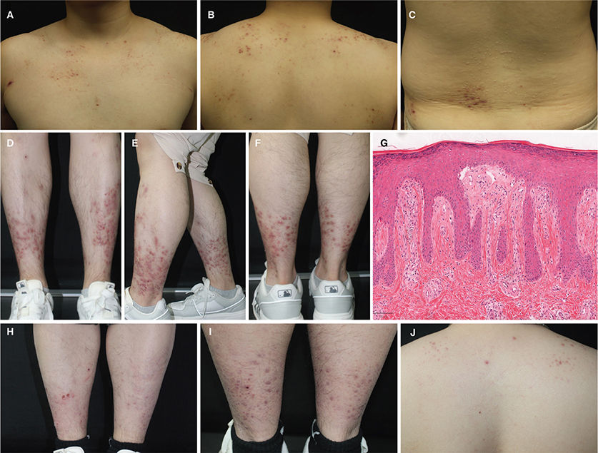
Fig. 1. Clinical and histopathologic features. Numerous firm papules and nodules with punctate scarring involving the trunk (A–C) and extremities (D–F) on initial presentation. Subepidermal blister with a mixed inflammatory infiltrate in the dermis (G) (HE, ×50). (H–J) Marked remission of cutaneous lesions after 20 weeks of Tofacitinib.
LITERATURE REVIEW
We reviewed the literature for reported cases of DEB treated with either dupilumab or JAK inhibitors, using the following search terms: ‘JAK inhibitor’, ‘tofacitinib’, ‘baricitinib’, ‘upadacitinib’, ‘abrocitinib’, ‘dupilumab’, and ‘dystrophic epidermolysis bullosa’. Articles without patient-specific VAS score data were excluded. A total of 53 patients with complete clinical data were included, as summarized in Table SI.
Of these patients, 18 were males and 32 were females, with an overall mean age of 25.48 years (range: 1–60) (sex and age not reported in 4 cases). Subtypes of DEB included EBP (n = 28), RDEB (n = 15), and DDEB (n = 11). The overall median baseline pruritus VAS score was 8 (IQR: 7–8.5). Both dupilumab and JAK inhibitors treatments resulted in significant reductions in pruritus. Among 27 patients treated with JAK inhibitors, the median baseline pruritus VAS score was 8 (IQR: 7–8), which improved to 2 ((IQR: 0–3.5) after treatment (median follow-up: 16 weeks). Similarly, in 27 patients treated with dupilumab, the median VAS score decreased from 8 (IQR: 7–8.75) to 3 (IQR: 2–4) (median follow-up: 17 weeks). To assess medication-specific differences in treatment response, we compared the median percentage reduction in pruritus VAS scores between JAK inhibitors and dupilumab. The median pruritus VAS score reduction rate in the JAK inhibitor group was superior to that in the dupilumab group (75% vs 59%, p = 0.029). In terms of gender differences, there was no significant difference in the median percentage reduction in pruritus VAS scores between males and females (67% vs 68%, p = 0.976) (Fig. 2). Notably, 2 patients who were non-responders to dupilumab achieved effective pruritus control after switching to a JAK inhibitor. Reported adverse events of JAK inhibitors were generally mild and included fatigue, recurrent otitis media, and mild hyperlipidaemia. No serious adverse events were documented with dupilumab in these cases.
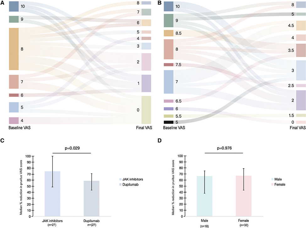
Fig. 2. (A) VAS scores change in patients with DEB treated with JAK inhibitors. (B) VAS scores change in patients with DEB treated with dupilumab. (C) Comparison of pruritus improvement in DEB patients treated with JAK inhibitors vs dupilumab (data derived from 54 combined cases in Table SI, including the current case). (D) Comparison of pruritus improvement between male and female patients with DEB (4 patients were excluded due to not specifying the characteristics).
DISCUSSION
Pruritus is a hallmark of DEB and has a profound impact on quality of life. Recent research has shown that pruritic lesions in DEB harbour infiltrates of GATA3-positive Th2 cells, which are likely to contribute to the intense itch (3). This provides a strong rationale for therapies that target the Th2 pathway. Dupilumab, by blocking IL-4/IL-13 signalling, addresses part of this pathway and can ameliorate itch. In contrast, JAK inhibitors broadly inhibit the JAK–STAT pathways used by multiple cytokines (including IL-4, IL-13, and IL-31), thereby exerting a more comprehensive antipruritic effect (4). Our findings, along with a recent real-world study by Hou et al., suggest that JAK inhibitors may offer superior efficacy compared with dupilumab for refractory pruritus in DEB (4). In clinical practice, dupilumab’s favourable safety profile make it a reasonable first-line biologic for severe pruritus in DEB, especially in paediatric cases. For patients who do not respond adequately to dupilumab, oral JAK inhibitors (such as baricitinib, upadacitinib, or tofacitinib) can provide dramatic itch relief, as demonstrated in our case and in the literature, but careful monitoring for adverse effects is recommended (4, 5).
In conclusion, both dupilumab and JAK inhibitors are effective therapies for the treatment of chronic pruritus in DEB, but current evidence suggests that JAK inhibitors may offer superior efficacy in pruritus control compared with dupilumab. Given the continuous therapeutic requirements of DEB, the long-term safety profile of JAK inhibitors requires further investigation. Additional rigorous studies are needed to validate the effectiveness observations and to establish comprehensive safety guidelines for the long-term use of JAK inhibitors in patients with DEB.
ACKNOWLEDGEMENT
Data availability: The data used to support the findings of this study are available from the corresponding author upon request.
IRB approval status: The participant has consented to the submission of this case report to the journal.
REFERENCES
- Papanikolaou M, Onoufriadis A, Mellerio JE, Nattkemper LA, Yosipovitch G, Steinhoff M, et al. Prevalence, pathophysiology and management of itch in epidermolysis bullosa. Br J Dermatol 2021; 184: 816–825. https://doi.org/10.1111/bjd.19496
- Snauwaert JJ, Yuen WY, Jonkman MF, Moons P, Naulaers G, Morren MA. Burden of itch in epidermolysis bullosa. Br J Dermatol 2014; 171: 73–78. https://doi.org/10.1111/bjd.12885
- Aala WJF, Hou PC, Hong YK, Lin YC, Lee YR, Tu WT, et al. Dominant dystrophic epidermolysis bullosa is associated with glycolytically active GATA3+ T helper 2 cells which may contribute to pruritus in lesional skin. Br J Dermatol 2024; 191: 252–260. https://doi.org/10.1093/bjd/ljae110
- Hou PC, Aala W Jr, Tu WT, McGrath JA, Hsu CK. Real-world experience of using dupilumab and JAK inhibitors to manage pruritus in epidermolysis bullosa pruriginosa. Skin Health Dis 2024; 4: e445. https://doi.org/10.1002/ski2.445
- Kwon IJ, Kim SE, Kim SC, Lee SE. Efficacy of oral JAK1 or JAK1/2 inhibitor for treating refractory pruritus in dystrophic epidermolysis bullosa: a retrospective case series. J Dermatol 2024; 51: 441–447. https://doi.org/10.1111/1346-8138.17079