RESEARCH LETTER
ABCD for Optical Coherence Tomography (OCT): An Acronym to Aid OCT Evaluation for Detecting Basal Cell Carcinoma
Myrthe M. G. MOERMANS1,2  , Tom WOLSWIJK1,2 and Klara MOSTERD1,2
1Dermatology Department, Maastricht University Medical Center+, PO Box 5800, 6229 HX Maastricht, The Netherlands, and 2Grow Research Institute for Oncology and Reproduction, Maastricht University, Maastricht. E-mail: myrthe.moermans@mumc.nl
Citation: Acta Derm Venereol 2025; 105: adv43589. DOI: https://doi.org/10.2340/actadv.v105.43589.
Copyright: 2025 © The Author(s). Published by MJS Publishing, on behalf of the Society for Publication of Acta Dermato-Venereologica. This is an Open Access article distributed under the terms of the Creative Commons Attribution-NonCommercial 4.0 International License (https://creativecommons.org/licenses/by-nc/4.0/).
Submitted: Apr 10, 2025; Accepted after revision: May 5, 2025; Published: Jun 2, 2025.
Competing interests and funding: The authors have no conflicts of interest to declare.
To the Editor,
As the incidence of basal cell carcinoma (BCC) continues to rise, the demand for non-invasive diagnosis and treatment increases (1, 2). Optical coherence tomography (OCT) provides a non-invasive diagnostic alternative to conventional biopsy, as OCT-guided diagnosis and treatment of lesions suspect for BCC is known to be non-inferior compared with a biopsy-guided approach (3). Sufficient training and a structured framework for evaluating OCT scans are essential for establishing a high-confidence OCT diagnosis, which is a necessity to avoid the need for biopsy. However, there is a lack of standardized OCT assessment and a shortage of fully trained OCT assessors, which complicates the integration of OCT into clinical practice.
Therefore, we aim to describe a step-by-step approach for OCT scan evaluation to facilitate and standardize OCT assessment and aid novice assessors. In analogy with the ABCD mnemonic acronym for nevi (4), we have embodied this systematic approach in the acronym “OCT-ABCD”: Anatomical layers, Boundaries, Characteristics, and Differential diagnosis. The use of a clinical algorithm, such as an acronym, can positively impact the learning of a sequential procedural task, making it easier to memorize consecutive steps and teach novice assessors (5). Moreover, we describe the most characteristic OCT features, as specified by the existing literature and based on expert consensus (6, 7).
Anatomical layers: All layers of the skin should be assessed in systematic order, from top to bottom. First the epidermis, together with the dermal–epidermal junction (DEJ), and finally the dermis. Even though clear BCC characteristics might attract the attention, it is important that this sequence is followed, to ensure no (subtle) abnormalities are overlooked. While assessing the consecutive layers, the normal anatomy and variations in healthy skin should be taken into account, which can be location- and patient-dependent.
Boundaries: To distinguish BCC from other diagnoses, it is important to trace and evaluate the integrity of the DEJ. If the DEJ is (multi-)focally non-traceable or disrupted, this could point in the direction of invasive lesions, e.g., cutaneous squamous cell carcinoma. Of note, features should never be classified on a single OCT slide as OCT features exist in a morphologic context. Therefore, attention must be given to the surroundings of the features and their interaction. For instance, a seemingly well-demarcated ovoid dermal structure on one slide may confluate with the epidermis on another slide, which signals the adnexal nature of the structure.
Characteristics: Highly specific BCC features can guide towards the diagnosis and help differentiate between subtypes (Fig. 1). For superficial BCC, attention should be given to the following features: bulging tumour nests originating from the epidermis (protrusions), including corresponding hyporeflective edges (dark rims). For nodular BCC, the epidermis is often atrophic because of mass effect from the dermal abnormalities. Besides, hyporeflective ovoid structures with hyporeflective clefting and hyperreflective peri-tumoral stroma can be recognized in the dermis, characterizing nodular BCC nests. Infiltrative BCCs are more difficult to detect, but can be recognized as multiple small hyporeflective ovoid structures, often referred to as “grape-like appearance”. They are not well defined and become increasingly indistinct in deeper dermal layers (6, 7).
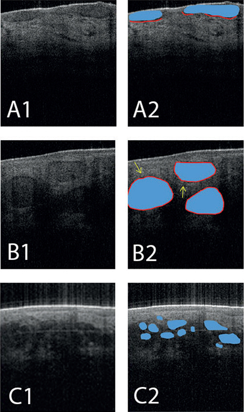
Fig. 1. Optical coherence tomography (OCT) images of different basal cell carcinoma (BCC) subtypes. (A1/2) OCT image of a superficial BCC. The blue demarcation represents hyporeflective protrusions of basaloid cell nests (blue) and the red line corresponds with the underlying dark rim. (B1/2) OCT image of a nodular BCC. The epidermis is atrophic because of the mass effect of the dermal abnormalities. The blue demarcation represents an ovoid structure with clefting (red line) and hyperreflective peri-tumoral stroma (yellow arrows). (C1/2) OCT image of an infiltrative BCC. The blue demarcation represents multiple small hyporeflective ovoid structures, which are not well defined, resembling a “grape-like appearance” and corresponding with a micronodular growth pattern.
Differential diagnosis: If all the previous steps are completed, a differential diagnosis can be established. It is important to correlate OCT and clinical features. Evaluation of the clinical presentation can improve interpretation of the anatomy and abnormalities on OCT (Wolswijk, Impact of Clinical and Dermoscopical Photographs on Accuracy of Remote OCT Assessment for BCC, Manuscript submitted). As an example, hyperkeratosis may complicate tracing the DEJ and can resemble BCC protrusions on OCT. However, keratosis on clinical evaluation is indicative of other (pre)malignancies such as actinic keratosis, Bowen’s disease, or squamous cell carcinoma (Fig. 2), which exhibit distinct OCT features. Besides, OCT assessors should be aware of common pitfalls: sometimes morphological characteristics of typical skin diseases can mimic BCC characteristics on OCT, and consequently point the OCT assessor in the wrong direction. Diagnoses such as dermal nevi, sebaceous gland hyperplasia, Bowen’s disease, and benign lichenoid keratosis are common pitfalls that may be differentiated from BCC on the basis of the correlation between their clinical and dermoscopic characteristics with OCT features (8).

Fig. 2. Optical coherence tomography (OCT) image of cutaneous squamous cell carcinoma (SCC). Optical coherence tomography (OCT) image of cutaneous squamous cell carcinoma shows irregular and thickened epidermis. The DEJ is difficult to locate and eventually is lost. The yellow dotted line represents the demarcation between the hyperkeratotic stratum corneum and the rest of the epidermis, which can easily be mistaken for the true DEJ (red dotted line).
DISCUSSION
Evaluating OCT scans is a continuous learning process that should be closely monitored and performance should be sufficient and consistent over time, before making OCT-based treatment decisions in clinical practice. Considering the shortage of trained OCT assessors, an OCT evaluation by a distant expert supervisor could increase BCC detection rates and facilitate the integration of OCT into clinical practice. In such a situation a structured transfer of information to a supervisor is essential and could be facilitated by a systematic approach, such as the ABCD-method.
REFERENCES
- Verkouteren JAC, Ramdas KHR, Wakkee M, Nijsten T. Epidemiology of basal cell carcinoma: scholarly review. Br J Dermatol 2017; 177: 359–372. https://doi.org/10.1111/bjd.15321
- Peris K, Fargnoli MC, Kaufmann R, Arenberger P, Bastholt L, Seguin NB, et al. European consensus-based interdisciplinary guideline for diagnosis and treatment of basal cell carcinoma—update 2023. Eur J Cancer 2023; 192: 113254. https://doi.org/10.1016/j.ejca.2023.113254
- Adan F, Nelemans PJ, Essers BAB, Brinkhuizen T, Dodemont SRP, Kessels J, et al. Optical coherence tomography versus punch biopsy for diagnosis of basal cell carcinoma: a multicentre, randomised, non-inferiority trial. Lancet Oncol 2022; 23: 1087–1096. https://doi.org/10.1016/S1470-2045(22)00347-3
- Friedman RJ, Rigel DS, Kopf AW. Early detection of malignant melanoma: the role of physician examination and self-examination of the skin. CA Cancer J Clin 1985; 35: 130–151. https://doi.org/10.3322/canjclin.35.3.130
- Radović T, Manzey D. The impact of a mnemonic acronym on learning and performing a procedural task and its resilience toward interruptions. Front Psychol 2019; 10: 2522. https://doi.org/10.3389/fpsyg.2019.02522
- Adan F, Mosterd K, Kelleners-Smeets NWJ, Nelemans PJ. Diagnostic value of optical coherence tomography image features for diagnosis of basal cell carcinoma. Acta Derm Venereol 2021; 101: adv00607. https://doi.org/10.2340/actadv.v101.421
- Fuchs CSK, Ortner VK, Mogensen M, Rossi AM, Pellacani G, Welzel J, et al. 2021 international consensus statement on optical coherence tomography for basal cell carcinoma: image characteristics, terminology and educational needs. J Eur Acad Dermatol Venereol 2022; 36: 772–778. https://doi.org/10.1111/jdv.17969
- Wolswijk T, Nelemans PJ, Adan F, Abdul Hamid M, Mosterd K. Pitfalls for differentiating basal cell carcinoma from non-basal cell carcinoma on optical coherence tomography: a clinical series. J Dermatol 2024; 51: 40–47. https://doi.org/10.1111/1346-8138.17020