SHORT COMMUNICATION
In Vitro Evidence Demonstrating the Nondepleting Mechanism of Action of Amlitelimab, an OX40 Ligand Monoclonal Antibody
James G. KRUEGER1*, Brian S. KIM2, Stephan WEIDINGER3, Yoko KATAOKA4, John T. O’MALLEY5, Karl YEN6, Cassie VAN KRINKS7 and Jose VELA5
1Laboratory for Investigative Dermatology, The Rockefeller University, New York, NY, USA, 2Department of Dermatology, Icahn School of Medicine at Mount Sinai, New York, NY, USA, 3Department of Dermatology and Allergy, University Hospital Schleswig-Holstein, Kiel, Germany, 4Department of Dermatology, Osaka Habikino Medical Center, Habikino, Japan; 5Sanofi, Cambridge, MA, USA, 6Sanofi, Rotkreuz, Switzerland, and 7Formerly Sanofi, Cambridge, UK. *E-mail: kruegej@rockefeller.edu
Citation: Acta Derm Venereol 2025; 105: adv43608. DOI: https://doi.org/10.2340/actadv.v105.43608.
Copyright: © 2025 The Author(s). Published by MJS Publishing, on behalf of the Society for Publication of Acta Dermato-Venereologica. This is an Open Access article distributed under the terms of the Creative Commons Attribution-NonCommercial 4.0 International License (https://creativecommons.org/licenses/by-nc/4.0/).
Submitted: Apr 11, 2025. Accepted after revision: Jul 19, 2025. Published: Aug 18, 2025.
Competing interests and funding: JGK: Consultant/honoraria: AbbVie, Aclaris, Allergan, Almirall, Amgen, Artax Biopharma, Arena, Aristea, Asana, Aurigene, Biogen Idec, Boehringer Ingelheim, Bristol Myers Squibb, Escalier, Galapagos, Janssen, Kyowa Kirin, Lilly, MoonLake Immunotherapeutics, Nimbus, Novartis, Pfizer, Sanofi, Sienna Biopharmaceuticals, Sun Pharma, Target-Derm, UCB, Valeant, and Ventyx. Grant support (to the Rockefeller University): AbbVie, Akros, Allergan, Amgen, Avillion, Biogen, Boehringer Ingelheim, Botanix, Bristol Myers Squibb, Exicure, Innovaderm, Incyte, Janssen, Kyowa Kirin, Lilly, Nimbus, Novan, Novartis, Parexel, Pfizer, Regeneron, UCB, and Vitae Pharmaceuticals. BSK: Co-founder of Alys Pharmaceuticals and Neurommune Therapeutics; he has served as a consultant for ABRAX Japan, AbbVie, Amgen, Attovia Therapeutics, Cara Therapeutics, Clexio Biosciences, Eli Lilly and Company, Escient Pharmaceuticals, Evommune, Galderma, Gilead, LEO Pharma, Micreos, Novartis, Pfizer, Recens Medical, Regeneron, Sanofi, Septerna, Teva Pharmaceuticals, and Triveni Bio; he has stock in ABRAX Japan, Alys Pharamaceuticals, Attovia Therapeutics, Locus Biosciences, Neurommune Therapeutics, Recens Medical, and Triveni Bio; he holds a patent for the use of JAK1 inhibitors for chronic pruritus. SW: AbbVie, Almirall, AstraZeneca, Galderma, GSK, Kymab Ltd (a Sanofi company), LEO Pharma, Lilly, Pfizer, Regeneron, and Sanofi. YK: Honoraria from AbbVie, Sanofi, Maruho, and Pfizer; research grants from AbbVie, Amgen, Eli Lilly Japan, LEO Pharma, Maruho, Otsuka Pharmaceutical, Sanofi, Taiho Pharmaceutical, and Pfizer. CVK: Former employee of Kymab Ltd (a Sanofi company) and Sanofi. JTOM, KY, and JV: Employees of Sanofi – may hold stock and/or stock options in the company.
This study was sponsored by Sanofi. Medical writing and editorial assistance were funded by Sanofi and provided by Anna Josephson, PhD, of IMPRINT Science, New York, NY, USA.
INTRODUCTION
The OX40 ligand (OX40L)/OX40 signalling pathway is a critical driver of atopic dermatitis (AD)-associated inflammation. OX40L, expressed on antigen-presenting cells (APCs), binds to OX40 on T cells, inducing T-cell proliferation and survival, while suppressing anti-inflammatory activity of regulatory T cells (Tregs). In AD, OX40L is upregulated on APCs as well as other cells involved in inflammation, potentiating the T-cell response and secretion of pro-inflammatory cyto-kines. Therefore, OX40L/OX40 interaction is a critical co-stimulatory signal that amplifies and sustains the immune response (1–4). Inhibition of OX40L/OX40 binding may attenuate the immune response and limit chronic inflammation (5).
Several monoclonal antibodies targeting the OX40L/OX40 signalling pathway have completed phase 2 trials in AD (4, 6, 7). Differences in molecular structure and binding among these antibodies may influence their safety and efficacy (4, 6, 7). Amlitelimab is a fully human, anti-OX40L human immunoglobulin G4 (hIgG4) S228P L235E (PE) antibody that binds to OX40L on APCs, preventing interaction with OX40 on activated T cells (3). The IgG4 backbone and the accompanying L235E mutation have been shown to suppress antibody-dependent cellular cytotoxicity (ADCC), a mechanism by which antibodies induce target-cell death through engagement of Fc receptors on effector cells, whereas the S228P mutation increases stability and prevents Fab-arm exchange (8, 9). In contrast, rocatinlimab and telazorlimab target OX40 on T cells and have an immunoglobulin G1 (IgG1) backbone, which has been shown to lead to reductions in T cells via increased ADCC (6, 7, 10). Results from the phase 2b STREAM-AD study demonstrated clinically meaningful improvements and off-therapy maintenance in AD with amlitelimab vs placebo (4). It is currently unknown whether ADCC occurs via blockade of OX40L on APCs with an IgG4 antibody and whether it differentially impacts OX40L-expressing cells, OX40-expressing pro-inflammatory effector T cells, and immune-suppressing OX40-expressing Tregs. The current study aimed to evaluate in vitro the potential nondepleting mechanism of action of amlitelimab and other monoclonal antibodies targeting the OX40L/OX40 pathway using 2 approaches: first, by assessing ADCC in cluster of differentiation (CD)4+ T cells with anti-OX40 vs anti-OX40L (amlitelimab) antibodies; and second, by evaluating ADCC activity in hOX40L-expressing cells with anti-OX40L antibodies with different backbones (IgG4 vs IgG1).
MATERIALS AND METHODS
An ADCC assay was established using activated CD4+ T cells as target cells and autologous natural killer (NK)-containing peripheral blood mononuclear cells (PBMCs [Cellero]; see Table SI for list of reagents and vendors) derived from healthy donors as effector cells. Target cells were labelled with CellTrace™ Violet (Thermo Fisher Scientific, Waltham, MA, USA) and co-cultured with effector cells in the presence of the following antibodies: anti-OX40L (amlitelimab), in-house-generated anti-OX40 hIgG1 [anti-OX40 hIgG1 (A)], in-house-generated fucosylated anti-OX40 hIgG1 [anti-OX40 hIgG1 (B)], and isotype control antibodies, hIgG4(PE) and hIgG1. Isotype control antibodies were matched to the antibodies being evaluated. Changes in live cell numbers were analysed using flow cytometry to assess ADCC against activated CD4+ effector T cells (CD4+CD25+Foxp3-) or Foxp3+ Tregs (CD4+CD25+Foxp3+CD127-) utilizing 3 independent donors with 2 replicates for each (Fig. S1).
OX40L was expressed in human embryonic kidney (HEK) cells (HEK hOX40L). Labelled target cells (HEK hOX40L) and effector cells (primary NK cells isolated from PBMCs obtained from leukoreduction chambers) were incubated with the following anti-OX40L antibodies: amlitelimab hIgG4(PE), anti-OX40L hIgG1 (C), anti-OX40L hIgG1 (D), and isotype controls, IgG4(PE) and IgG1. The 2 anti-OX40L hIgG1antibodies had similar binding affinities to Fc gamma receptors (FcγRs), but different antigen binding site sequences compared with one another, with anti-OX40L hIgG1 (D) having a similar sequence to amlitelimab. Antibody titres were matched across the 5 antibodies. OX40L surface expression on HEK cells was confirmed by flow cytometry with fluorescently labelled amlitelimab. HEK hOX40L cell lysis by NK cells was assessed using the DELFIA™ cell cytotoxicity kit (Revivity Life and Analytical Sciences, Llantrisant, Wales), which utilizes fluorescence to evaluate ADCC activity. Three independent donors were used (Fig. S2).
RESULTS
In ADCC co-cultures of activated OX40-expressing CD4+ T cells with NK-cell–containing PBMCs as effector cells, the percentages of OX40-expressing activated CD4+ T cells and Foxp3+ Tregs were decreased in the presence of anti-OX40 IgG1 antibodies (anti-OX40 hIgG1 [A] and anti-OX40 hIgG1 [B]; range: effector T cells, 17.0–43.5%; Tregs, 4.8–14.9%). In contrast, the percentages of activated CD4+ T cells and Foxp3+ Tregs remained unchanged with amlitelimab and isotype controls IgG4(PE) and IgG1 (range: effector T cells, 45.7–48.8%; Tregs, 12.8–17.2%), indicating absence of ADCC activity (Fig. 1). The percentage difference in activated CD4+ T cells between anti-OX40 IgG1 antibodies and amlitelimab across various concentrations ranges from 4.7% to 63.5%, and in Foxp3+ Tregs; the difference ranges from 0.9% to 63.0% (Fig. 1).
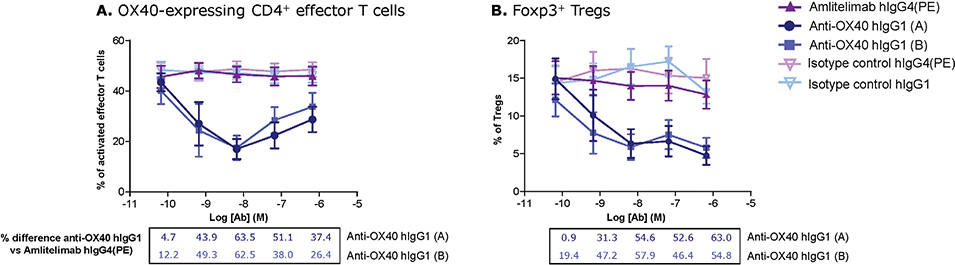
Fig. 1. ADCC activity against activated OX40-expressing CD4+ effector T cells and Foxp3+ Tregs by amlitelimab and anti-OX40 IgG1 antibodies. Flow cytometry analysis quantifying (A) the percentage of activated CD4+ effector T cells and (B) Foxp3+ Tregs following incubation with amlitelimab hIgG4(PE), anti-OX40 hIgG1 (A), anti-OX40 hIgG1 (B) and isotype controls at various concentrations, with the percentage difference between CD4+ T cells and Tregs with anti-OX40 hIgG1 antibodies compared with amlitelimab hIgG4(PE). Ab: antibody; ADCC: antibody-dependent cellular cytotoxicity; CD: cluster of differentiation; h: human; Ig: immunoglobulin; PE: S228P L235E; Treg: regulatory T cell.
Following co-culture of hOX40L-transfected HEK cells with purified NK cells, specific lysis of hOX40L-HEK cells increased significantly in the presence of anti-OX40L IgG1 antibodies (anti-OX40L hIgG1 [C] and anti-OX40L hIgG1 [D], percentage specific lysis [standard error of the mean (±SEM)], 58.4% [±5.5] and 59.3% [±3.0], respectively). Conversely, cell lysis was not significantly increased with amlitelimab (22.7% [±6.5]), with percentages similar to those observed with isotype control hIgG4(PE) (16.7% [±4.1]) and isotype control hIgG1 (17.4% [±3.2]), suggesting amlitelimab did not mediate ADCC against hOX40L-expressing target cells (Fig. 2).
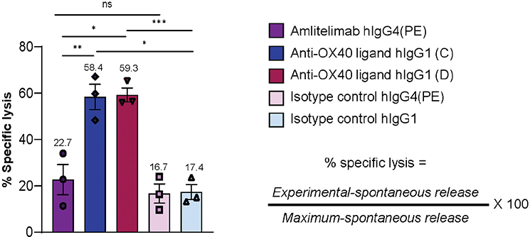
Fig. 2. ADCC activity against hOX40L-expressing HEK cells by anti-OX40L antibodies. Percentage of specific lysis of hOX40L-expressing HEK cells following ADCC assay with anti-OX40L antibodies quantified by detection of fluorescent label. Data plotted are mean ± SEM; n = 3 from 3 independent donors; *p ≤ 0.05, **p ≤ 0.01, ***p ≤ 0.0001. RM one-way ANOVA with Tukey’s post hoc multiple comparison test. ADCC: antibody-dependent cellular cytotoxicity; ANOVA: analysis of variance; h: human; HEK: human embryonic kidney; lg: immunoglobulin; ns: not significant; PE: S228P L235E; RM: repeated measures; SEM: standard error of the mean.
DISCUSSION
Results of this in vitro study indicate that amlitelimab, an anti-OX40L hIgG4(PE) monoclonal antibody, does not deplete OX40-expressing T cells or OX40L-expressing cells via ADCC. In contrast, anti-OX40 hIgG1 and anti-OX40L hIgG1 antibodies exhibited ADCC activity against CD4+ T cells and Tregs, and OX40L-expressing cells, respectively. The differences observed align with prior studies demonstrating that IgG4 antibodies exhibit decreased binding affinity to Fc gamma receptors (FcγRs) on effector cells, reducing their ability to deplete immune cells via ADCC. Furthermore, PE mutations (S228P and L235E) within the IgG4 backbone of amlitelimab reduce Fab-arm exchange, improve stability, and diminish residual FcγR binding, respectively (8, 9, 11).
AD is a chronic inflammatory skin disease characterized by dysregulated immune responses often driven by T-cell activation (2, 12). Current therapeutic approaches target specific cytokines or intracellular signalling pathways downstream of T-cell activation (13). By binding to OX40L on APCs to prevent interaction with OX40 at the level of T-cell co-stimulation, amlitelimab prevents activation and expansion of a broader range of pathogenic T-cell subsets involved in AD to modulate the immune response without substantial T-cell depletion (3, 4). T cells are essential for normal immune function. Their depletion may affect the ability to respond to infections. (2, 7, 12, 14). Tregs help regulate immune responses and can act to prevent excessive immune system activation. Treg depletion could potentially lead to unregulated T-cell activation and exacerbated inflammation (1, 2, 12, 15). Furthermore, in AD, the OX40L/OX40 pathway potentiates chronic inflammation by suppressing Treg function (2). Targeting OX40L/OX40 signalling while preserving OX40-expressing cells, including Tregs, and OX40L-expressing cells, such as innate lymphoid type 2 cells, potentially allows for restoration of immune homeostasis. In turn, this may attenuate the amplification of the inflammatory process involved in cytokine production and immune dysregulation central to AD chronicity (2, 4, 12).
This in vitro study provides insights into the nondepleting mechanism of amlitelimab; however, several limitations should be acknowledged. While HEK cells engineered to express OX40L were used as a proxy for APCs, they likely do not fully mimic APC biology. Alternative cell types may serve as better physiological models. Experiments were conducted with a limited number of donors, which may affect generalizability of the findings. Although the effect of an anti-OX40L IgG4 antibody on T-cell depletion was evaluated, additional studies are needed to better understand the effects of OX40L/OX40 pathway modulation with amlitelimab on cytokine production. This may further elucidate how targeting OX40L and maintaining T-cell populations shapes the immune response in AD.
Notably, multiple trials evaluating anti-OX40L and anti-OX40 antibodies in AD are ongoing and will provide further insight into their efficacy and safety profiles (12). The availability of multiple therapies would enable physicians and patients to select treatments tailored to individual needs.
ACKNOWLEDGEMENTS
The authors would like to acknowledge Janina Nahler, Eric Li, and Ester Huang, former employees of Sanofi, for their contributions to the design and execution of the experiments, as well as Ellen Lin, of Sanofi, Cambridge, MA, USA, for her valuable input throughout the study.
REFERENCES
- Iriki H, Takahashi H, Amagai M. Diverse role of OX40 on T cells as a therapeutic target for skin diseases. J Invest Dermatol 2023; 143: 545–553. https://doi.org/10.1016/j.jid.2022.11.009
- Sadrolashrafi K, Guo L, Kikuchi R, Hao A, Yamamoto RK, Tolson HC, et al. An OX-Tra’Ordinary tale: the role of OX40 and OX40L in atopic dermatitis. Cells 2024; 13. https://doi.org/10.3390/cells13070587
- Weidinger S, Bieber T, Cork MJ, Reich A, Wilson R, Quaratino S, et al. Safety and efficacy of amlitelimab, a fully human nondepleting, noncytotoxic anti-OX40 ligand monoclonal antibody, in atopic dermatitis: results of a phase IIa randomized placebo-controlled trial. Br J Dermatol 2023; 189: 531–539. https://doi.org/10.1093/bjd/ljad240
- Weidinger S, Blauvelt A, Papp KA, Reich A, Lee CH, Worm M, et al. Phase 2b randomized clinical trial of amlitelimab, an anti-OX40 ligand antibody, in patients with moderate-to-severe atopic dermatitis. J Allergy Clin Immunol 2025; 155: 1264–1275. https://doi.org/10.1016/j.jaci.2024.10.031
- Saghari M, Gal P, Gilbert S, Yateman M, Porter-Brown B, Brennan N, et al. OX40L inhibition suppresses KLH-driven immune responses in healthy volunteers: a randomized controlled trial demonstrating proof-of-pharmacology for KY1005. Clin Pharmacol Ther 2022; 111: 1121–1132. https://doi.org/10.1002/cpt.2539
- Guttman-Yassky E, Simpson EL, Reich K, Kabashima K, Igawa K, Suzuki T, et al. An anti-OX40 antibody to treat moderate-to-severe atopic dermatitis: a multicentre, double-blind, placebo-controlled phase 2b study. Lancet 2023; 401: 204–214. https://doi.org/10.1016/S0140-6736(22)02037-2
- Rewerska B, Sher LD, Alpizar S, Pauser S, Pulka G, Mozaffarian N, et al. Phase 2b randomized trial of OX40 inhibitor telazorlimab for moderate-to-severe atopic dermatitis. J Allergy Clin Immunol Glob 2024; 3: 100195. 63 https://doi.org/10.1016/j.jacig.2023.100195
- Silva JP, Vetterlein O, Jose J, Peters S, Kirby H. The S228P mutation prevents in vivo and in vitro IgG4 Fab-arm exchange as demonstrated using a combination of novel quantitative immunoassays and physiological matrix preparation. J Biol Chem 2015; 290: 5462–5469. https://doi.org/10.1074/jbc.M114.600973
- Crescioli S, Correa I, Karagiannis P, Davies AM, Sutton BJ, Nestle FO, et al. IgG4 characteristics and functions in cancer immunity. Curr Allergy Asthma Rep 2016; 16: 7. https://doi.org/10.1007/s11882-015-0580-7
- Zahavi D, AlDeghaither D, O’Connell A, Weiner LM. Enhancing antibody-dependent cell-mediated cytotoxicity: a strategy for improving antibody-based immunotherapy. Antib Ther 2018; 1: 7–12. https://doi.org/10.1093/abt/tby002
- Rispens T, Huijbers MG. The unique properties of IgG4 and its roles in health and disease. Nat Rev Immunol 2023; 23: 763–778. https://doi.org/10.1038/s41577-023-00871-z
- Guttman-Yassky E, Croft M, Geng B, Rynkiewicz N, Lucchesi D, Peakman M, et al. The role of OX40 ligand/OX40 axis signalling in atopic dermatitis. Br J Dermatol 2024; 191: 488–496. https://doi.org/10.1093/bjd/ljae230
- Chovatiya R, Paller AS. JAK inhibitors in the treatment of atopic dermatitis. J Allergy Clin Immunol 2021; 148: 927–940. https://doi.org/10.1016/j.jaci.2021.08.009
- Guttman-Yassky E, Pavel AB, Zhou L, Estrada YD, Zhang N, Xu H, et al. GBR 830, an anti-OX40, improves skin gene signatures and clinical scores in patients with atopic dermatitis. J Allergy Clin Immunol 2019; 144: 482–493 e487. https://doi.org/10.1016/j.jaci.2018.11.053
- Martin A, Tisch RM, Getts DR. Manipulating T cell-mediated pathology: targets and functions of monoclonal antibody immunotherapy. Clin Immunol 2013; 148: 136–147. https://doi.org/10.1016/j.clim.2013.04.011