RESEARCH LETTER
Paxbp1 is Indispensable for the Maintenance of Hair Follicle Homeostasis
Cong HUANG1,2, Wenting LI3, Shizheng ZHAO4, Wei ZHANG4 and Bo YU1,2*
1Department of Dermatology, Peking University Shenzhen Hospital, Shenzhen, Guangdong, China, 2Shenzhen Key Laboratory for Translational Medicine of Dermatology, Shenzhen Peking University - The Hong Kong University of Science and Technology Medical Center, Shenzhen, Guangdong, China, 3The Digestive and Reproductive System Cancers Precise Prevention Engineering Research Center of Jiangsu Province, Institute of Medicinal Biotechnology, Jiangsu College of Nursing, Huai’ an, Jiangsu, China, and 4Biomedical Research Institute, Shenzhen Peking University-the Hong Kong University of Science and Technology Medical Center, Shenzhen, Guangdong, China. *E-mail: drboyu_derm@126.com
Citation: Acta Derm Venereol 2025; 105: adv43648. DOI: https://doi.org/10.2340/actadv.v105.43648.
Copyright: © 2025 The Author(s). Published by MJS Publishing, on behalf of the Society for Publication of Acta Dermato-Venereologica. This is an Open Access article distributed under the terms of the Creative Commons Attribution-NonCommercial 4.0 International License (https://creativecommons.org/licenses/by-nc/4.0/).
Submitted: Apr 17, 2025; Accepted after revision: Apr 29, 2025. Published: May 19, 2025.
Competing interests and funding: The authors have no conflicts of interest to declare.
This work was supported by grants from the National Natural Science Foundation of China (82103726), Guangdong Basic and Applied Basic Research Foundation (2023A1515010575 and 2025A1515010947), Shenzhen Science and Technology Program (JCYJ20210324110008023 and JCYJ20230807095809019), Shenzhen Sanming Project (SZSM202311029), and Shenzhen Key Medical Discipline Construction Fund (SZXK040), Shenzhen High-level Hospital Construction Fund, and Peking University Shenzhen Hospital Scientific Research Fund (KYQD2024378).
To the Editor,
Paxbp1, an evolutionarily conserved nuclear protein, is broadly expressed in multiple tissues and cell types. Recent studies have identified Paxbp1 as an important regulator of muscle and immune system development (1–4). It has been reported that loss of Paxbp1 in adult mouse muscle satellite cells leads to regeneration failure in muscle (3). Additionally, mutations in PAXBP1 are closely correlated with developmental delay and myopathic hypotonia in the clinic (2), revealing its crucial role in muscle function maintenance. Recently, our mouse studies demonstrated that T-cell-specific Paxbp1 deletion leads to marked thymic atrophy (4). These insights highlight the essential roles of Paxbp1 in tissue development. However, its role in the skin and skin appendages remains largely unexplored.
To investigate the role of Paxbp1 in skin development, we generated a skin-specific Paxbp1 knockout (Paxbp1 KO) by crossing the Paxbp1 floxed allele with the K14-Cre (5). Histological analysis revealed that epidermal Paxbp1 deletion leads to extremely severe skin structure disruption, because the mice born with Paxbp1 deficiency showed marked separation at the dermo–epidermal junction and significantly thinner epidermal thickness (5). Besides, H&E-stained skin sections from Paxbp1 KO and control mice revealed a significant change in the number of hair follicle architectures (Fig. 1A). Additionally, a dramatically reduced number of hair follicles in Paxbp1 KO mice were detected by quantitative microscopy (Fig. 1B). Thus, mice lacking epidermal Paxbp1 may lead to obvious hair loss.
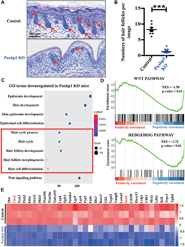
Fig. 1. Absence of Paxbp1 leads to a reduction in the number of hair follicles. (A) Haematoxylin & eosin staining of sections from abdominal skins of newborn Control and Paxbp1 KO (homozygous) animals. Red arrows mark hair follicle architectures in the skin section of Control and Paxbp1 KO (homozygous) animals. Scale bar = 100 μm. (B) Quantification data of hair follicle numbers in different groups (n = 8 for each group). (C) GO term enrichment analysis of downregulated genes in Paxbp1 KO epidermis. Please note that signalling pathways related to hair follicle development and function are highlighted in a red box. (D) The Wnt and Hedgehog signalling pathways associated with Paxbp1 in Control and Paxbp1 KO mice were explored by GSEA (gene set enrichment analysis). (E) Heatmap showing differentially expressed genes (DEGs) associated with hair follicle development and function in Control and Paxbp1 KO epidermis. Three independent RNA-Seq experiments are shown in each group. ***p < 0.001, compared with the indicated controls.
To further investigate the potential mechanisms underlying the abnormal development of hair follicles caused by Paxbp1 deletion, we conducted an RNA sequencing analysis using epidermis from control and Paxbp1 KO mice. Global transcriptome analysis revealed profound gene expression differences between the 2 groups (5). Subsequently, gene ontology (GO) term analysis and gene set enrichment analysis (GSEA) were performed to explore the potential biological functions in which these DEGs could be involved. As expected, the GO term analysis revealed that biological processes, including epidermis development, skin development, and epidermal cell differentiation were significantly enriched in the DEGs between control and Paxbp1 KO epidermis (5). Interestingly, downregulated DEGs were also enriched in processes related to hair cycle process and hair follicle development in the mRNA profiles of Paxbp1 KO mice (Fig. 1C). Moreover, the GSEA analysis indicated that the Paxbp1 KO group was correlated with Wnt and Hedgehog signalling pathways (Ref 5, Fig. 1D), both of which play crucial roles in regulating hair follicle development and function (6, 7). Collectively, these results prompted us to further conduct a more detailed comparison of the hair follicle development-related genes at the transcriptome level. Indeed, the heatmap analysis showed that a number of DEGs specific to hair follicle development and function were evidently altered in Control and Paxbp1 KO mice, with the mRNA levels of genes including Shh, Lhx2, Foxi3, Edar, etc., markedly downregulated in the Paxbp1 KO group (Fig. 1E).
Moreover, we performed qPCR analysis to examine the altered genes indicated by the RNA-Seq analysis. The qPCR results confirmed that genes related to hair follicle development and function, including Shh, Lhx2, Foxi3, Edar, etc., were significantly decreased in Paxbp1-deficient mice (Fig. 2A). Accordingly, hair follicle development, evaluated by Lhx2 and Sox9 immunohistochemical staining, was significantly blocked in Paxbp1 KO mice compared with control littermates (Fig. 2B). These results suggest that the Paxbp1-deficient murine exhibits obvious hair loss by altering molecules involved in hair follicle cycling and maintenance.
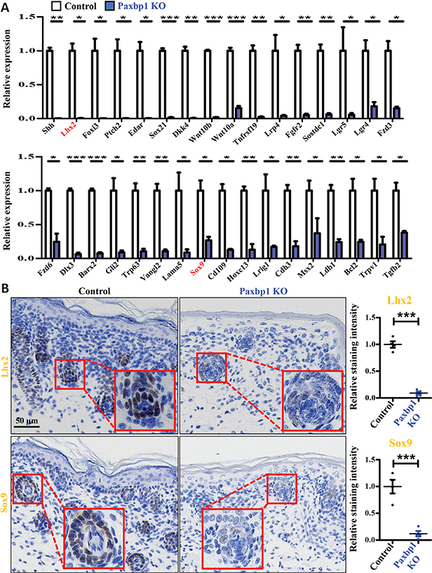
Fig. 2. Hair follicle development is dysregulated in Paxbp1 KO epidermis. (A) Real-time quantitative PCR (RT-qPCR) analysis was performed to validate the differentially expressed genes (DEGs) related to hair follicle development and function in Control and Paxbp1 KO mice (n = 3). (B) Immunohistochemistry staining for hair follicle development-related molecules, including Lhx2 and Sox9, was performed using skin tissues of Control and Paxbp1 KO mice. Nuclei are counterstained with haematoxylin. Scale bar = 50 μm. Red arrows indicate the positive staining. Image-Pro Plus 6.0 software was used to analyse the relative staining intensity of these proteins in Control and Paxbp1 KO epidermis (n = 4 for each group). ***p < 0.001, **p < 0.01, *p < 0.05, compared with the indicated controls.
One limitation of our study is that mice heterozygous for the floxed Paxbp1 allele appeared normal, when compared with control siblings (5). This could be explained by genetic compensatory mechanisms (8). To avoid inefficient deletion of the Paxbp1 gene, we focused our subsequent study on Paxbp1 KO (homozygous) and control mice. In addition, further studies are expected to shed light on the precise mechanism of Paxbp1 in hair follicle development. For example, whether those downregulated genes correlated with hair follicle function are directly targeted by Paxbp1 or not remains unexplored. Thus, chromatin immunoprecipitation sequencing (ChIP-Seq) will be helpful to identify the direct targets of Paxbp1 in future studies.
In conclusion, our data reveal a critical role of Paxbp1 in modulating hair follicle development in a murine model. Future studies assessing Paxbp1 activity in hair follicles may provide a novel model and potential therapeutic approach to treat alopecia.
ACKNOWLEDGEMENTS
IRB approval status: All animal experiments were approved by the Committee for the Ethics of Animal Experiments, Shenzhen Peking University-The Hong Kong University of Science and Technology Medical Center.
REFERENCES
- Diao Y, Guo X, Li Y, Sun K, Lu L, Jiang L, et al. Pax3/7BP is a Pax7- and Pax3-binding protein that regulates the proliferation of muscle precursor cells by an epigenetic mechanism. Cell Stem Cell 2012; 11: 231–241. https://doi.org/10.1016/j.stem.2012.05.022
- Alharby E, Albalawi AM, Nasir A, Alhijji SA, Mahmood A, Ramzan K, et al. A homozygous potentially pathogenic variant in the PAXBP1 gene in a large family with global developmental delay and myopathic hypotonia. Clin Genet 2017; 92: 579–986. https://doi.org/10.1111/cge.13051
- Zhou S, Han L, Weng M, Zhu H, Heng Y, Wang G, et al. Paxbp1 controls a key checkpoint for cell growth and survival during early activation of quiescent muscle satellite cells. Proc Natl Acad Sci U S A 2021; 118: e2021093118. https://doi.org/10.1073/pnas.2021093118
- Li W, Yang Y, Liu S, Zhang D, Ren X, Tang M, et al. Paxbp1 is indispensable for the survival of CD4 and CD8 double-positive thymocytes. Front Immunol 2023; 14: 1183367. https://doi.org/10.3389/fimmu.2023.1183367
- Huang C, Liu S, Li W, Zhao S, Ren X, Zhuo F, et al. Paxbp1 is indispensable for the maintenance of epidermal homeostasis. J Invest Dermatol 2025; 145: 864–875. https://doi.org/10.1016/j.jid.2024.08.012
- Rishikaysh P, Dev K, Diaz D, Qureshi WM, Filip S, Mokry J. Signaling involved in hair follicle morphogenesis and development. Int J Mol Sci 2014; 15: 1647–1670. https://doi.org/10.3390/ijms15011647
- Frech S, Forsthuber A, Korosec A, Lipp K, Kozumov V, Lichtenberger BM. Hedgehog signaling in papillary fibroblasts is essential for hair follicle regeneration during wound healing. J Invest Dermatol 2022; 142: 1737–1748. https://doi.org/10.1016/j.jid.2021.11.026
- El-Brolosy MA, Stainier DYR. Genetic compensation: a phenomenon in search of mechanisms. PLoS Genet 2017; 13: e1006780. https://doi.org/10.1371/journal.pgen.1006780