QUIZ SECTION
A Thick-walled Pink Bulla with a Firm Nodule Underneath: A Quiz
Moe KANAYA1, Yasuhiro MITSUI1,2* , Chinatsu SHOBATAKE2, Satoka TATEBAYASHI3 and Takashi NAKANISHI3
1Department of Dermatology, Nara Prefecture General Medical Center, Nara, Japan, 2Department of Dermatology, Nara Medical University School of Medicine, Kashihara, Japan, 3Department of Plastic Surgery, Nara Prefecture General Medical Center, Nara, Japan. *E-mail: nagaratenroku@yahoo.co.jp
Citation: Acta Derm Venereol 2025; 105: adv43674. DOI: https://doi.org/10.2340/actadv.v105.43674.
Copyright: 2025 © The Author(s). Published by MJS Publishing, on behalf of the Society for Publication of Acta Dermato-Venereologica. This is an Open Access article distributed under the terms of the Creative Commons Attribution-NonCommercial 4.0 International License (https://creativecommons.org/licenses/by-nc/4.0/).
Published: May 16, 2025.
Competing interests and funding: The authors have no conflicts of interest to declare.
A 20-year-old man was referred for treatment of a cutaneous “cyst” on his left upper arm. A few months previously, the lesion had grown substantially, and he had no history of trauma. Physical examination revealed a thick-walled asymptomatic pink bulla, measuring 47 × 28 mm in size (Fig. 1), with palpation revealing a firm mass within. A greyscale ultrasound image showed a heterogeneous hyperechoic mass with scattered punctate echogenic foci, along with posterior acoustic shadowing. The overlying dermis contained anechoic areas (Fig. 2A). Color Doppler imaging exhibited intratumoral hypervascularity (Fig. 2B).
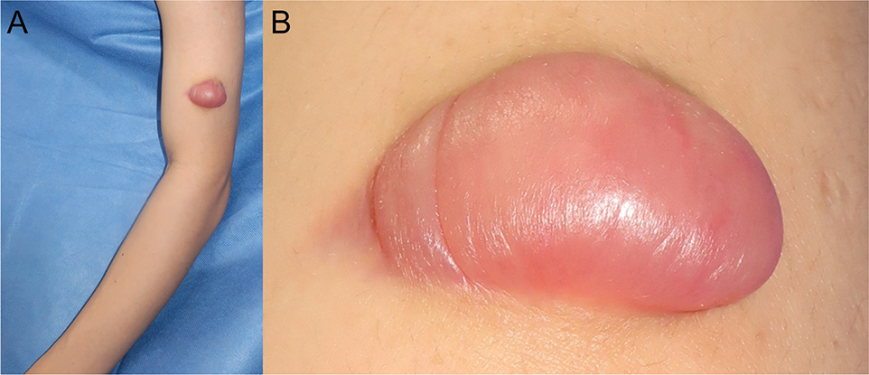
Fig. 1. Clinical presentation. (A) Photograph demonstrating a 4-cm pink bulla on the left upper arm of a 20-year-old man. (B) Close-up view.

Fig. 2. Greyscale and colour Doppler ultrasound images. (A) Greyscale ultrasound image showing an oval-shaped heterogeneous hyperechoic mass involving the dermis and subcutis (asterisk) with scattered hyperechoic punctate foci and posterior acoustic shadowing. The dermis contains anechoic areas (arrowheads). (B) Colour Doppler imaging showing intratumoral hypervascularity.
What is your diagnosis?
1: Keloid
2: Epidermoid cyst
3: Bullous pilomatricoma
4: Dermatofibrosarcoma protuberans
See next page for answer.
ANSWERS TO QUIZ
A Thick-walled Pink Bulla with a Firm Nodule Underneath: A Commentary
Diagnosis: Bullous pilomatricoma
Excisional biopsy revealed a dermal and hypodermal nodule consisting of basophilic and eosinophilic shadow cells (Fig. 3A). The overlying dermis showed separation of collagen bundles in an oedematous stroma (Fig. 3B). Higher magnification showed scattered calcifications within the nodule (Fig. 3C). Accordingly, a diagnosis of bullous pilomatricoma was established.

Fig. 3. Histopathology. (A) Histopathology of a resected specimen revealing a well-circumscribed nodule involving the dermis and subcutis composed of basophilic and eosinophilic shadow cells with scattered foci of calcification (haematoxylin–eosin [HE] stain, original magnification ×10). (B) Markedly oedematous dermis with separation of collagen bundles (HE, ×200). (C) Close-up view of calcification and shadow cells (HE, ×400). Scale bars=5 mm (A), 200 μm (B), and 50 μm (C).
Bullous pilomatricoma, also called anetodermic pilomatricoma, is an uncommon variant that presents clinically as a semi-transparent wrinkled blister with a firm nodule underneath (1). The lesions occur mostly in children and young adults (1). The surface coloration varies: most are red, and the others are blue, purple, or normal skin colour (1). While pilomatricoma mainly affect the head and neck (2), the bullous variant occurs predominantly on the upper arm and shoulders (1). Theories have been proposed to explain the mechanism of bullae formation, including mechanical irritation, obstruction of lymphatic vessels resulting in congestion and leakage of lymphatic fluids (1), and degradation of elastic fibres and collagen by matrix metalloproteinases-9 and -12 (3). In our case, the patient reported no history of trauma, and immunohistochemistry for D2-40 did not show lymphatic vessels lining dermal spaces.
Bullous pilomatricoma can be misdiagnosed as keloid and dermatofibrosarcoma protuberans (DFSP) (4). In the present case, a cyst was initially suspected. To avoid misdiagnosis, enhanced awareness of bullous pilomatricoma and palpation of the mass within a blister is critical. The clinical context of its mimickers may help exclude the differential diagnoses. Keloid usually occurs in areas of previous trauma and its development is associated with genetic and environmental factors. DFSP commonly affects the trunk and leg (5). For clinical diagnosis of an epidermoid cyst, identification of a punctum is useful, and compression of the cyst accentuates the appearance of the punctum (6).
Ultrasound may help distinguish bullous pilomatricoma from other differential diagnoses. On ultrasound, pilomatricomas most often appear as hyperechoic nodules with posterior acoustic shadowing and show vascularity on colour Doppler imaging (5). As for the bullous variant, the tumour shares sonographic features with the common type, and the stroma, consisting of -remarkably oedematous connective tissue, can show anechoic lagoons (7), similar to those seen in our case. In contrast, epidermoid cysts typically present as hypoechoic nodules with posterior acoustic enhancement and lack vascularity (5). DFSP mainly presents as a homogeneous, isoechoic/hypoechoic mass with vascularity (5). Keloid presents as a hypoechoic mass with or without vascularity and rarely shows hyperechoic spots representing calcification (8).
In conclusion, we present a case of bullous pilomatricoma with sonographic evaluation. This variant is uncommon, and our report will enhance its awareness. Ultrasound and colour Doppler may help in the identification of pilomatricoma before invasive treatment.
REFERENCES
- Chen SY, Wu F, Qian Y, Zhu L, Tu YT, Huang CZ. Pilomatricoma with bullous appearance: a case report and review of literature. Int J Dermatol 2011; 50: 615–618. https://doi.org/10.1111/j.1365-4632.2010.04586.x
- Julian CG, Bowers PW. A clinical review of 209 pilomatricomas. J Am Acad Dermatol 1998; 39: 191–195. https://doi.org/10.1016/S0190-9622(98)70073-8
- Motegi SI, Uehara A, Fujiwara C, Sekiguchi A, Ishikawa O. Pilomatricoma with bullous-like/anetodermic appearance: possibly associated with matrix metalloproteinases. J Dermatol 2018; 45: 505–506. https://doi.org/10.1111/1346-8138.14209
- Yu N, Zhang M, Chen C, Liang Z, Meng T, Pan D, et al. Giant pilomatrixoma with bullous appearance on the back: a case report and discussion of misdiagnosis. J Cosmet Dermatol 2022; 21: 4245–4248. https://doi.org/10.1111/jocd.14934
- Zhu H, Wu S. Integrating vascularity into the pattern classification of pilomatricomas on ultrasound provides a more competent approach for discriminative evaluation. Skin Res Technol 2024; 30: e13585. https://doi.org/10.1111/srt.13585
- Borg Grech S, Mintoff D, Scerri L. The punctum sign in epidermal cyst. Clin Exp Dermatol 2024; 49: 275–276. https://doi.org/10.1093/ced/llad380
- Rodríguez Bandera AI, Stewart N, Herranz Pinto P. High-frequency ultrasonography of bullous pilomatricoma. Skin Res Technol 2020; 26: 608–609. https://doi.org/10.1111/srt.12828
- Lobos N, Wortsman X, Valenzuela F, Alonso F. Color Doppler ultrasound assessment of activity in keloids. Dermatol Surg 2017; 43: 817–825. https://doi.org/10.1097/DSS.0000000000001052