SHORT COMMUNICATION
Sexually Transmitted Psoriasis-like Kerion of the Pubis and Vulva
Stefano VERALDI1, Paolo BORTOLUZZI2, Francesca GERMINIASI2 and Gianluca NAZZARO2
1Dermatological Centre in Milan, Milan, and 2Department of Pathophysiology and Transplantation, Università degli Studi, IRCCS Foundation, Ca’Granda Ospedale Maggiore Policlinico, Milan, Italy. E-mail: stefano.veraldi@dcim.it
Citation: Acta Derm Venereol 2025; 105: adv43744. DOI: https://doi.org/10.2340/actadv.v105.43744.
Copyright: © 2025 The Author(s). Published by MJS Publishing, on behalf of the Society for Publication of Acta Dermato-Venereologica. This is an Open Access article distributed under the terms of the Creative Commons Attribution-NonCommercial 4.0 International License (https://creativecommons.org/licenses/by-nc/4.0/).
Submitted: Apr 30, 2025; Accepted after revision: Jun 2, 2025; Published: Jun 16, 2025.
INTRODUCTION
Fungal infections of the pubis and vulva may be divided into 2 different clinical presentations: tinea pubis and kerion (1–21). Tinea pubis, also named pubic tinea (4), tinea genitalis (11, 13, 19), tinea in the genital area (13), and pubogenital tinea (13, 18, 19), is characterized clinically by roundish lesions, with well-defined, erythematous, vesicular borders and an inner area with mild erythema, scales and crusts. Kerion, or kerion celsi, also named trichophytic granuloma (1), Majocchi’s granuloma (2, 12, 15), cystic tinea profunda (3), and tinea profunda (3, 5, 12), is characterized by inflammatory, infiltrated plaques, with follicular pustules. A case of sexually transmitted psoriasis-like kerion of the pubis and vulva is reported here.
CASE REPORT
A 29-year-old Caucasian woman was admitted with a clinical diagnosis of psoriasis located on the pubis and vulva. The patient stated that she was in good general health and that she was not on therapy with systemic drugs. She also declared that her dermatitis had appeared 4 months earlier. It was treated at another dermatological centre with topical calcipotriol and corticosteroids; however, both drugs caused a worsening of the dermatitis.
Dermatological examination showed the presence of 2 lesions: the first lesion was located in the centre of the pubis: it was erythematous-squamous, infiltrated, roundish, 1.5 cm in diameter; the second lesion involved the mons veneris and labia majora and was erythematous-squamous, infiltrated, with tiny follicular pustules. Pubic hairs were of different lengths (Fig. 1). The patient complained of mild itching. No other lesions were observed elsewhere.
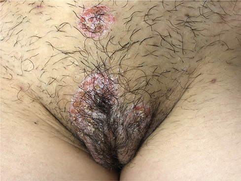
Fig. 1. Psoriasis-like kerion of the pubis and vulva.
General physical examination did not reveal anything pathological; in particular, no inguinal lymphadenopathy was detected.
Laboratory tests were within normal ranges. The patient refused a biopsy. Bacteriological examinations were negative. Mycological cultures were positive for T. mentagrophytes from pubic and vulval lesions, and negative from inguinal and intergluteal folds.
A thorough medical history revealed that the patient’s boyfriend, who worked as a veterinarian, had been suffering from tinea inguinalis for months. We therefore decided to carry out in our patient laboratory tests for syphilis, HIV, and hepatitis C (negative results), swabs for Herpes Simplex Virus types 1 and 2 (negative results), urethral swabs for real-time PCR for C. trachomatis, M. genitalium, and N. gonorrhoeae (negative results) and microscopical examinations and cultures for N. gonorrhoeae (negative results). Vaginal swabs for fungi and bacteria were also negative.
Mycological examinations of the inguinal folds of the patient’s boyfriend were positive for T. mentagrophytes. It is likely that he was infected by cats or dogs he repeatedly touched in his job.
Final diagnosis was sexually transmitted psoriasis-like kerion of the pubis and vulva. The patient was treated with itraconazole (200 mg/day for 3 weeks) and isoconazole cream (1 application/day for 3 weeks). No side effects were reported or observed. -Complete clinical remission was observed 18 days after the beginning of the therapy. Mycological examinations were negative at day 21 of therapy. Follow up (4 months) was negative.
DISCUSSION
Kerion of the pubis and vulva is often caused by T. mentagrophytes (3, 6–10,12, 17, 20, 21) (also T. interdigitale [11, 13, 19] and Arthroderma/T. benhamiae [13, 14, 18, 21]). Other involved species are T. erinacei (7), T. rubrum (3, 13, 15, 19, 21), T. tonsurans (13), T. verrucosum (5, 13), and M. canis (1, 13). Mixed fungal/bacterial infections have also been reported: T. mentagrophytes + E. coli + P. aeruginosa (4) and T. mentagrophytes + P. aeruginosa + Klebsiella sp. (20).
Predisposing factors include contacts with animals such as mice (13), rats (13, 14), guinea pigs (18), cats (5, 13) and dogs (2); travels in endemic areas (11, 13); sexual intercourse, as in our patient (11, 15, 17–19, 21); poor personal hygiene (19); hyperhidrosis (19); sharing clothes (underwear, bed linens, towels) (19); shaving (13, 15, 17, 18, 20); waxing (12); and autoinoculation from other infected areas (19). Chronic use of topical (2, 15, 19) and oral (3) corticosteroids is also considered as a predisposing factor. Except for very rare cases in children (6, 8), kerion in the pubic and genital area occurs much more frequently in women (13).
The clinical picture of kerion of the pubis and vulva is characterized by alopecia (5, 6, 10–12), lymphadenopathy (1, 6, 8, 15), pain (11, 21) and fever (15).
Itraconazole (2, 4, 7, 10, 11, 13, 15) and terbinafine (9, 12, 13, 15, 17) are the 2 most frequently used antifungals. As previously mentioned, we successfully used itraconazole. Other drugs used are fluconazole (3, 13) griseofulvin (1, 6, 8), and amphotericin B (3). Some authors have added systemic corticosteroids (2, 6, 11).
In conclusion, we have described a case of kerion -located on the pubis and vulva. The infection was acquired by the patient through intercourse with her boyfriend, who in turn had been infected by cats or dogs during his work as a veterinarian. The case we have reported is in our opinion interesting for several reasons: (a) the atypical clinical presentation of kerion; (b) the confirmation that kerion of the pubis and vulva can be considered as a true sexually transmitted infection, and (c) once again, the great adaptive capacity of an originally zoophilic species, such as T. mentagrophytes.
REFERENCES
- Margolis DJ, Weinberg JM, Tangoren IA, Cheney RT, Johnson BL Jr. Trichophytic granuloma of the vulva. Dermatology 1998; 197: 69–70. https://doi.org/10.1159/000017961
- Chang SE, Lee DK, Choi JH, Moon KC, Koh JK. Majocchi’s granuloma of the vulva caused by Trichophyton mentagrophytes. Mycoses 2005; 48: 382–384. https://doi.org/10.1111/j.1439-0507.2005.01147.x
- Kobayashi M, Ishida E, Yasuda H, Yamamoto O, Tokura Y. Tinea profunda cysticum caused by Trichophyton rubrum. J Am Acad Dermatol 2006; 54: S11–13. https://doi.org/10.1016/j.jaad.2005.03.058
- Barile F, Filotico R, Cassano N, Vena GA. Pubic and vulvar inflammatory tinea due to Trichophyton mentagrophytes. Int J Dermatol 2006; 45: 1375–1377. https://doi.org/10.1111/j.1365-4632.2006.02793.x
- Grunewald S, Paasch U, Gräser Y, Glander HJ, Simon JC, Nenoff P. Vernarbende Tinea profunda des Mons pubis durch Trichophyton verrucosum. Hautarzt 2006; 57: 811–813. https://doi.org/10.1007/s00105-005-1066-7
- Annabel M, Hubert L, Jacques C, Fabienne L, Emmanuelle L, Gérard L. Traitement de 2 cas de kérions par griséofulvine et corticoïdes oraux. Arch Pediatr 2009; 16: 1464–1466. https://doi.org/10.1016/j.arcped.2009.07.023
- Feldmann R, Riegler E, Willinger B, Steiner A, Breier F. 54-jährige Frau mit perigenitalen follikulären Pusteln. Pilzfollikulitis durch Trichophyton erinacei. J Dtsch Dermatol Ges 2011; 9: 249–251. https://doi.org/10.1111/j.1610-0387.2010.07613.x
- Hali F, Khadir K, Benchikhi H. Kérion du pubis chez une adolescente. Presse Med 2013; 42: 1149–1150. https://doi.org/10.1016/j.lpm.2012.09.013
- Shreberk-Hassidim R, Ramot Y, Maly A, Horev L, Zlotogorski A. Kerion celsi of the vulva: an unusual location. J Mycol Med 2014; 24: 359–360. https://doi.org/10.1016/j.mycmed.2014.06.050
- Bougrine A, Villeneuve-Tang C, Bouffard D, Rouleau D, Chartier S. Kerion of the vulva caused by Trichophyton mentagrophytes. J Cutan Med Surg 2014; 18: 206–209. https://doi.org/10.2310/7750.2013.13101
- Luchsinger I, Bosshard PP, Kasper RS, Reinhardt D, Lautenschlager S. Tinea genitalis: a new entity of sexually transmitted infection? Case series and review of the literature. Sex Transm Infect 2015; 91: 493–496. https://doi.org/10.1136/sextrans-2015-052036
- Bakardzhiev I, Chokoeva A, Tchernev G, Wollina U, Lotti T. Tinea profunda of the genital area: successful treatment of a rare skin disease. Dermatol Ther 2016; 29: 181–183. https://doi.org/10.1111/dth.12311
- Ginter-Hanselmayer G, Nenoff P, Kurrat W, Propst E, Durrant-Finn U, Uhrlaß S, et al. Tinea im Genitalbereich. Eine diagnostische und therapeutische Herausforderung. Hautarzt 2016; 67: 689–699. https://doi.org/10.1007/s00105-016-3848-5
- Hiernickel C, Wiegand C, Schliemann S, Seyfarth F, Jung K, Elsner P, et al. Trichophyton Spezies von Arthroderma benhamiae. Klinisch therapeutische Aspekte eines neuen Erregers in der Dermatologie. Hautarzt 2016; 67: 706–711. https://doi.org/10.1007/s00105-016-3837-8
- Rallis E, Katoulis A, Rigopoulos D. Pubic Majocchi’s granuloma unresponsive to itraconazole successfully treated with oral terbinafine. Skin Appendage Disord 2016; 1: 111–113. https://doi.org/10.1159/000438706
- Gracia-Cazaña T, Milagro A, Rezusta A, Gilaberte Y. Querion de Celso de la vulva: una enfermedad común en una localización infrecuente. Rev Iberoam Micol 2017; 34: 123–125. https://doi.org/10.1016/j.riam.2016.11.003
- Dickison P, Forward E, Fischer G. Fungal kerion of the vulva. Australas J Dermatol 2018; 59: 333–334. https://doi.org/10.1111/ajd.12777
- Wagenknecht D, Simon JC, Treudler R, Nenoff P. Pubogenital tinea after intimate shaving with isolation of Trichophyton benhamiae. J Dtsch Dermatol Ges 2018; 16: 596–598. https://doi.org/10.1111/ddg.13500
- Thakur R, Kushwaha P, Kalsi AS, Singh P. Tinea genitalis in a rural tertiary care hospital of Western U.P. India. Indian J Clin Exp Dermatol 2018; 4: 266–273. https://doi.org/10.18231/2581-4729.2018.0056
- Saadia A, Reckhow J, Rozenblat M, Last O. Kerion of the pubis and vulva with bacterial superinfection: a rare occurrence. BMJ Case Rep 2019; 12: e230320. https://doi.org/10.1136/bcr-2019-230320
- Kupsch C, Czaika VA, Deutsch C, Gräser Y. Trichophyton mentagrophytes – ein neuer Genotyp des zoophilen Dermatophyten verursacht sexuell übertragbare Infektionen. J Dtsch Dermatol Ges 2019; 17: 493–501. https://doi.org/10.1111/ddg.13776_g