SHORT COMMUNICATION
The Regression Heterogeneity Pattern of Infantile Haemangiomas after Treatment with Propranolol: A Multicentre Retrospective Study
Kaizhi ZHANG1#, Maierhaba TUERSUNTUOHETI1#, Wei SHAN2, Fan HU3 and Yi JI1*
1Division of Oncology, Department of Pediatric Surgery, West China Hospital of Sichuan University, Chengdu, 2Department of Pediatric Surgery, Sichuan Women and Children’s Hospital, Chengdu, and 3Department of Vascular & Interventional Radiology, West China Second University Hospital of Sichuan University, Chengdu, China. E-mail: jijiyuanyuan@163.com
#These authors contributed equally to this work.
Citation: Acta Derm Venereol 2025; 105: adv44079. DOI: https://doi.org/10.2340/actadv.v105.44079.
Copyright: © 2025 The Author(s). Published by MJS Publishing, on behalf of the Society for Publication of Acta Dermato-Venereologica. This is an Open Access article distributed under the terms of the Creative Commons Attribution-NonCommercial 4.0 International License (https://creativecommons.org/licenses/by-nc/4.0/).
Submitted: Jun 5, 2025. Accepted after revision: Sep 5, 2025. Published: Sep 23, 2025.
Competing interests and funding: The authors have no conflicts of interest to declare.
This work was supported by the National Natural Science Foundation of China (grant numbers 82473553 and 82273556), the Excellent Youth Foundation of Sichuan Province of China (grant number 2025NSFJQ0070), the Project of “0–1” of Sichuan University (grant number 2022SCUH0033), the Med-X Center for Informatics Funding Project (grant number YGJC004), the 1.3.5 Project for Disciplines of Excellence Clinical Research Incubation Project of West China Hospital of Sichuan University (grant number 2023HXFH004), and the 1.3.5 Project for Disciplines of Excellence Clinical Research Interdisciplinary Innovation Project of West China Hospital of Sichuan University (grant number ZYJC21060).
INTRODUCTION
Infantile haemangioma (IH) is among the most common benign tumours, occurring in 4.5% of the infant population, and can be accompanied by a risk of dysfunction, ulceration, and disfigurement (1–4). Studies have reported that segmental IHs on the head and face may lead to PHACE syndrome (posterior fossa, haemangioma, arterial lesions, cardiac abnormalities, and eye abnormalities), segmental IHs in the lumbosacral and perineal regions may lead to LUMBAR syndrome (lower body haemangioma, urogenital anomalies, ulceration, myelopathy, bony deformities, anorectal abnormalities, and renal abnormalities), and segmental IHs on the hands and feet may have a “biker glove” pattern (5–7). These findings indicate that there may be different patterns for IHs in different regions. Clinically, regression differences are observed for perioral, hand, and anogenital IHs involving 2 opposite regions after treatment with propranolol. To date, this phenomenon has not been specifically studied. Therefore, the current study aimed to identify the clinical characteristics and regression features of these IHs.
MATERIALS AND METHODS
This was a multicentre retrospective study that included 3 tertiary medical centres. Approval was obtained from the local institutional review boards at each participating site. The data (photographs, basic information, clinical characteristics, and complications) of patients with IHs from January 2017 to June 2023 were obtained. Given that only deidentified data were collected, the need for informed consent was waived. The inclusion criteria were as follows: (i) age at treatment initiation of 1 to 6 months and (ii) perioral, hand, or anogenital IHs involving 2 opposite regions and treated with oral propranolol for at least 6 months. The exclusion criteria were as follows: (i) patients with deep IHs and (ii) patients with incomplete clinical data.
Patients with 2-sided IHs were defined as those with IHs involving 2 opposite regions (external vs internal). External side included the dorsal side of the hand, the region above and/or below the lip, and the outside of the natural closure of the lip (if the other side of the IH was within the inside of the natural closure of the lip), extending vertically from the mons pubis along the natural closure of the bilateral labia majora outside the sacral concavity or from the root of the penis on both sides to the area outside the sacral concavity. Internal side included the volar side of the hand, the outside of the natural closure of the lip (if the other side of the IH was above and/or below the lip), and the inside of the natural closure of the lip, extending vertically from the mons pubis along the natural closure of the bilateral labia majora to the area between the sacral concavity or from the root of the penis on both sides to the area between the sacral concavity (Fig. 1). Regression heterogeneity was defined as a difference of ≥ 0.5 in the Hemangioma Activity Score (HAS) between IHs on external and internal sides after treatment with propranolol at month 6 (Fig. 2). HAS assessment was performed according to the methods of Janmohamed et al. (8). Satisfactory regression was defined as complete or nearly complete regression of IH lesions.
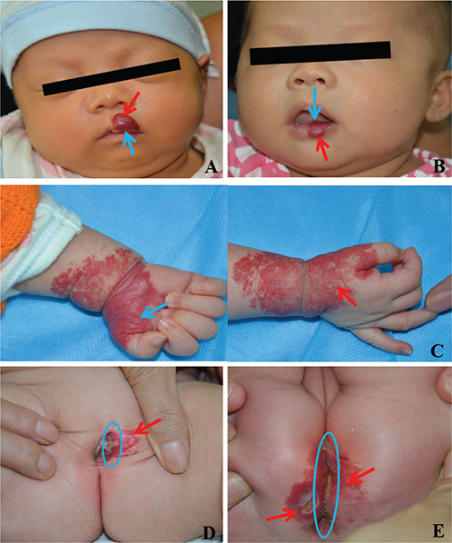
Fig. 1. Schematic diagrams of external and internal sides of IHs. (A–E): Red arrows refer to the external side, and blue arrows and circles refer to the internal side in patients with IH.
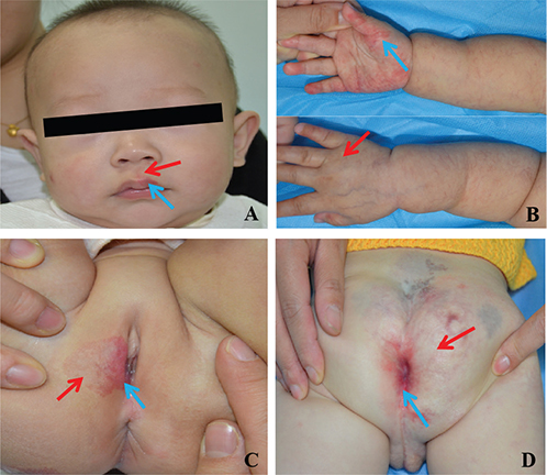
Fig. 2. Pictures of IHs in perioral, hand, and anogenital regions exhibiting regression heterogeneity. Regression of tumours on the external side (red arrows) was better than that of tumours on the internal side (blue arrows).
The primary outcome measure was the presence or absence of regression heterogeneity pattern of IHs. The secondary outcome measures were the time to achieve satisfactory regression of IHs and the presence of ulceration. All patients followed a standardized treatment regimen with propranolol (administered orally at 1.0 mg/kg/day in 3 doses over 1 week and then increased to 2.0 mg/kg/day in 3 doses from week 2 onwards). The data of all the patients were collected and reviewed by professionals at each centre. Finally, the photographs and other data were reviewed and summarized again by 2 professionals. Disagreements were resolved via discussion among the research team.
Frequencies (percentages) and medians (interquartile ranges, IQRs) were used to present the clinical data. For qualitative variables, the χ2 test or Fisher’s exact test was used for comparisons between groups. Quantitative variables were analysed by using Student’s t-test or the Mann‒Whitney U test. Multivariate logistic analysis was conducted for variables for which p < 0.1. SPSS 26.0 software was used for statistical analysis (IBM Corp, Armonk, NY, USA). P < 0.05 was considered to indicate statistical significance, and the odds ratio (OR) and 95% confidence interval (CI) were calculated.
RESULTS
A total of 75 patients with IHs were included in this study. There were 57 cases (76.0%) with regression heterogeneity; the IHs were located in the perioral region in 64.1% of cases, the hand region in 90% of cases, and the anogenital region in 88.5% of cases. Anogenital (p = 0.049) and segmental and indeterminate (p = 0.047) 2-sided IHs more commonly exhibited regression heterogeneity. Additional information on the patients is provided in Table I.
Regarding the regression heterogeneity pattern of IHs, the average HAS after treatment with propranolol at month 6 was significantly greater for the internal side than for the external side (1.97 ± 0.68 vs 1.02 ± 0.62, p < 0.001). The regression of all external tumours was better than that of internal tumours after treatment with propranolol at month 6. Three patients with regression heterogeneity did not have satisfactory regression times during follow-up. The median times for satisfactory regression of the remaining IHs with regression heterogeneity in external and internal sides were 7.3 and 16.5 months, respectively (p < 0.001). Additionally, ulceration was more common in external side for IHs with regression heterogeneity (p = 0.006).
DISCUSSION
The regression heterogeneity pattern of IHs was not accidental, and its manifestation resembled the “biker glove” (7), indicating that IHs in certain locations may exhibit unique biological behaviour and that regression heterogeneity was a significant manifestation of this particularity. Our findings revealed that regression heterogeneity was associated with a high incidence of anogenital and nonlocalized 2-sided IHs, indicating that anatomical location and morphological subtype were critical factors in determining whether regression heterogeneity occurs. Regarding the regression heterogeneity, IHs on the internal side generally need to be treated over a longer term than those on the external side. These observations underscore the imperative need for individualized treatment strategies for IHs and provide new insights into the mechanism of IH regression. Additionally, explaining the differences in the regression of IHs to patients’ parents can help alleviate their anxiety.
Regression heterogeneity may be associated with differences in tissue structure, the microenvironment, and the response to oral propranolol between IHs on the external and internal sides and mosaic genetic patterns. Clinically, we did not observe regression heterogeneity patterns of IHs on the nose or ears, but they were evident in the IHs on the hands. It is plausible that 2-sided IHs on the feet may also exhibit regression heterogeneity. Future studies with larger sample sizes and prospective designs are needed to confirm these patterns.
This study has several limitations. First, this was a retrospective study, and the photographs obtained were not standardized, which may have introduced variability in assessing the regression heterogeneity of IHs. Second, because there were no cases of 2-sided IHs on the feet at the 3 medical institutions, it was impossible to analyse the regression heterogeneity of such IHs. Finally, the small sample size for certain subgroups (patients with IHs on the hands, n = 10) may have limited the statistical power and affected the generalizability of subgroup-specific findings, particularly in detecting subtle associations between clinical factors and regression heterogeneity patterns.
In conclusion, this study identified a regression heterogeneity pattern in 2-sided IHs on the perioral, hand, and anogenital regions after treatment with propranolol. The internal side of these IHs required a significantly longer treatment duration for satisfactory regression. These findings emphasize the importance of individualized treatment strategies, provide new insights into the mechanism of IH regression, and establish a bridge between basic research and clinical practice.
ACKNOWLEDGEMENTS
The authors are indebted to all the patients in this study.
IRB approval status: This study was approved by the Ethics Committee of West China Hospital of Sichuan University, Sichuan Women and Children’s Hospital, and West China Second University Hospital (number: 2017--414).
Data availability statement: The data that support the findings of this study are available from the corresponding author upon reasonable request.
REFERENCES
- Munden A, Butschek R, Tom WL, Marshall JS, Poeltler DM, Krohne SE, et al. Prospective study of infantile haemangiomas: incidence, clinical characteristics and association with placental anomalies. Br J Dermatol 2014; 170: 907–913. https://doi.org/10.1111/bjd.12804
- Krowchuk DP, Frieden IJ, Mancini AJ, Darrow DH, Blei F, Greene AK, et al. Clinical practice guideline for the management of infantile hemangiomas. Pediatrics 2019; 143: e20183475. https://doi.org/10.1542/peds.2018-3475
- Rodríguez Bandera AI, Sebaratnam DF, Wargon O, Wong LF. Infantile hemangioma. Part 1: Epidemiology, pathogenesis, clinical presentation and assessment. J Am Acad Dermatol 2021; 85: 1379–1392. https://doi.org/10.1016/j.jaad.2021.08.019
- Macca L, Altavilla D, Di Bartolomeo L, Irrera N, Borgia F, Li Pomi F, et al. Update on treatment of infantile hemangiomas: what’s new in the last five years. Front Pharmacol 2022; 13: 879602. https://doi.org/10.3389/fphar.2022.879602
- Zhang K, Xiang S, Zhou J, Qiu T, Lan Y, Ji Y. PHACE syndrome: looking backward and forward. Orphanet J Rare Dis 2025; 20: 345. https://doi.org/10.1186/s13023-025-03899-7
- Metry D, Copp HL, Rialon KL, Iacobas I, Baselga E, Dobyns WB, et al. Delphi consensus on diagnostic criteria for LUMBAR syndrome. J Pediatr 2024; 272: 114101. https://doi.org/10.1016/j.jpeds.2024.114101
- Weitz NA, Bayer ML, Baselga E, Torres M, Siegel D, Drolet BA, et al. The “biker-glove” pattern of segmental infantile hemangiomas on the hands and feet. J Am Acad Dermatol 2014; 71: 542–547. https://doi.org/10.1016/j.jaad.2014.04.062
- Janmohamed SR, de Waard-van der Spek FB, Madern GC, de Laat PC, Hop WC, Oranje AP. Scoring the proliferative activity of haemangioma of infancy: the Haemangioma Activity Score (HAS). Clin Exp Dermatol 2011; 36: 715–723. https://doi.org/10.1111/j.1365-2230.2011.04080.x