QUIZ SECTION
Erythematous Follicular Papules on the Thighs: A Quiz
Shin IINUMA1,2, Takahiro KOBAYASHI1, Akemi ISHIDA-YAMAMOTO2 and Yasuyuki FUJITA2
1Department of Dermatology, Japanese Red Cross Kitami Hospital, North 6, East 2, Kitami, 090-8666, Japan, and 2Department of Dermatology, Asahikawa Medical University, Asahikawa, Japan. E-mail: iinuma@asahikawa-med.ac.jp
Citation: Acta Derm Venereol 2025; 105: adv44139. DOI: https://doi.org/10.2340/actadv.v105.44139.
Copyright: © 2025 The Author(s). Published by MJS Publishing, on behalf of the Society for Publication of Acta Dermato-Venereologica. This is an Open Access article distributed under the terms of the Creative Commons Attribution-NonCommercial 4.0 International License (https://creativecommons.org/licenses/by-nc/4.0/).
Published: Aug 11, 2025
Competing interests and funding: The authors have no conflicts of interest to declare
A 68-year-old Japanese woman presented with a 1-year history of pruritic papules on the thighs and facial erythema. Physical examination revealed erythematous, keratotic, follicular papules symmetrically distributed along the lateral aspects of the thighs (Fig. 1). Additional findings included erythema on the forehead, eyelids, cheeks, and periungual regions. She reported no systemic symptoms such as fever, photosensitivity, Raynaud phenomenon, arthralgia, muscle pain, or weakness. There was no relevant family history of dermatological conditions. Laboratory investigations, including complete blood count, liver and renal function panels, serum creatine kinase, aldolase, and complement levels, were all within normal limits. Antinuclear antibody testing was negative. Skin biopsy from the right thigh showed a hyperkeratotic mound with parakeratotic foci, mild vacuolar interface alteration, superficial perivascular lymphocytic infiltration, and dermal mucin deposition (Fig. 2). The patient was initially treated with topical corticosteroids (diflucortolone valerate 0.1% cream, applied twice daily); however, the cutaneous lesions demonstrated minimal improvement over the following weeks.
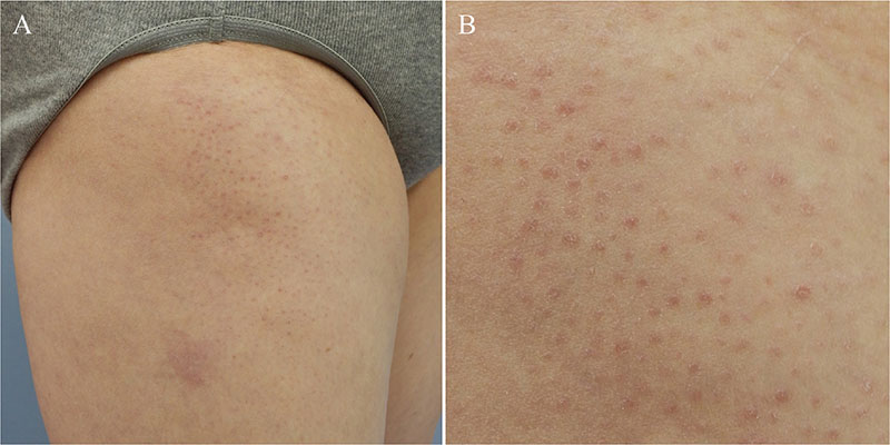
Fig. 1. Clinical presentation. (A) Erythematous, keratotic follicular papules symmetrically distributed along the lateral aspect of the right thigh. (B) Close-up view of the lesions shown in (A).

Fig. 2. Histopathology of the lesion. (A) Skin biopsy from the thigh showing a follicular hyperkeratotic mound. The boxed area corresponds to (B). (B) Hyperkeratotic mound with parakeratotic foci, mild vacuolar interface changes, and superficial perivascular lymphocytic infiltration (haematoxylin–eosin stain; scale bar: 100 µm). (C) Dermal mucin deposition (Alcian blue stain; scale bar: 100 µm).
What is your diagnosis?
1: Systemic lupus erythematosus
2: Dermatomyositis
3: Pityriasis rubra pilaris
4: Porokeratosis
See next page for answer.
ANSWERS TO QUIZ
Erythematous Follicular Papules on the Thighs: A Commentary
Diagnosis: Dermatomyositis (Wong-type dermatomyositis)
A myositis-specific antibody panel was subsequently performed as part of the diagnostic workup and revealed a positive anti-transcription intermediary factor 1-gamma (TIF1-γ) antibody (75 U/mL; reference value < 32 U/mL), as determined by enzyme-linked immunosorbent assay. Based on the clinical presentation, histopathological features, and serological findings, a diagnosis of Wong-type dermatomyositis (DM) was established. Comprehensive screening for underlying malignancies and interstitial lung disease was also undertaken, with no abnormalities identified.
Three months after the initial visit, the patient developed new-onset muscle pain and limb weakness. Laboratory testing showed elevated serum creatine kinase (CK) levels (307 U/L; reference range, 41–153 U/L), raising concerns regarding muscle involvement. In light of these findings, oral prednisolone was initiated at 0.6 mg/kg/day (total 40 mg/day), along with methotrexate at a dose of 8 mg weekly. The cutaneous and muscular symptoms gradually improved with this regimen, and the patient remains under regular follow-up with ongoing tapering of corticosteroids.
Wong-type DM is a rare clinical variant of DM, first described by Wong et al., and is characterized by erythematous, hyperkeratotic follicular papules that may resemble the lesions observed in pityriasis rubra pilaris (PRP) (1). This variant has been reported in both paediatric and adult populations, and the frequency of associated myositis is comparable to that of classic DM (2). PRP-like lesions often coexist with characteristic cutaneous manifestations of DM, such as heliotrope rash or Gottron papules, and may precede, coincide with, or follow the onset of myositis (3). In this case, the patient was clinically amyopathic at the initial presentation, delaying the definitive diagnosis and highlighting the diagnostic challenge posed by this DM subtype.
Histopathologically, the features of Wong-type DM overlap with those of PRP and classic DM. PRP-like changes include compact orthokeratosis within follicular and non-follicular epidermal invaginations, while DM-related findings typically comprise vacuolar interface dermatitis and dermal mucin deposition (2). Recent reports have also proposed that columnar dyskeratosis – defined as nonfollicular epidermal invaginations containing keratotic plugs with scattered dyskeratotic cells – may serve as a histological hallmark of Wong-type DM (3). However, this finding was not observed in the present case, underscoring the histopathological heterogeneity of the condition.
In this case, the differential diagnoses included systemic lupus erythematosus (SLE), PRP, and porokeratosis. SLE was considered less likely in view of the absence of systemic symptoms and a negative antinuclear antibody test. PRP was initially suspected due to the presence of follicular keratotic papules but was excluded on the basis of histopathological findings showing vacuolar interface changes and dermal mucin deposition. Porokeratosis was ruled out due to the absence of a cornoid lamella and an uncharacteristic lesion distribution.
To date, Wong-type DM has not been definitively linked to myositis-specific autoantibodies or systemic complications. Nevertheless, myositis-specific autoantibodies are wellestablished markers associated with defined clinical phenotypes of DM and are valuable in predicting extracutaneous involvement, including interstitial lung disease and malignancy. Among these, anti-TIF1-γ antibody-positive DM is associated with an increased risk of malignancy, particularly ovarian cancer, with this elevated risk concentrated within a 3-year window surrounding disease onset (4). Wong-type DM has also been reported in the context of paraneoplastic syndromes, with some cases occurring in association with internal malignancies (5). In contrast, malignancy screening in this patient was unremarkable at the time of diagnosis. However, close follow-up is ongoing, particularly during the first 3 years after disease onset, given the recognized oncogenic potential linked to the anti-TIF1-γ antibody.
Given the rarity of Wong-type DM, no standardized treatment protocols are currently available, and therapeutic approaches are generally adapted from those used for classic DM. In this case, combination therapy with oral prednisolone and methotrexate resulted in marked improvement in both skin and muscle symptoms, suggesting that Wong-type DM may respond well to conventional immunosuppressive treatment.
This case underscores the importance of recognizing Wong-type DM as a distinct clinical variant that may present with atypical features, particularly in amyopathic patients. The presence of anti-TIF1-γ antibodies should prompt thorough malignancy screening and close longterm surveillance. Further research is needed to elucidate the relationship between autoantibody profiles and clinical variants of DM and to inform evidence-based guidelines for the diagnosis and management of Wong-type DM.
REFERENCES
- Wanzenberg A, Sami N. Wong-type dermatomyositis: Literature review of a rare variant. J Clin Rheumatol 2024; 30: 326–331. https://doi.org/10.1097/RHU.0000000000002143
- Mutasim DF, Egesi A, Spicknall KE. Wong-type dermatomyositis: a mimic of many dermatoses: Wong-type dermatomyositis. J Cutan Pathol 2016; 43: 781–786. https://doi.org/10.1111/cup.12733
- Umanoff N, Fisher A, Carlson JA. Wong-type dermatomyositis showing porokeratosis-like changes (columnar dyskeratosis): a case report and review of the literature. Dermatopathol 2015; 2: 1–8. https://doi.org/10.1159/000371573
- Oldroyd A, Sergeant JC, New P, McHugh NJ, Betteridge Z, Lamb JA,. The temporal relationship between cancer and adult onset anti-transcriptional intermediary factor 1 antibody-positive dermatomyositis. Rheumatology (Oxford) 2019; 58: 650–655. https://doi.org/10.1093/rheumatology/key357
- Xue K, Diao L, Zhao X, Li H, Zhu L, Cao H. Wong-type dermatomyositis associated with parotid gland tumor and anti-TIF1-γ antibody. J Dtsch Dermatol Ges 2022; 20: 1645–1647. https://doi.org/10.1111/ddg.14915