QUIZ SECTION
A Linear Elevation of the Skin Along the Periphery of the External Nostrils: A Quiz
Minako TANIMURA, Yasuaki IKUNO*, Akihiko YAMAGUCHI*, Toshifumi TAKAHASHI and Noriki FUJIMOTO
Department of Dermatology, Shiga University of Medical Science, Setatsukinowa, Otsu, Shiga 520-2192, Japan. *E-mails: yassan@belle.shiga-med.ac.jp; ymgch@belle.shiga-med.ac.jp
Citation: Acta Derm Venereol 2025; 105: adv44155. DOI: https://doi.org/10.2340/actadv.v105.44155.
Copyright: © 2025 The Author(s). Published by MJS Publishing, on behalf of the Society for Publication of Acta Dermato-Venereologica. This is an Open Access article distributed under the terms of the Creative Commons Attribution-NonCommercial 4.0 International License (https://creativecommons.org/licenses/by-nc/4.0/).
Published: Jan 3, 2025
A 57-year-old Japanese man noticed a linear elevation of the skin along the periphery of the external nostrils, which had rapidly enlarged over the first 4 months after its onset. He presented with an indurated linear elevation of the skin along the periphery of the external nostrils (Fig. 1). He had psoriasis vulgaris and was treated with oral cyclosporin 200 mg/day.
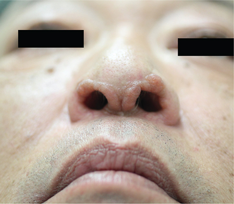
Fig. 1. A linear elevation of the skin along the periphery of the external nostrils. Overview.
Laboratory examinations revealed elevated serum soluble IL-2 receptor (1030 U/mL). Peripheral blood cell counts revealed an increased number of white blood cells (9400 /µL), but no atypical lymphocytes. Computed tomography scans revealed no lymphadenopathy or organ involvement. Histological and immunohistopathological examination of the lesion showed a proliferation of atypical lymphocytes, positive for CD3, CD4, CD8, and CD25, and negative for CD1a and CD30 on the dermis (Fig. 2A-G).
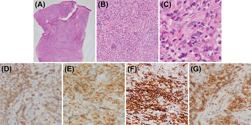
Fig. 2. Histopathological image demonstrates proliferation of atypical lymphocytes on the dermis. Haematoxylin eosin, (A) x1, (B) x100, and (C) x400. Immunohistochemical staining revealed the atypical lymphocytes were positive for (D, x400) CK3, (E, x400) CD4, (F, x400) CD8, and (G, x400) CD25.
What is your diagnosis?
1: Adult xanthogranuloma
2: Verruca vulgaris
3: Lymphomatoid papulosis
4: Adult T-cell leukaemia-lymphoma
See next page for answer.
ANSWERS TO QUIZ
A Linear Elevation of the Skin Along the Periphery of the External Nostrils: A Commentary
Diagnosis: Adult T-cell leukaemia-lymphoma
Particle agglutination and Western blotting tests revealed positivity of human T-lymphotropic virus type I (HTLV-1). Southern blot analysis of the biopsy specimen revealed monoclonal rearranged band in Jγ lesion. Also, southern blot analyses of HTLV-1 provirus integration revealed polyclonally expanded HTLV-1 infected cells. Taken together, we diagnosed him with adult T-cell leukaemia-lymphoma (ATLL). We withdrew oral cyclosporine and treated him with oral etretinate 30 mg daily. The lesion had gradually improved and was completely cured 267 days after the treatment (Fig. 3). During the course, no other organ involvements had been revealed by computed tomography scans of the entire body. No relapse has been observed on oral etretinate for 12 years.
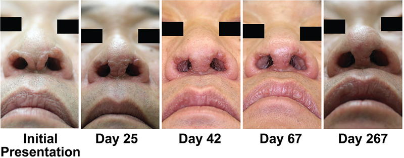
Fig. 3. Time course of the skin lesion after the initiation of oral etretinate. The skin lesion had gradually improved and was completely cured 267 days after the treatment. The number of days indicates days after the initiation of oral etretinate.
ATLL is an aggressive peripheral T-cell malignant lymphoma caused by HTLV-1 infection, which is endemic globally, mainly in south-western Japan and the Caribbean basin. ATLL is classified into acute, lymphoma, chronic, and smouldering types (1). Cutaneous lesions are reported to occur frequently in ATLL patients (39–72%), and they are often the first symptoms of the smouldering types, followed by the leukaemic phase over months or years (2–3). Among the smouldering types, ATLL with involvement limited to skin was classified into the cutaneous-type ATLL (3). The patient was smouldering-type ATLL based on the criteria and presented with only a cutaneous lesion; therefore, our case was cutaneous-type ATLL.
The cutaneous lesions of ATLL have been reported to be polymorphous, including patches, papules, plaques, nodules, tumours, erythroderma, and purpura (2). Our case manifested extremely rare linear elevation of the skin along the periphery of the external nostrils, which may consist of coalescing papules. In our case, it was extremely difficult to diagnose ATLL by clinical appearance of the skin only. Therefore, dermatologists should keep in mind that ATLL manifests as polymorphous cutaneous lesions and include ATLL as the differential diagnosis in endemic areas of HTLV-1.
The differential diagnosis for this presentation includes adult xanthogranuloma (XG), verruca vulgaris (VV), and lymphomatoid papulosis (LyP). Diagnosis relies on a comprehensive assessment of clinical courses, laboratory examinations, and histopathological and immunohistochemical findings. XG is a benign type of histiocytosis rarely seen in adults. Cutaneous forms can present as solitary or multiple yellowish, orange-red, or tan-hued papules. Some cases of cutaneous-form adult XG have been reported; however, each case is not plaque-like linear elevation like our case, but a solitary nodule (4). VV are very common warts, which are caused by infection with human papillomavirus. VV presents with firm, well-circumscribed, small papules or nodules with a hyperkeratotic and verrucous surface, and can involve the face (5). Although VV may also present linear lesions as in our case by dissemination, VV can be distinguished by the clinical findings with verrucous keratinization and petechiae, and pathological findings such as epidermal hyperkeratosis, papillomatosis, and changes in the granular layer. LyP is a benign, chronic, and recurrent papulonodular dermatitis, belonging to the CD30-positive cutaneous lymphoproliferative disorders involving T-cells and NK-cells. LyP presents with recurrent crops of disseminated pruritic pink papules, ranging from a single to a few to hundreds of lesions over the trunk and/or limbs, which subsequently become post-inflammatory hyperpigmented macules and eventually atrophic varioliform scars in the process of spontaneous resolution. If the papules coalesce, LyP manifests as a plaque-like variant (6). Although there are no reports of LyP with linear manifestation around the external nostrils, plaque-like LyP can manifest as in our case. In our case, the distinctive histopathological findings of CD3, CD4, CD8, and CD25 positivity along with several tests of HTLV-1 discriminated ATLL from LyP.
The smouldering type with skin manifestations has a poor prognosis (median survival time approximately 14 to 24 months), and the prognosis of cutaneous-type ATLL appears to be worse than that of the smouldering type without skin manifestation; therefore, the cutaneous-type ATLL was classified (7–8). Recently, the use of oral etretinate in combination with other treatments has been reported to be effective in cutaneous-type ATLL (3, 9). However, long-term remission after complete response has not been reported, except for the reported case with 6-year remission achieved by oral etretinate and PUVA. In our case, complete response and 12-year remission were achieved through the use of oral etretinate only. The predictive factors for retinoid responsiveness to every type of lymphoma are largely unknown; therefore, further investigations are needed to elucidate the positive predictor of retinoid efficacy in cutaneous-type ATLL.
ATLL is caused by monoclonally expanded HTLV-1-infected cells. Among a polyclonal population of HTLV-1 infected cells, monoclonal or oligoclonal expansion is considered to lead to clinical ATLL onset. Recently, a prospective cohort study of HTLV-1 carriers, using next-generation sequencing, revealed that individuals with polyclonal pattern of HTLV-1 showed no clinical progression, whereas those with monoclonal or oligoclonal patterns showed clinical progression (10). In our case, HTLV-1-infected cells may have expanded polyclonally in the skin lesion before monoclonal HTLV-1 infected cells dominantly increased, suggesting a better prognosis than a monoclonal or oligoclonal pattern. Therefore, the clonality analysis by Southern blotting may be the predictor of prognosis in cutaneous-type ATLL.
In conclusion, we present an extremely rare case of a linear elevation of the skin along the periphery of the external nostrils by cutaneous-type ATLL. Even in a case of unique presentations, we should include ATLL as the differential diagnosis in endemic area of HTLV-1.
The patient provided written consent for the use of his photographs and medical records.
REFERENCES
- Shimoyama M. Diagnostic criteria and classification of clinical subtypes of adult T-cell leukaemia-lymphoma: a report from the Lymphoma Study Group (1984–87). Br J Haematol 1991; 79: 428–437. https://doi.org/10.1111/j.1365-2141.1991.tb08051.x
- Miyashiro D, Sanches JA. Cutaneous manifestations of adult T-cell leukemia/lymphoma. Semin Diagn Pathol 2020; 37: 81–91. https://doi.org/10.1053/j.semdp.2019.07.010
- Inozume T, Matsue H, Furuhashi M, Nakamura Y, Mitsui H, Ando N, et al. Successful use of etretinate for long-term management of a patient with cutaneous-type adult T-cell leukaemia/lymphoma. Br J Dermatol 2005; 153: 1239–1241. https://doi.org/10.1111/j.1365-2133.2005.06974.x
- Tan LC, Tan KB, Aw CW. Unusual presentation of adult xanthogranuloma: a case report. Singapore Med J 2014; 55: e25–e27. https://doi.org/10.11622/smedj.2013207
- Ma Y, Huo W, Hong YX, Chen HD, Gao XH. Successful clearance of facial common warts by local hyperthermia: report of two cases. Dermatol Ther 2012; 25: 386–388. https://doi.org/10.1111/j.1529-8019.2012.01470.x
- Martinez-Cabriales SA, Walsh S, Sade S, Shear NH. Lymphomatoid papulosis: an update and review. J Eur Acad Dermatol Venereol 2020; 34: 59–73. https://doi.org/10.1111/jdv.15931
- Setoyama M, Katahira Y, Kanzaki T. Clinicopathologic analysis of 124 cases of adult T-cell leukemia/lymphoma with cutaneous manifestations: the smouldering type with skin manifestations has a poorer prognosis than previously thought. J Dermatol 1999; 26: 785–790. https://doi.org/10.1111/j.1346-8138.1999.tb02093.x
- Johno M, Ohishi M, Kojo Y, et al. Cutaneous manifestations of adult T-cell leukemia/lymphoma. Gann Monogr Cancer Res 1992; 39: 33–41.
- Yonekura K, Takeda K, Kawakami N, Kanzaki T, Kanekura T, Utsunomiya A. Therapeutic efficacy of etretinate on cutaneous-type adult T-cell leukemia-lymphoma. Acta Derm Venereol 2019; 99: 774–776. https://doi.org/10.2340/00015555-3196
- Firouzi S, Farmanbar A, Nakai K, Iwanaga M, Uchimaru K, Utsunomiya A, et al. Clonality of HTLV-1-infected T cells as a risk indicator for development and progression of adult T-cell leukemia. Blood Adv 2017; 1: 1195–1205. https://doi.org/10.1182/bloodadvances.2017005900