ORIGINAL REPORT
Treatment Survival and Reasons for Discontinuation in Patients with Recalcitrant Folliculitis Decalvans
Lena FARNINA, Simone CAZZANIGA, Robert E. HUNGER, Kristine HEIDEMEYER, Pierre A. de VIRAGH and S. Morteza SEYED JAFARI
Department of Dermatology, Inselspital, Bern University Hospital, Bern, Switzerland
Due to the risk of scarring and the psychological impact on affected patients, folliculitis decalvans (FD) requires prompt and focused care to control inflammation and prevent disease progression. This study aimed to provide a comprehensive long-term evaluation of treatment outcomes in difficult-to-treat cases of FD by analysing the effectiveness of various therapies and identifying reasons for treatment switches or discontinuation. The single-centre, retrospective cohort study collected information on patient characteristics and management strategies, focusing on different treatment groups and reasons for discontinuation or switching. Eighteen patients with biopsy-confirmed FD were included in the study because of their recalcitrant course (38.9% females, median age 33.0 years), with a median follow-up period of 1.5 years. During the study period, all patients received at least one prescription for topical therapies, primarily non-antibiotic disinfectants, topical corticosteroids, topical antibiotics, and topical dapsone. Systemic antibiotics were prescribed for 88.9% of patients, predominantly tetracyclines and a combination of rifampicin and clindamycin. Non-biological systemic therapies, excluding steroids, were used in 61.1% of patients, with isotretinoin being the most common (27.8%). Among immunomodulatory drugs, apremilast was prescribed to 11.1% of patients. Overall, the highest treatment discontinuation rates were observed with systemic antibiotics (risk ratio: 1.63; 95% confidence interval: 1.46–1.82), followed by systemic steroids. The treatment of patients with severe FD requires a personalized, multifaceted approach, typically involving a combination of local and systemic therapies. Antibiotics are often used as a first-line treatment, but they are associated with a high rate of discontinuation. This highlights the urgent need for effective immunomodulatory treatments, either as alternatives or as adjuncts to current options.
SIGNIFICANCE
This study emphasizes the urgent need for prompt and personalized treatment of folliculitis decalvans (FD) due to its potential for scarring and its significant psychological impact. By analyzing long-term outcomes in difficult-to-treat cases of FD, the study reveals high discontinuation rates of systemic antibiotics, which are commonly used as a first-line therapy. These results emphasize the limitations of current treatments and the need for more effective immunomodulatory options. This research advances the understanding of FD management by emphasizing a multifaceted approach that combines local and systemic therapies to better control inflammation, prevent disease progression, and ultimately improve patient quality of life.
Key words: discontinuation; efficacy; folliculitis decalvans; treatment survival.
Citation: Acta Derm Venereol 2025; 105: adv44185. DOI: https://doi.org/10.2340/actadv.v105.44185.
Copyright: © 2025 The Author(s). Published by MJS Publishing, on behalf of the Society for Publication of Acta Dermato-Venereologica. This is an Open Access article distributed under the terms of the Creative Commons Attribution-NonCommercial 4.0 International License (https://creativecommons.org/licenses/by-nc/4.0/).
Submitted: Jun 20, 2025. Accepted after revision: Sep 29, 2025. Published: Nov 11, 2025.
Corr: S. Morteza Seyed Jafari, MD, PhD, Department of Dermatology, Inselspital, Bern University Hospital, Bern, Switzerland. E-mail: Morteza.jafari@insel.ch
Competing interests and funding: RH: Advisory boards and consulting for UCB, Novartis, and Galderma. SMSJ: Advisory boards and speaker at educational events for LEO Pharma, Eli Lilly, Novartis, and Pfizer. PdV: Eli Lilly, Pfizer. The other authors declare that they have no competing interests for the study.
This research received no external funding.
INTRODUCTION
Folliculitis decalvans (FD) is a primary neutrophilic scarring alopecia that accounts for 6% and 1%, respectively, of alopecia patients in the public and private sectors (1). Its aetiology is not fully understood (2–4). A familial association has been reported, suggesting a genetic background (5). FD causes inflammation of the scalp resulting in scarring alopecia. It preferentially affects young and middle-aged males and is characterized by pustules, papules, scarring, haemorrhagic crusts, and erosions (6). FD hair follicles have a specific heterogeneous follicular bacterial microbiota signature. In addition, these patients appear to have an impaired immune response (7).
Because of the risk of scarring and the psychological consequences for affected patients, FD deserves full attention and immediate care to stop the inflammatory process and halt disease progression. However, its poorly understood pathogenesis often makes successful treatment very difficult and frustrating. It has been suggested that microbial “superantigens” or cytotoxins that bind to major histocompatibility complex class II molecules may stimulate T cells, leading to excessive inflammation, but “escape” detection by the host immune system, and play a role in the pathogenesis (8). Thus, dysregulated host–pathogen interactions are assumed to be the trigger for an excessive immune response. Staphylococcus aureus is often cultured from typical FD-affected hair follicles, and its eradication is one of the primary goals of first-line antibiotic treatment. Unfortunately, gram-negative bacteria can also be isolated and bacterial biofilms can develop, increasing treatment resistance, promoting relapse, and requiring different treatment strategies (7).
There is a notable paucity of data on the efficacy of treatments specifically targeting FD (9, 10). However, some evidence for a faulty immune response has emerged from case series of patients with refractory FD treated with adalimumab and/or ixekizumab after failure to respond to conventional therapies (11, 12). In addition, other case series have shown that Janus kinase (JAK) inhibitors, such as baricitinib, may be an effective treatment option for refractory FD (13).
Drug survival, defined as the interval between initiation and ending of a drug, is an indicator of therapeutic success in the case of continuous treatment (14, 15). A short drug survival may reflect either a lack of efficacy, tolerability, or safety or, conversely, treatment-free remission. The current study aimed to provide a comprehensive understanding of the treatment outcomes in patients with FD by examining various therapies and identifying the reasons for switching or discontinuing each treatment. This approach sought to offer clearer guidance on achieving safe and effective disease control.
MATERIALS AND METHODS
This was a single-centre retrospective cohort study that included the patients with difficult-to-treat FD seen at the University Hospital of Bern, Switzerland, between July 2016 and May 2024. Patients without complete documentation, regular follow-up information, and informed consent were not considered for the study. The study was approved by the local ethical committee.
Collected data
For patients meeting the inclusion and exclusion criteria, the following information was collected: demographics, coexisting comorbidities, age at first complaint and at first diagnosis, clinical findings, and prescribed treatments. In addition, changes in prescribed treatments and reasons for discontinuation were recorded at follow-up.
Statistical analysis
Data were presented as medians with interquartile ranges (IQR) or numbers with percentages for continuous and categorical variables respectively. A longitudinal analysis of the risk of therapy discontinuation at follow-up was performed by using generalized estimating equations (GEE) with Poisson distribution and exchangeable correlation structure. Effects were expressed in terms of risk ratios (RR) along with 95% confidence intervals (CI) and p-values. Increased RR were interpreted as follows: > 1 and < 1.5 = low risk; ≥ 1.5 and < 2 = moderate risk; ≥ 2 high risk (16). Discontinuation rates were derived from GEE and expressed as number of cases per 100 patient-months with their 95% CIs.
The trajectories of switching between different classes of therapies were shown using a directed graph representation. Switches were weighted based on their absolute frequency multiplied by a factor that diminishes over time from the baseline. This factor was an exponential decay function of the median switching time. Using Edmonds’ algorithm (17), we computed the maximum spanning tree that connects all classes based on the most relevant trajectories of switching. This tree accounted for both frequency and temporal sequence of switching and highlighted the most important network patterns and hubs.
All tests were considered statistically significant at p-values < 0.05. Analyses were performed with SPSS v.26.0 (IBM Corp, Armonk, NY, USA) and MATLAB v.9.1 (The MathWorks Inc, Natick, MA, USA).
RESULTS
A total of 18 consecutive patients (38.9% female) diagnosed with recalcitrant biopsy-proven FD were included in the study. The median age at first complaint was 27.5 years (IQR: 24.0–36.0), while the age at first diagnosis was 33.0 years (IQR: 28.0–46.0). Among other known comorbidities, the most frequent was diabetes mellitus (11.1%) (Table I).
| Sex, n (%) | |
| Male | 11 (61.1) |
| Female | 7 (38.9) |
| Age at first complaint, years, median (IQR) | 27.5 (24.0–36.0) |
| Age at first diagnosis, years, median (IQR) | 33.0 (28.0–46.0) |
| Other known concomitant diseases | |
| Metabolic syndrome | 1 (5.6) |
| Hypertension | 1 (5.6) |
| Diabetes mellitus | 2 (11.1) |
| Liver disease | 1 (5.6) |
| Gastrointestinal disease | 1 (5.6) |
| Other | 2 (11.1) |
| Complaintsa | |
| Hair loss | 7 (38.9) |
| Pain | 7 (38.9) |
| Pruritus | 11 (61.1) |
| Burning | 1 (5.6) |
| Redness | 2 (11.1) |
| Scaling | 7 (38.9) |
| Skin lesions | 13 (72.2) |
| Secretion | 4 (22.2) |
| aMultiple complaints, clinical findings, and localizations were possible. IQR: interquartile range. |
|
The main complaints at baseline were skin lesions (72.2%) and pruritus (61.1%), although scaling, hair loss, and pain were also frequent (38.9%). The main clinical findings were scarring (77.8%), erythema (72.2%), crusting (66.7%), tufting (50.0%), and scaling (38.9%).
At baseline, hair loss/lesions were predominantly located in the vertex (55.6%) or occipital (50.0%) areas, with a patchy pattern in 84.6% of patients.
Treatments prescribed
During a median of 1.5 years (IQR: 0.8–3.8), all patients had at least 1 prescription for topical therapies, particularly non-antibiotic disinfectants (88.9%), topical corticosteroids (88.9%), topical antibiotics (66.7%), and topical dapsone (55.6%) (Table II). Systemic antibiotics were prescribed to 88.9% of patients, mainly tetracyclines (77.8%) and rifampicin plus clindamycin (61.1%). Systemic steroids were used in 11.1% of patients, while other non-biological systemic therapies were used in 61.1% of patients, mainly isotretinoin (27.8%). Among immunomodulatory drugs, apremilast was used in 11.1% of patients. When considering combinations of therapies, topicals plus systemic antibiotics were used by 83.3% of patients, followed by topicals plus other systemic therapies (55.6%).
Treatment discontinuation
Overall, the observed discontinuation rate during the study period was 31.0 per 100 patient-months (95% CI: 25.9–37.1). A longitudinal analysis of the risk of therapy discontinuation at follow-up is presented in Table III. Topical antibiotics (RR: 1.15; 95% CI: 1.01–1.31), systemic steroids (RR: 1.41; 95% CI: 1.26–1.59), dapsone (RR: 1.29; 95% CI: 1.06–1.56), and mycophenolate mofetil (RR: 1.48; 95% CI: 1.17–1.88) showed a significant low increased risk of discontinuation. A moderate increased risk was observed for rifampicin plus clindamycin (RR: 1.61; 95% CI: 1.30–1.99), tetracyclines (RR: 1.54; 95% CI: 1.31–1.81), and other systemic therapies (RR: 1.59; 95% CI: 1.24–2.04). When considering the class of treatment prescribed, only systemic antibiotics and systemic steroids showed a significant increased risk of discontinuation, while immunomodulatory drugs (apremilast) had a barely significant reduced risk of discontinuation (RR: 0.86; 95% CI: 0.74–1.01).
| Item | RR (95% CI) | p-value* |
| Topical antibiotics | 1.15 (1.01–1.31) | 0.03 |
| Topical disinfectants | 1.25 (0.97–1.61) | 0.09 |
| Topical corticosteroids | 1.09 (0.87–1.35) | 0.46 |
| Topical dapsone | 1.10 (0.84–1.44) | 0.50 |
| Topical isotretinoin | 1.15 (0.79–1.67) | 0.48 |
| Other topical therapiesa | 1.49 (1.25–1.78) | < 0.001 |
| Rifampicin + clindamycin | 1.61 (1.30–1.99) | < 0.001 |
| Tetracyclines | 1.54 (1.31–1.81) | < 0.001 |
| Systemic steroids | 1.41 (1.26–1.59) | < 0.001 |
| Acitretin | 0.97 (0.72–1.31) | 0.84 |
| Isotretinoin | 0.93 (0.70–1.22) | 0.59 |
| Dapsone | 1.29 (1.06–1.56) | 0.01 |
| Mycophenolate mofetil | 1.48 (1.17–1.88) | 0.001 |
| Other systemic therapiesb | 1.59 (1.24–2.04) | < 0.001 |
| Apremilast | 1.05 (0.79–1.40) | 0.72 |
| Topical therapies | 1.24 (0.78–1.97) | 0.36 |
| Systemic antibiotics | 1.63 (1.46–1.82) | < 0.001 |
| Systemic steroids | 1.49 (1.28–1.73) | < 0.001 |
| Other systemic therapies | 1.21 (0.95–1.53) | 0.12 |
| Immunomodulatory drugs (apremilast) | 0.86 (0.74–1.01) | 0.06 |
| aIncluding minoxidil and local injection of cortisone. bIncluding other systemic antibiotics, zinc salts, and vitamins/supplements. CI: confidence interval, RR: risk ratio. *Generalized estimating equations with Poisson distribution including therapies. |
||
Reasons for discontinuation are given in Table SI. Topical therapies, systemic antibiotics, systemic steroids, and other systemic therapies were mainly discontinued because of partial response (37.2%, 63.6%, 40.0%, and 42.3% respectively).
Trajectories of switching
The trajectories of switching between different classes of therapy are shown in Fig. 1. The most relevant primary switching pattern was from topicals plus systemic antibiotics to topicals alone (18.1% of the total switches), although a smaller proportion of patients also switched initially from no therapy to topicals plus systemic antibiotics (2.8%). The main relevant secondary trajectory was from topicals alone to topicals plus other systemic therapies (4.2%). Immunomodulatory drugs (e.g., apremilast) were mainly used in combination with other therapies as a third-line treatment. The main hubs of the network, from which most switching patterns originated, were topicals plus other systemic therapies and topicals alone.
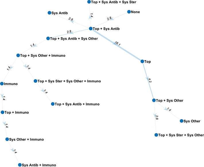
Fig. 1. Maximum spanning tree capturing the most relevant trajectories of switching. Numbers on connecting lines represent the percentage over the total number of switches (n = 72). Line widths are proportional to switching frequencies multiplied by a factor that diminishes over time from the baseline. Antib: antibiotics, Immuno: immunomodulatory drugs, Sys: systemic, Ster: steroids, Top: topical therapies.
DISCUSSION
FD often results in significant frustration and diminished quality of life for patients. The primary goal of treatment is early and effective intervention to halt the progression of scarring alopecia. However, all current treatments are off-label, and there is currently no long-term effective treatment for recalcitrant FD (18, 19). Treatment selection depends on disease severity as defined by size of affected area, lesion localization, inflammation level, symptom intensity, patients’ comorbidities, as well as the cost and accessibility of treatment options. Conventional treatment approaches for FD typically involve a combination of topical and oral antibiotics, isotretinoin, and oral, topical, or intralesional corticosteroids (20). The approach to changing treatment has evolved in recent years with the availability of more effective and better tolerated drugs such as biologics. Therapies such as tumour necrosis factor (TNF) inhibitors have shown significant success in managing this sometimes difficult-to-treat disease (20).
In line with previous studies, most of our patients received combination therapy with both topical and systemic antibiotics, specifically rifampicin in combination with clindamycin, or tetracyclines. Oral antibiotics, particularly tetracyclines and the combination of clindamycin and rifampicin, have greater evidence of efficacy in reducing inflammation and alleviating clinical symptoms (18). Their efficacy in managing FD symptoms likely stems from the combined antibiotic and anti-inflammatory actions of these treatments (18).
In addition, some data have been published on the treatment of FD with isotretinoin or other retinoids in combination with antibiotics or with antibiotics and corticosteroids (21, 22). In our patients, both topical and systemic isotretinoin were also often used. In addition to inhibiting sebaceous lipid production and modification of sebaceous gland function, isotretinoin has a direct inhibitory effect on the immune system (21–24). It reduces the expression of several pro-matrix metalloproteinases and thus inhibits the migration of neutrophils into the skin (21, 25). It also reduces the expression of toll-like receptor 2, which mediates the immune response to gram-positive bacteria (21, 26). Therefore, this treatment can be considered as an interesting option for patients with FD (21).
In some cases, dapsone has also been given topically or systemically. Dapsone has immunoregulatory, anti-microbial, and anti-inflammatory properties; it inhibits myeloperoxidase, an enzyme essential to the oxidative burst produced by neutrophils and there is evidence that dapsone may alter neutrophil chemotaxis. Thus, it appears to be a suitable treatment for FD (21). Dapsone, especially topical dapsone, has shown good tolerability and high patient compliance. It may therefore be considered as a useful treatment regimen to reduce the frequency of outbreaks (27).
Based on recent case reports and small case series, systemic treatments such as small molecules or biologics could be used in refractory cases after standard therapies are exhausted or no longer effective (20), as summarized in Table IV. Currently, most clinical experience in the literature involves anti-TNFα agents, particularly adalimumab. Although nearly all studies report significant clinical improvement and symptoms such as pruritus and pain in a remarkable proportion of patients, the degree and rapidity of response are heterogeneous (9, 11, 12, 28–35). This variability may reflect differences in disease severity, clinical presentation, and dosing regimens, but it may also reflect heterogeneous molecular pathways of the disease, or only indirect or partial disease modulation by the therapeutic molecule, resulting in varying efficacy. Alternatively, these findings suggest that patients with FD might require higher doses of adalimumab, similar to those used in hidradenitis suppurativa (33). In addition, patients in studies often relapsed quickly, suggesting that sustained disease control requires prolonged treatment (31, 35). While discontinuation leads to recurrence, clinical improvement is observed upon re-initiation (32, 34). Apremilast (a phosphodiesterase 4 inhibitor) has also been reported as a possible alternative in previous reports (36–38). In the current study, apremilast was prescribed in 11.1% of cases. Inhibition of phosphodiesterase 4 alters the cytokine composition and produces an anti-inflammatory state, which is likely to lead to control of inflammation. As neutrophils are the predominant inflammatory cell in FD, the inhibitory effect of apremilast on neutrophil activity may explain its beneficial effect (36). Given the heterogeneity of FD, apremilast may not be successful for everyone. However, it has been confirmed as a valuable addition to the treatment options for difficult cases, both in our experience and in the experience of others (37, 38). Furthermore, JAK inhibitors may represent a promising new approach for treating scarring alopecias refractory to standard therapies by targeting key inflammatory pathways (39) (Table IV). Jerjen et al. (40) and Moussa et al. (13) demonstrated the beneficial effects of tofacitinib and baricitinib, respectively, in the management of FD. Last but not least, the potential effect of IL-17 inhibitors has also been reported in some additional case studies (12, 41).
| Author (year) | Study type | Treatment (dosing regimen)a | Durationb | Treatment response |
| TNF-alpha blockers | ||||
| Mihaljević et al. (2012) (30) | Case-report (45-year-old male patient) | Infliximab (5 mg/kg, intervals as in psoriasis patients) | 12 months | After 3 infusions, lesions remitted rapidly and no recurrence was seen after 12 months |
| Kreutzer et al. (2014) (31) | Case series (3 female patients)c | Adalimumab (Humira®, 40 mg subcutaneously every 2 weeks) | 2–3 months | Marked remission or stabilization was observed |
| Shireen et al. (2018) (28) | Case report (23-year-old male patient) | Adalimumab (biosimilar, 80 mg at week 0 and week 2 and then 40 mg at weeks 3, 4, 6, 8, 10, 12, 14, and 16, then 40 mg every 4 weeks) | 6 months | Improved condition with hair regrowth; no recurrence of scalp lesions or pustules |
| Alhameedy et al (2019) (35) | Case report (54-year-old female patient) | Adalimumab (Humira®, 160 mg at week 0, 80 mg at week 2, then 40 mg every week) | 3 months | Inflammation was repressed, and no new lesions appeared |
| Hoy et al. (2022) (9) | Case report (42-year-old male patient) | Certolizumab pegol (400 mg during the first 3 SC injections, then 200 mg every 2 weeks) | 33 months | Patient-reported outcomes resolved strikingly, with markedly reduced scalp inflammation |
| Lobato-Berezo et al. (2022) (12) | Case series (3 male patients) | Adalimumab (biosimilar, 160 mg at week 0, 80 mg at week 2, then 40 mg every 2 weeks) | 6–13 months | One worsened, 1 mildly improved, and 1 greatly improved |
| Iorizzo et al. (2022) (32) | Case series (6 female and 17 male patients) | Adalimumab (160 mg at week 0, 80 mg at week 2, and 80 mg every other week) | 6–24 months | All patients showed clinical improvement from month 1, maintained throughout treatment |
| Dupont et al. (2023) (33) | Case series (6 female and 5 male patients) | Nine patients: infliximab (5 mg/kg, every 4–8 weeks); 2 patients: adalimumab (40 mg every 2 weeks); 1 patient: infliximab and then adalimumab 6 months later | 12 months | At the end of follow-up, 5 patients were considered responders |
| Alsantali et al. (2023) (11) | Case report (40-year-old-male patient) | Adalimumab (Humira®, 80 at week 0, then 40 mg every other week) | 10 months | No new areas of scarring alopecia or inflammation have appeared since starting adalimumab |
| Ramos et al (2024) (29) | Case report (39-year-old-male patient) | Adalimumab (160 mg at week 0, 80 mg at week 2, then 40 mg every week) | 15 months | Asymptomatic patient with no active lesions |
| Andrade et al. (2025) (34) | Case report (33-year-old-male patient) | Adalimumab (dosage/interval as in hidradenitis suppurativa patients) | 13 weeks | The patient showed marked improvement |
| JAK inhibitors | ||||
| Jerjen et al. (2020) (40) | Case series (1 female and 2 male patient) | Tofacitinib (2.5–5 mg/day) | 10–16 months | Improvement observed in all patients |
| Moussa et al. (2022) (13) | Case series (2 female and 2 male patients) | Baricitinib (3.4 or 6.8 mg/day) | 5–15 months | All patients showed symptom improvement and reduced inflammation |
| Other small molecules and biologics | ||||
| Ismail et al. (2020) (41) | Case report (30-year-old female patient) | Secukinumab (300 mg at weeks 0, 1, 2, 3, and 4, then 300 mg every 4 weeks) | 7 months | Significant improvement in her condition with a reduction of inflammation |
| Fässler et al. (2020) (36) | Case report (28-year-old-male patient) | Apremilast (N/A) | 25 weeks | Rapid improvement of lesions and pain, but flare-up occurred after 7-week discontinuation |
| Lobato-Berezo et al. (2022) (12) | Case report (26-year-old-male) | Ixekizumab (160 mg at week 0, 80 mg at weeks 2, 4, 6, 8, 10, 12, then 80 mg every 4 weeks from week 16) | 12 weeks | Severe worsening |
| Dethier et al. (2024) (37) | Case report (56-year-old-male patient) | Apremilast (N/A) | 44 months | Significant clinical improvement in was seen by month 2, with complete remission at month 7; after stopping apremilast at month 18, no recurrence occurred until 26 months later |
| de Viragh et al. (2024) (38) | Case series (9 patients) | Apremilast (various dosing regimen) | N/A | Treatment failure was observed in 3 patients; in another case, an initially positive response was reversed by relapse during treatment. Remission was only maintained in 3 patients with sustained therapy at the conventional dose of 30 mg twice daily, accompanied by other anti-inflammatory therapies. One patient could reduce the dose to 30 mg once daily every other day, but FD relapsed at 30 mg twice a week. Finally, 1 patient shows sustained remission |
| aThe patients had undergone various systemic antibiotic and/or anti-inflammatory treatments prior to commencing small-molecule or biologic therapy. In some studies concurrent therapies were also noted. bTreatment duration and/or observed period. cTwo cases of folliculitis decalvans and 1 case of therapy-refractory lichen planopilaris. FD: folliculitis decalvans; N/A: not available. |
||||
In our study, the primary switching pattern was from topicals plus systemic antibiotics to topicals alone, while the main secondary trajectory was from topicals alone to topicals plus other systemic therapies. Taking all treatments into account, the highest probability of discontinuation was observed in patients with systemic antibiotics – whether due to good response, partial response, no response, or adverse effects – observed in those using systemic antibiotics, particularly rifampicin and clindamycin (RR:1.61) and tetracyclines (RR: 1.54). Given the growing problem of antibiotic resistance, it is important to explore therapeutic alternatives that minimize the repeated use of systemic antibiotics and prolong periods of inflammation-free status (27). In contrast, inflammation-modulating treatments such as apremilast had a reduced risk of discontinuation (RR: 0.86), making it a more stable treatment option in patients requiring continuous therapy. Besides other treatments such as TNF-alpha inhibitors, apremilast appears to be one of the possible options given these data, suggesting its potential for long-term treatment at reasonable costs. However, in nearly all cases with good responses, apremilast was used alongside local and/or systemic anti-inflammatory treatments (37, 38).
In the study, the median time from the first complaint to the diagnosis of FD was over 5.5 years. Considering the significant impact this scarring form of alopecia can have on patients’ quality of life, including emotional distress and irreversible hair loss, this delay highlights a critical window of opportunity that must not be overlooked. Early recognition and targeted treatment during the active inflammatory phase are critical to prevent irreversible scarring and optimize clinical outcomes. While clinical and histological evaluations remain essential, non-invasive diagnostic tools – such as trichoscopy and reflectance confocal microscopy (42) – have recently shown promise in folliculitis decalvans by improving diagnostic accuracy and facilitating earlier intervention. Incorporating these methods into routine practice could help reduce diagnostic delays and allow timely, tailored therapeutic strategies.
There are several limitations to this study. First, the small sample size limits the generalization of our results. The single-centre, retrospective design may also introduce selection bias and limit the applicability of the results to other settings. In addition, the length of follow-up varied considerably among patients, which may affect the consistency of the data. The study relied on medical records, which may not capture all relevant patient information or reasons for discontinuation. In addition, the lack of standardized treatment and assessment protocols may have influenced the choice and combination of therapies, adding another layer of variability.
This study highlights the need for tailored and holistic approaches to the treatment of recalcitrant cases of FD. The high dropout rates and frequent treatment changes highlight the challenges of managing this chronic disease. Future research should focus on larger, multi-centre studies to validate these findings and explore new therapeutic options to improve treatment efficacy and patient outcomes. The results also suggest the potential benefit of developing standardized treatment guidelines to reduce variability in treatment choice and improv, and standardized questionnaires/documentation in future studies would allow for a more comprehensive understanding of the impact of primary cicatricial alopecia and its treatment on patients’ lives. By addressing these limitations and building on current evidence, we can advance the management of primary cicatricial alopecia and ultimately improve outcomes for affected patients.
ACKNOWLEDGEMENTS
Ethical approval/ethics statement: The study was approved by the local ethical committee.
Data availability statement: The data supporting the results of this study are presented in the current paper.
REFERENCES
- Vañó-Galván S, Saceda-Corralo D, Blume-Peytavi U, Cucchía J, Dlova NC, Gavazzoni Dias MFR, et al. Frequency of the types of alopecia at twenty-two specialist hair clinics: a multicenter study. Skin Appendage Disord 2019; 5: 309–315. https://doi.org/10.1159/000496708
- Moreno-Arrones OM, Del Campo R, Saceda-Corralo D, Jimenez-Cauhe J, Ponce-Alonso M, Serrano-Villar S, et al. Folliculitis decalvans microbiologic signature is specific for disease clinical phenotype. J Am Acad Dermatol 2021; 85: 1355–1357. https://doi.org/10.1016/j.jaad.2020.10.073
- Jahns AC, Lundskog B, Nosek D, Killasli H, Emtestam L, Alexeyev OA. Microbiology of folliculitis decalvans: a histological study of 37 patients. J Eur Acad Dermatol Venereol 2015; 29: 1025–1026. https://doi.org/10.1111/jdv.12448
- Chiarini C, Torchia D, Bianchi B, Volpi W, Caproni M, Fabbri P. Immunopathogenesis of folliculitis decalvans: clues in early lesions. Am J Clin Pathol 2008; 130: 526–534. https://doi.org/10.1309/NG60Y7V0WNUFH4LA
- Sarkis A, Cortez de Almeida RF, Lemes LR, Obadia DL, Machado CJ, Müller-Ramos P, et al. Folliculitis decalvans in women: a retrospective multicentre study of 150 patients. J Eur Acad Dermatol Venereol 2024; 38: e66–70. https://doi.org/10.1111/jdv.19434
- Kashikar Y, Saoji V, Madke B, Chandak MS, Meghe S. Successful management of folliculitis decalvans. Cureus 2024; 16: e52881. https://doi.org/10.7759/cureus.52881
- Moreno-Arrones OM, Garcia-Hoz C, Del Campo R, Roy G, Saceda-Corralo D, Jimenez-Cauhe J, et al. Folliculitis decalvans has a heterogeneous microbiological signature and impaired immunological response. Dermatology 2023; 239: 454–461. https://doi.org/10.1159/000529301
- Otberg N, Kang H, Alzolibani AA, Shapiro J. Folliculitis decalvans. Dermatol Ther 2008; 21: 238–244. https://doi.org/10.1111/j.1529-8019.2008.00204.x
- Hoy M, Böhm M. Therapy-refractory folliculitis decalvans treated with certolizumab pegol. Int J Dermatol 2022; 61: e26–28. https://doi.org/10.1111/ijd.15914
- Rambhia PH, Conic RRZ, Murad A, Atanaskova-Mesinkovska N, Piliang M, Bergfeld W. Updates in therapeutics for folliculitis decalvans: a systematic review with evidence-based analysis. J Am Acad Dermatol 2019; 80: 794–801.e1. https://doi.org/10.1016/j.jaad.2018.07.050
- Alsantali A, Baghdadi R, Alghamdi Y, Abbas EB, Ghazi R. Folliculitis decalvans managed with adalimumab: a case report. Clin Case Rep 2023; 11: e8193. https://doi.org/10.1002/ccr3.8193
- Lobato-Berezo A, Pujol RM. Refractory folliculitis decalvans treated with biologics: case series in 4 situations. Dermatol Ther 2022; 35: e15778. https://doi.org/10.1111/dth.15778
- Moussa A, Asfour L, Eisman S, Bhoyrul B, Sinclair R. Successful treatment of folliculitis decalvans with baricitinib: a case series. Australas J Dermatol 2022; 63: 279–281. https://doi.org/10.1111/ajd.13786
- Kromer C, Wilsmann-Theis D, Gerdes S, Philipp S, Schaarschmidt ML, Schmieder A, et al. Drug survival and reasons for drug discontinuation in palmoplantar pustulosis: a retrospective multicenter study. J Dtsch Dermatol Ges 2019; 17: 503–516. https://doi.org/10.1111/ddg.13834
- Schulte-Oestrich L, Cazzaniga S, Hunger RE, Seyed Jafari SM. Treatment survival in patients with hidradenitis suppurativa. JEADV Clin Pract 2025; 4: 751–758. https://doi.org/10.1002/jvc2.70027
- Moore SF, Price S, Konya J, Blummers S, Walter FM, Neal RD, et al. Risk factors for pancreatic cancer in electronic health records: an umbrella review of systematic reviews and meta-analyses. EClinicalMedicine 2025; 85: 103297. https://doi.org/10.1016/j.eclinm.2025.103297
- Edmonds J. Optimum branchings. J Res Natl Inst Stand Technol Section B. 1967; 71B: 233–240. https://doi.org/10.6028/jres.071B.032
- Vañó-Galván S, Molina-Ruiz AM, Fernández-Crehuet P, Rodrigues-Barata AR, Arias-Santiago S, Serrano-Falcón C, et al. Folliculitis decalvans: a multicentre review of 82 patients. J Eur Acad Dermatol Venereol 2015; 29: 1750–1757. https://doi.org/10.1111/jdv.12993
- Waśkiel-Burnat A, Starace M, Iorizzo M, Katoulis A, Apalla Z, Asfour L, et al. Management of folliculitis decalvans: the EADV task force on hair diseases position statement. J Eur Acad Dermatol Venereol 2025; 39: 1385–1394 https://doi.org/10.1111/jdv.20687
- Fakhoury T, Urban K, Ettefagh L, Nami N. Recalcitrant folliculitis decalvans treatment outcomes with biologics and small molecule inhibitors. Cutis 2024; 113: E32–34. https://doi.org/10.12788/cutis.1023
- Tietze JK, Heppt MV, von Preußen A, Wolf U, Ruzicka T, Wolff H, et al. Oral isotretinoin as the most effective treatment in folliculitis decalvans: a retrospective comparison of different treatment regimens in 28 patients. J Eur Acad Dermatol Venereol 2015; 29: 1816–1821. https://doi.org/10.1111/jdv.13052
- Gemmeke A, Wollina U. Folliculitis decalvans of the scalp: response to triple therapy with isotretinoin, clindamycin, and prednisolone. Acta Dermatovenerol Alp Pannonica Adriat 2006; 15: 184–186.
- Aksoy B, Hapa A, Mutlu E. Isotretinoin treatment for folliculitis decalvans: a retrospective case-series study. Int J Dermatol 2018; 57: 250–253. https://doi.org/10.1111/ijd.13874
- Harries MJ, Paus R. Scarring alopecia and the PPAR-gamma connection. J Invest Dermatol 2009; 129: 1066–1070. https://doi.org/10.1038/jid.2008.425
- Zouboulis CC. Isotretinoin revisited: pluripotent effects on human sebaceous gland cells. J Invest Dermatol 2006; 126: 2154–2156. https://doi.org/10.1038/sj.jid.5700418
- Dispenza MC, Wolpert EB, Gilliland KL, Dai JP, Cong Z, Nelson AM, et al. Systemic isotretinoin therapy normalizes exaggerated TLR-2-mediated innate immune responses in acne patients. J Invest Dermatol 2012; 132: 2198–2205. https://doi.org/10.1038/jid.2012.111
- Melián-Olivera A, Burgos-Blasco P, Selda-Enríquez G, Suárez-Valle A, Miguel-Gómez L, Vañó-Galván S, et al. Topical dapsone for folliculitis decalvans: a retrospective cohort study. J Am Acad Dermatol 2022; 87: 150–151. https://doi.org/10.1016/j.jaad.2021.07.004
- Shireen F, Sudhakar A. A case of isotretinoin therapy-refractory folliculitis decalvans treated successfully with biosimilar adalimumab (Exemptia). Int J Trichology 2018; 10: 240–241. https://doi.org/10.4103/ijt.ijt_34_18
- Ramos J, Silva AM, António AM, Alves J. Recalcitrant folliculitis decalvans successfully controlled with adalimumab. An Bras Dermatol 2024; 99: 480–482. https://doi.org/10.1016/j.abd.2024.02.001
- Mihaljević N, von den Driesch P. Successful use of infliximab in a patient with recalcitrant folliculitis decalvans. J Dtsch Dermatol Ges 2012; 10: 589–590. https://doi.org/10.1111/j.1610-0387.2012.07972.x
- Kreutzer K, Effendy I. Therapy-resistant folliculitis decalvans and lichen planopilaris successfully treated with adalimumab. J Dtsch Dermatol Ges 2014; 12: 74–76. https://doi.org/10.1111/ddg.12224
- Iorizzo M, Starace M, Vano-Galvan S, Piraccini BM, Reygagne P, Rudnicka L, et al. Refractory folliculitis decalvans treated with adalimumab: a case series of 23 patients. J Am Acad Dermatol 2022; 87: 666–669. https://doi.org/10.1016/j.jaad.2022.02.044
- Dupont A, Eyraud A, Milpied B, De Bataille S, Cassassa E, Darrigade AS, et al. Efficacy and safety of tumour necrosis factor-α antagonists for folliculitis decalvans: a retrospective case-series pilot study. Acta Derm Venereol 2023; 103: adv3713. https://doi.org/10.2340/actadv.v103.3713
- Andrade A, Scuotto CRC, Da Silva DLF. Refractory folliculitis decalvans: new therapeutic horizons? Int J Trichology 2025; 17: 66–68. https://doi.org/10.4103/ijt.ijt_83_23
- Alhameedy MM, Alsantali AM. Therapy-recalcitrant folliculitis decalvans controlled successfully with adalimumab. Int J Trichology 2019; 11: 241–243. https://doi.org/10.4103/ijt.ijt_92_19
- Fässler M, Radonjic-Hoesli S, Feldmeyer L, Imstepf V, Pelloni L, Yawalkar N, et al. Successful treatment of refractory folliculitis decalvans with apremilast. JAAD Case Rep 2020; 6: 1079–1081. https://doi.org/10.1016/j.jdcr.2020.08.019
- Dethier C, Azirar S, Verluyten L, Boonen H, Grosber M, Gutermuth J. Response to Fässler et al.’s “Successful treatment of refractory folliculitis decalvans with apremilast”. JAAD Case Rep 2025; 57: 122–123. https://doi.org/10.1016/j.jdcr.2024.10.036
- de Viragh PA, Seyed Jafari SM. Response to Dethier et al., “Experience in nine additional cases of apremilast for therapy-resistant folliculitis decalvans”. JAAD Case Rep 2025; 57: 102. https://doi.org/10.1016/j.jdcr.2024.12.001
- Agarwal P, Ajuchi O, Lukowiak TM, Rao BK. Off-label use of biologics and Janus kinase (JAK) inhibitors for scarring alopecias: a narrative review. Arch Dermatol Res 2025; 317: 803. https://doi.org/10.1007/s00403-025-04301-z
- Jerjen R, Meah N, Trindade de Carvalho L, Wall D, Sinclair R. Folliculitis decalvans responsive to tofacitinib: a case series. Dermatol Ther 2020; 33: e13968. https://doi.org/10.1111/dth.13968
- Ismail FF, Sinclair R. Successful treatment of refractory folliculitis decalvans with secukinumab. Australas J Dermatol 2020; 61: 165–166. https://doi.org/10.1111/ajd.13190
- Piscazzi F, Franceschini C, Narcisi A, Valenti M, Rossi A, Ardigò M. Non-invasive assessment and management of folliculitis decalvans by trichoscopy and reflectance confocal microscopy. Dermatol Pract Concept 2024; 14: e2024167. https://doi.org/10.5826/dpc.1403a167