RESEARCH LETTER
Melanoma during Ruxolitinib Treatment for Myelofibrosis
Lingzhi ZHONG, Houmin LI and Guangdong WEN*
Department of Dermatology, Peking University People’s Hospital, Beijing, China. *E-mail: 163vip2008@163.com
Citation: Acta Derm Venereol 2025; 105: adv44196. DOI: https://doi.org/10.2340/actadv.v105.44196.
Copyright: © 2025 The Author(s). Published by MJS Publishing, on behalf of the Society for Publication of Acta Dermato-Venereologica. This is an Open Access article distributed under the terms of the Creative Commons Attribution-NonCommercial 4.0 International License (https://creativecommons.org/licenses/by-nc/4.0/).
Submitted: Jun 21, 2025. Accepted after revision: Jul 1, 2025. Published: Jul 19, 2025
Competing interests and funding: The authors have no conflicts of interest to declare.
To the Editor,
Hydroxyurea and ruxolitinib are commonly used to treat myeloproliferative disorders and long-term treatment increases the risk of developing non-melanoma skin cancers, while there are relatively few reports of melanoma occurrence. A 65-year-old female presented to the dermatology clinic with a lesion on the left ribs for 1 year. It was initially a brown macule with no obvious discomfort. It gradually grew larger and raised 2 months ago and broke and bled 1 week ago. The patient was treated with oral hydroxyurea for myelofibrosis (MF) for 4 months, and the treatment switched to oral ruxolitinib (30 mg/day) 3 months ago. Physical examination revealed a black and double spherical mass on the left ribs, measuring approximately 0.5 cm×1 cm, with a ruptured surface and black-brown macules around it (Fig. 1). Dermoscopy showed a raised dark-brown mass with bleeding in the centre, and stains around it, with a false reticular pattern and some areas appearing blue. Based on the patient’s medical history, physical examination, and dermoscopy result, the diagnosis of melanoma required high suspicion. Therefore, the dosage of ruxolitinib was gradually reduced and the patient was admitted to hospital for expanded resection surgery.
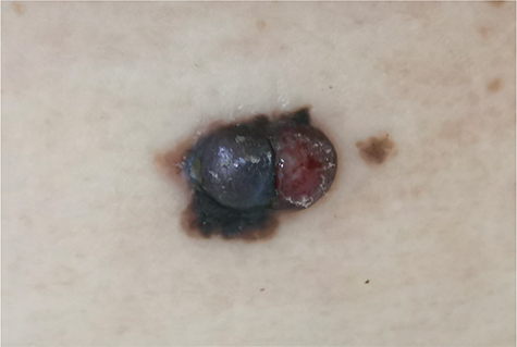
Fig. 1. Physical examination revealed a black and double spherical mass on the left ribs, measuring approximately 0.5 cm×1 cm, with a ruptured surface and black brown macules around it.
Postoperative histopathological results conformed the diagnosis of malignant melanoma (nodular type) with moderate cellular atypia (Fig. 2A–B). The main tumour size was 1 cm×1 cm×0.3 cm, with focal invasion of the epidermis. The Breslow thickness was about 2.1 mm, and there was no clear intravascular tumour thrombus or nerve invasion. There was also surrounding seborrheic keratosis, with a diameter of 0.3 cm (Fig. 2C). Immunohistochemistry showed tumour cells positive for S-100, Melan A, HMB45, p53 (scattered minority+, wild-type expression), and BRAF (strong+) (Fig. 2D–H). The proliferative index measured by Ki-67 was over 20% (Fig. 2I). High-throughput sequencing revealed tumour gene mutation of BRAF p.V600E with abundance of 48.36% (Class I). Postoperative PET-CT showed that there were no metabolically active lesions or metastases.
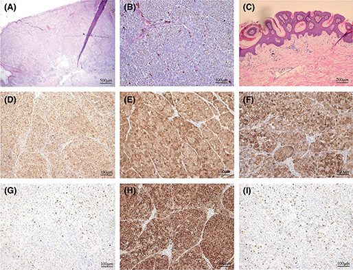
Fig. 2. Histopathology conformed malignant melanoma (nodular type). (A, B) There was moderate cellular atypia (H&E, A: ×40, B: ×200). (C) There was also surrounding seborrheic keratosis, with a diameter of 0.3 cm (H&E×100). Immunohistochemistry (×200) showed tumour cells positive for (D) S-100, (E) Melan A, (F) HMB45, (G) p53 (scattered minority+, wild-type expression), (H) BRAF (strong+). (I) The Ki-67 was over 20% of tumour cells.
Considering the diagnosis of melanoma (IIB, T3bN0M0), the use of ruxolitinib was discontinued, and 9 million units of recombinant human interferon alpha 2b injection were given subcutaneously 3 times a week. One week later, the drug dose was increased to 18 million units 3 times a week. The patient’s condition remained stable after 1-year of follow-up.
Previous ruxolitinib clinical trials in polycythaemia vera (PV) and MF have reported the occurrence of non-melanoma skin cancers (NMSCs) (1, 2). A 5-year phase 3 study of ruxolitinib for MF showed 17.1% of patients on ruxolitinib went on to have basal cell carcinomas or squamous cell carcinomas (SCCs) compared with only 2.7% of patients on best available therapy (2). A 10-year retrospective cohort study indicated that SCC risk is increased in patients with PV or MF taking ruxolitinib; the adjusted hazard ratio for NMSCs after ruxolitinib was 2.69 (95% CI 1.03–7.02) (3). There have been some case reports of patients with PV or MF taking ruxolitinib who developed aggressive SCC (4, 5), while there are relatively few reports of melanoma occurrence. One case of lentigo maligna melanoma and another case of metastatic undifferentiated pleomorphic sarcoma were reported in patients with MF taking ruxolitinib (5).
Here we report a case of melanoma during ruxolitinib treatment for MF. This case suggests that regular skin examinations are recommended for patients with myeloproliferative disorders receiving treatment with ruxolitinib. If suspicious skin masses are found, timely biopsy or excision surgery and a comprehensive examination should be performed, and ruxolitinib should be discontinued in a timely fashion. Interferon can be considered as a treatment option for patients with MF and melanoma. The repeat observation of skin cancers with aggressive features during JAK inhibitor treatment suggests that these medications may promote cutaneous malignant transformation in high-risk patients. Therefore, it is recommended to further test the relationship between JAK kinase and skin cancer risk.
ACKNOWLEDGEMENT
Ethics statement: The patient in this manuscript has given written informed consent to the publication of her case details.
Data availability statement: Data are available on request from the authors.
REFERENCES
- Verstovsek S, Vannucchi AM, Griesshammer M, Masszi T, Durrant S, Passamonti F, et al. Ruxolitinib versus best available therapy in patients with polycythemia vera: 80-week follow-up from the RESPONSE trial. Haematologica 2016; 101: 821–829. https://doi.org/10.3324/haematol.2016.143644
- Harrison CN, Vannucchi AM, Kiladjian JJ, Al-Ali HK, Gisslinger H, Knoops L, et al. Long-term findings from COMFORT-II, a phase 3 study of ruxolitinib vs best available therapy for myelofibrosis. Leukemia 2016; 30: 1701–1707. https://doi.org/10.1038/leu.2016.148
- Lin JQ, Li SQ, Li S, Kiamanesh EF, Aasi SZ, Kwong BY, et al. A 10-year retrospective cohort study of ruxolitinib and association with nonmelanoma skin cancer in patients with polycythemia vera and myelofibrosis. J Am Acad Dermatol 2022; 86: 339–344. https://doi.org/10.1016/j.jaad.2021.10.004
- Gambichler T, Stockfleth E, Susok L. Aggressive cutaneous squamous cell carcinoma in a hydroxyurea- and ruxolitinib-pretreated patient with polycythaemia vera. J Eur Acad Dermatol Venereol 2022; 36 Suppl 1: 63–65. https://doi.org/10.1111/jdv.17406
- Blechman AB, Cabell CE, Weinberger CH, Duckworth A, Leitenberger JJ, Zwald FO, et al. Aggressive skin cancers occurring in patients treated with the Janus kinase inhibitor ruxolitinib. J Drugs Dermatol 2017; 16: 508–511.