SHORT COMMUNICATION
Euthyroid Pretibial Mucinous Dermatoses: A Systematic Review of Current Evidence and the Controversy Surrounding Their Classification
Ghassan BARNAWI1#, Raquel LAZAROWITZ2#, Ammar SAED ALDIEN, JD2, Tammam ALANEZI3 and Ivan V. LITVINOV4*
1Division of Dermatology, McGill University, Montreal, QC, Canada, 2Faculty of Medicine and Health Sciences, McGill University, Montreal, QC, Canada, 3Department of Dermatology, Medical University of South Carolina, Charleston, SC, USA, and 4Department of Specialized Medicine, St. Mary’s Hospital Centre, Montreal, QC, Canada. *E-mail: Ivan.litvinov@mcgill.ca
#Authors contributed equally to the manuscript.
Citation: Acta Derm Venereol 2025; 105: adv44322. DOI: https://doi.org/10.2340/actadv.v105.44322.
Copyright: © 2025 The Author(s). Published by MJS Publishing, on behalf of the Society for Publication of Acta Dermato-Venereologica. This is an Open Access article distributed under the terms of the Creative Commons Attribution-NonCommercial 4.0 International License (https://creativecommons.org/licenses/by-nc/4.0/).
Submitted: Jul 6, 2025. Accepted after revision: Aug 19, 2025. Published: Oct 22, 2025.
Competing interests and funding: The authors have no conflicts of interest to declare.
INTRODUCTION
Cutaneous mucinoses are broadly classified as primary or secondary, and further as localized or generalized based on clinical distribution (1). Pretibial myxoedema, classically seen in Graves’ disease, is a typical example of secondary localized mucinosis (1). However, reports have described pretibial myxoedema-like lesions in euthyroid individuals with no history of Graves’ disease – particularly in those with other thyroid dysfunction, morbid obesity, venous stasis, or chronic lower extremity lymphoedema (2). This has led to the proposed diagnoses of obesity-associated lymphoedematous mucinosis (OALM) and stasis mucinosis, though some authors suggest these may represent a spectrum of the same condition (2). We herein describe a representative case followed by a systematic review of the literature on euthyroid pretibial mucinous dermatoses.
CASE PRESENTATION
A 63-year-old man with a history of psoriasis, primary hypo-thyroidism, and morbid obesity (BMI > 40) presented to the emergency department with progressive bilateral lower extremity oedema and shortness of breath. He was diagnosed with acute decompensated congestive heart failure, pulmonary oedema, and anasarca. Dermatological examination revealed generalized psoriatic plaques on the trunk and extremities, along with grade +3 bilateral pitting oedema and multiple translucent to erythematous, dyspigmented soft papulonodular lesions with signs of impetiginization on the anterior shins (Fig. 1). The lower legs were diffusely indurated and exhibited a peau d’orange texture (Fig. 1). Thyroid autoantibody testing revealed a normal anti-thyroid peroxidase antibody titre (6.10 IU/mL), and thyrotropin receptor antibody levels (TRAb) were not elevated. The patient had no signs exophthalmos, and no history of hyperthyroidism or prior treatment for Graves’ disease.
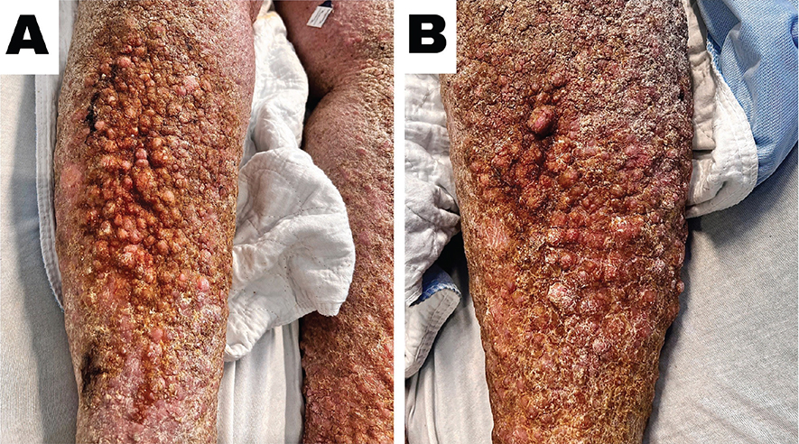
Fig. 1. (A) Right leg, (B) Left leg. Clinical presentation shows indurated, erythematous to translucent papulonodular lesions on the shins with overlying peau d’orange texture and signs of impetiginization. The findings are consistent with overlapping features of obesity-associated lymphoedematous mucinosis (OALM) and stasis mucinosis.
A clinical diagnosis of pretibial mucinosis, likely multifactorial due to chronic lower limb oedema, morbid obesity, and venous stasis, was made. The patient was also diagnosed with bacteraemia and treated with systemic antibiotics. Cutaneous management included ultrapotent topical corticosteroids, compression therapy, and intravenous diuretics for volume overload. Over the course of 4 weeks, he demonstrated significant improvement in oedema and skin lesions. However, he was lost to follow-up after discharge. His presentation was consistent with a euthyroid pretibial mucinosis, most likely representing overlapping features of OALM and stasis mucinosis.
DISCUSSION
We conducted a systematic review to summarize existing evidence on this underrecognized entity. A comprehensive search of PubMed, Embase, Web of Science, and MEDLINE was conducted from inception to 20 May 2025, following the PRISMA guidelines (Fig. S1). The study was registered in PROSPERO (CRD420251081183). Study quality was assessed using the Oxford Centre for Evidence-Based Medicine 2011 criteria.
A total of 24 studies met the inclusion criteria, describing 38 patients (Table SI). The mean age was 65 (± 13) years, and 50% were female. All patients were euthyroid at the time of diagnosis. Only 3/38 had any clinical or serologic evidence suggestive of subclinical Graves’ disease despite euthyroid status. This was identified as having 1 of the following: abnormal TRAb, absent response to thyrotropin-releasing hormone (TRH) stimulation test, or exophthalmos, either at presentation or on follow-up. The remaining 92% (35/38) had no lifetime history or signs of Graves’.
The most common clinical presentation was bilateral, chronic, gradually progressive lower leg oedema – ranging from pitting to non-pitting – accompanied by indurated skin with translucent, erythematous, or pigmented papules, nodules, or plaques, often with an overlying peau d’orange texture. A unilateral presentation was described in 3 patients. Morbid obesity was the most common risk factor, present in 55% (21/38) of cases, followed by venous stasis and other known contributors to chronic lower extremity oedema or secondary lymphatic obstruction.
Common histopathological findings among OALM and stasis mucinosis were the presence of an atrophic epidermis overlying an oedematous fibromyxoid stroma, with abundant mucin deposition in the papillary dermis, splaying of collagen fibres, and stellate fibroblasts. Other common histopathological features of venous stasis were also noted in those with stasis mucinosis. The most effective management strategies involved addressing underlying risk factors such as obesity, chronic oedema, or venous stasis, with interventions like lifestyle modification, weight reduction, and gradual compression therapy. Mid-to-high potency topical corticosteroids and monthly intramuscular triamcinolone injections were commonly used for cutaneous symptoms and led to clinical improvement.
The development of mucinous skin changes in obesity-associated and stasis mucinosis is multifactorial (3, 4). In obesity, lymphatic compression, venous insufficiency, and chronic inflammation lead to fluid retention and tissue hypoxia, which stimulate fibroblasts to produce glycosaminoglycans (GAGs) such as hyaluronic acid (3). In venous stasis, sustained venous hypertension and capillary leakage trigger inflammation and metalloproteinase activity, further promoting mucin accumulation (3, 4).
Somach et al. (5) described histopathological features that help distinguish OALM/stasis mucinosis from Graves’ dermopathy. In Graves’ dermopathy, mucin is confined to the reticular dermis, with preservation of the superficial papillary dermis and a zone of normal-appearing collagen.
In summary, OALM and stasis mucinosis are underrecognized mimics of Graves’ dermopathy. Clinicians should consider these entities in patients with bilateral lower extremity oedema and mucinous skin changes in the absence of thyroid disease. Early recognition and targeted management addressing underlying risk factors and cutaneous symptoms are essential to improving outcomes.
REFERENCES
- Rongioletti F. Mucinoses. In: Clinical and pathological aspects of skin diseases in endocrine, metabolic, nutritional and deposition disease. New York: Springer New York; 2010. p. 139–152. https://doi.org/10.1007/978-1-60761-181-3_18
- Rongioletti F. New and emerging conditions of acquired cutaneous mucinoses in adults. J Eur Acad Dermatol Venereol 2022; 36: 1016–1024. https://doi.org/10.1111/jdv.17983
- Pai SB, Pai K, Rao ACK, Bharath SV. Obesity-associated lymphedematous mucinosis in two patients: an emerging lifestyle disease. Indian J Dermatol Venereol Leprol 2024, July 20 [Online ahead of print]. https://doi.org/10.25259/IJDVL_1316_2023
- Hirt PA, MacQuhae FE, Cho-Vega JH, Kirsner RS. Stasis mucinosis: insights into euthyroid localized mucinosis. Wounds 2019; 31: E58–E560.
- Somach SC, Helm TN, Lawlor KB, Bergfeld WF, Bass J. Pretibial mucin: histologic patterns and clinical correlation. Arch Dermatol 1993; 129: 1152–1156. https://doi.org/10.1001/archderm.1993.01680300080012