QUIZ SECTION
An Elderly Man with a Solitary Hypopigmented Annular Plaque on the Chest: A Quiz
Junzhu XU and Wenting HU*
Department of Dermatology, Hangzhou Third People’s Hospital, 38 Xihu Ave, Hangzhou 310009, PR China. *E-mail: sendnote@126.com
Citation: Acta Derm Venereol 2025; 105: adv44335. DOI: https://doi.org/10.2340/actadv.v105.44335.
Copyright: © 2025 The Author(s). Published by MJS Publishing, on behalf of the Society for Publication of Acta Dermato-Venereologica. This is an Open Access article distributed under the terms of the Creative Commons Attribution-NonCommercial 4.0 International License (https://creativecommons.org/licenses/by-nc/4.0/).
Published: September 1, 2025
A 76-year-old man presented with a 10-year history of an erythematous lesion on the left chest (Fig. 1A). Previous treatments with topical triamcinolone acetonide and terbinafine cream yielded no significant improvement. Physical examination revealed a hypopigmented patch on the left anterior chest, surrounded by annular erythema. Periodic acid–Schiff staining showed no fungal elements. Histopathology demonstrated atypical lymphocytes confined to the epidermis and reactive lymphohistiocytic cells in the dermis (Fig 1B). Immunohistochemistry was positive for CD4 (Fig. 1C), but negative for CD8 and CD30 in the epidermis (Fig. 1D). T-cell receptor (TCR) gene rearrangement analysis revealed a clonal rearrangement of the TCR γ chain.
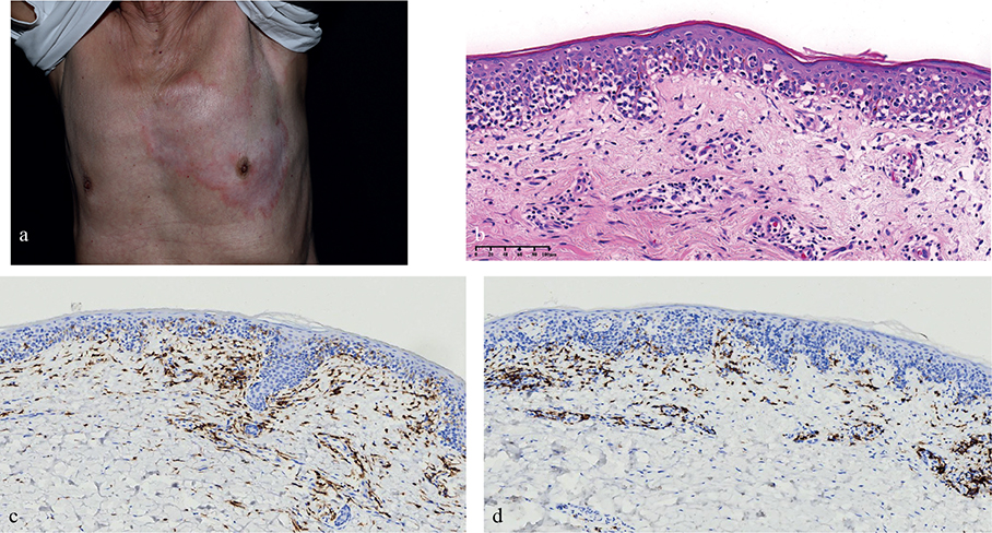
Fig. 1. (A) Hypopigmented patches on the left anterior chest, surrounded by annular erythema. (B) Histopathology (H&E stain, ×100): The epidermis shows atrophy or mild acanthosis, with prominent epidermotropism of atypical lymphocytes arranged in a Pagetoid pattern. The neoplastic cells exhibit hyperchromatic, cerebriform nuclei and infiltrate the epidermis as single cells or small clusters. (C) Immunohistochemical staining for CD3 (×40) revealed diffuse positivity in the epidermis. (D) Immunohistochemical staining for CD8 (×40) was negative in the epidermis.
What is your diagnosis?
1: Classic mycosis fungoides
2: Annular lichenoid dermatitis of youth
3: Pagetoid reticulosis
4: Plaque psoriasis
See next page for answer.
ANSWERS TO QUIZ
An Elderly Man with a Solitary Hypopigmented Annular Plaque on the Chest: A Commentary
Diagnosis: Pagetoid reticulosis
Based on the clinical and histopathological findings, the patient was diagnosed with pagetoid reticulosis (also known as Woringer–Kolopp disease), a rare, localized variant of mycosis fungoides (MF) that classically presents as a solitary, slowly progressive plaque, most commonly on the distal extremities (1). Histologically, WKD is characterized by marked epidermal hyperplasia with prominent pagetoid spread of atypical epidermotropic T lymphocytes. The neoplastic cells typically display a CD3⁺/CD8⁺ phenotype, although CD4⁺, CD4⁻/CD8⁻, and other immunophenotypic variants have been reported (2), highlighting the importance of comprehensive immunophenotyping and T-cell receptor gene rearrangement studies for accurate diagnosis.
Hypopigmented variants of pagetoid reticulosis are exceedingly rare and have not been clearly described in the literature. In our case, the hypopigmented annular plaque could easily have been misdiagnosed as vitiligo, psoriasis, or tinea versicolor. This underscores the importance of correlating clinical features with histopathology and immunohistochemistry, especially when encountering persistent, treatment-resistant skin lesions. In the differential diagnosis, conditions such as classic MF, annular lichenoid dermatitis of youth, and inflammatory dermatoses should be considered. Features favouring pagetoid reticulosis include its solitary, well-circumscribed nature, slow progression, and distinctive pagetoid pattern without widespread dermal infiltration. Treatment with narrowband Ultraviolet B phototherapy was initiated and has shown favourable response to date.
REFERENCES
- Osto M, Afify O, Musa A, Ahmed U, Rehman R, Mehregan D. Woringer–Kolopp disease (localized pagetoid reticulosis): a systematic review. Int J Dermatol 2023; 62: 312–321. https://doi.org/10.1111/ijd.16224
- Mourtzinos N, Puri PK, Wang G, Liu ML. CD4/CD8 double negative pagetoid reticulosis: a case report and literature review. J Cutan Pathol 2010; 37: 491–496. https://doi.org/10.1111/j.1600-0560.2009.01323.x