CORRESPONDENCE
Is TRIM11 Immunohistochemical Positivity Specific for the Diagnosis of CRTC1::TRIM11 Cutaneous Tumours?
Shangshen CHEN1,2, Yeqiang LIU2, Mingyuan XU2 and Wei TAN1,2*
1Huan Kui College of Nanchang University, Nanchang, China, and 2Department of Pathology, Shanghai Skin Disease Hospital, School of Medicine, Tongji University, Shanghai, China. *E-mail: 2411233@tonfji.edu.cn
Citation: Acta Derm Venereol 2025; 105: adv44357. DOI: https://doi.org/10.2340/actadv.v105.44357.
Copyright: © 2025 The Author(s). Published by MJS Publishing, on behalf of the Society for Publication of Acta Dermato-Venereologica. This is an Open Access article distributed under the terms of the Creative Commons Attribution-NonCommercial 4.0 International License (https://creativecommons.org/licenses/by-nc/4.0/).
Submitted: Jul 9, 2025. Accepted after revision: Jul 25, 2025. Published: Aug 7, 2025.
To the Editor,
We read with great interest the article by Koga et al. (1). This represents the first study detailing dermatoscopic features of this tumour and reporting its occurrence on the sole, significantly enriching the clinical understanding of CRTC1::TRIM11 cutaneous tumours (CTCT). However, we have concerns regarding the pathological findings and diagnostic criteria presented.
In terms of histopathology, the HE staining of the tumour shows irregular elongation of the overlying epidermis with keratosis, nodular lesions in the deep dermis with fibrosis, and aggregated tumour cells that are oval to spindle-shaped with mild nuclear atypia. This differs from the morphological characteristics of CTCT reported in previous literature (2–4). The lack of in-depth discussion and analysis of this discrepancy may affect readers’ comprehensive understanding of the pathological features of this tumour.
Regarding the diagnostic criteria, the study detected positive SOX10 and TRIM11 through immunohistochemistry, and excluded EWSR1 fusion by break-apart fluorescence in situ hybridization (FISH). Based on our research experience, TRIM11 positivity cannot be regarded as a reliable basis for diagnosing CTCT cutaneous tumours. We encountered a case where TRIM11 immunohistochemistry showed diffuse positivity after HE staining, FISH verified the presence of TRIM11 fusion, and transcriptome sequencing further confirmed the CTCT (Fig. 1A, B). However, in another patient with similar clinical and pathological manifestations, although TRIM11 immunohistochemistry was positive, FISH detection did not identify TRIM11 fusion, and transcriptome sequencing also failed to detect the CRTC1::TRIM11 or EWSR1 fusion genes. This clearly indicates that TRIM11 positivity is not specific (Fig. 1C, D). Additionally, we performed TRIM11 immunohistochemistry on different types of melanomas and clear cell sarcomas, which are easily confused with CTCT tumours. The results showed that all samples were diffusely positive, further confirming the non-specificity of TRIM11 immunohistochemistry in diagnosis (Fig. 1E, F).
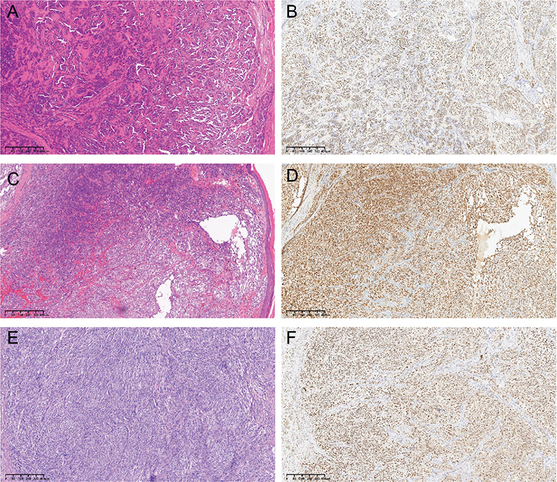
Fig. 1. Histological features and TRIM11 immunohistochemistry results of 3 patients. (A) HE staining and (B) TRIM11 IHC of a CTCT patient. (C) HE staining and (D) TRIM11 IHC of a melanoma patient with negative CRTC1::TRIM11 fusion. (E) HE staining and (F) TRIM11 IHC of a clear cell sarcoma patient, with EWSR1::ATF1 fusion revealed by RNA-seq. HE: haematoxylin–eosin; IHC: immunohistochemistry; CTCT: CRTC1::TRIM11 cutaneous tumour.
Based on these findings, we concur that HE morphology and TRIM11 IHC positivity can serve as important clues prompting consideration of a CTCT diagnosis. However, for definitive diagnosis, FISH detection confirming a TRIM11 rearrangement is more specific, and transcriptome sequencing confirming the CRTC1::TRIM11 fusion gene should be considered the diagnostic gold standard.
ACKNOWLEDGEMENT
Funding sources: This work was supported by the National Natural Science Foundation of China (grant number 82473508, 82173396); the Natural Science Foundation of Shanghai (grant number 23ZR1456100); and the Project of Clinical Research Incubation Program of Shanghai Skin Disease Hospital (NO. lcfy2022-01).
REFERENCES
- Koga M, Koga K, Shibayama Y, Imafuku S. Dermoscopic features of a CRTC1::TRIM11 cutaneous tumour: a case report and literature review. Acta Derm Venereol 2025; 105: adv43689. https://doi.org/10.2340/actadv.v105.43689
- Hanna J, Ko JS, Billings SD, Boivin F, Beaudoux O, Pissaloux D, et al. Cutaneous melanocytic tumor with CRTC1::TRIM11 translocation: an emerging entity analyzed in a series of 41 cases. Am J Surg Pathol 2022; 46: 1457–1466. https://doi.org/10.1097/PAS.0000000000001952
- Kashima J, Motoi T, Nishimaki M, Hayashi Y, Ogawa M, Kato I, et al. A case report of cutaneous melanocytoma with CRTC1-TRIM11 fusion: is CMCT distinct from clear cell sarcoma of soft tissue? Pathol Int 2019; 69: 496–501. https://doi.org/10.1111/pin.12826
- Kalmykova AV, Baranovska-Andrigo V, Michal M. Update on cutaneous mesenchymal tumors in the 5th edition of WHO classification of skin tumors with an emphasis on new fusion-associated neoplasms. Virchows Arch 2024; 485: 777–792. https://doi.org/10.1007/s00428-024-03925-2
Reply to: Is TRIM11 Immunohistochemical Positivity Specific for the Diagnosis of CRTC1::TRIM11 Cutaneous Tumours?
Monji KOGA, Kaori KOGA, Yoshitsugu SHIBAYAMA and Shinichi IMAFUKU
Department of Dermatology, Faculty of Medicine, Fukuoka University, Fukuoka, Japan, and Department of Dermatology, Fukuoka Sanno Hospital, Fukuoka, Japan. E-mail: mob3mob3@yahoo.co.jp
We partially agree with the commentary by Chen et al. The diagnosis of CTCT should be comprehensive and must not rely solely on a single immunohistopathological stain. Meanwhile, we believe that the results of TRIM11 immunohistochemical staining can serve as a reference, although the significance has not yet been fully established.
In our case, we believed that clear cell sarcoma and melanoma could initially be excluded based on the morphology in haematoxylin and eosin (H&E) stained images. Additionally, we performed EWSR1 break-apart fluorescence in situ hybridization (FISH) and performed immunohistochemical stains, such as PRAME, to confirm the negative results. Eventually, we established the diagnosis based on all these findings, including TRIM11 immunostaining.
According to the World Health Organization (WHO) diagnostic criteria, differentiating clear cell sarcoma is essential for diagnosing CTCT, although the confirmation of the fusion gene through genetic testing is not stated as necessary.
We would like to emphasize that the most significant diagnostic findings are obtained from H&E stained images.