SHORT COMMUNICATION
Self-healing Juvenile Cutaneous Mucinosis: an Extremely Rare and Alarming but Harmless Cutaneous Disease of Childhood
Lucie HARPAIN1  , Adrian TANEW2 , Mathias DRACH1 , and Sonja RADAKOVIC1*
1University Department of Dermatology, Medical University of Vienna, Vienna, and 2Private Practice, Vienna, Austria. *E-mail: sonja.radakovic@meduniwien.ac.at
Citation: Acta Derm Venereol 2025; 105: adv44491. DOI: https://doi.org/10.2340/actadv.v105.44491.
Copyright: © 2025 The Author(s). Published by MJS Publishing, on behalf of the Society for Publication of Acta Dermato-Venereologica. This is an Open Access article distributed under the terms of the Creative Commons Attribution-NonCommercial 4.0 International License (https://creativecommons.org/licenses/by-nc/4.0/).
Submitted: Jul 28, 2025. Accepted after revision: Aug 19, 2025. Published: Oct 7, 2025.
Competing interests and funding: The authors have no conflicts of interest to declare.
INTRODUCTION
Self-healing juvenile cutaneous mucinosis (SHJCM) is an extremely rare paediatric dermatological disorder, first described in the French literature by Colomb et al. in 1973 (1). It represents a rare variant of lichen myxoedematosus, also known as papular mucinosis (2). Since its first description, approximately 30 additional cases have been reported. Although SHJCM predominantly affects children, some adult cases with more heterogeneous clinical and histopathological presentations have been described (3). SHJCM is characterized by the eruptive appearance of itchy papules and painful subcutaneous nodules on the face, dorsum of the hand, and periarticular regions. The rash may be preceded by a prodromal stage of fever, asthenia, arthralgia, and muscle pain (4). The aetiopathogenesis of SHJCM remains unknown, but infections may be a possible trigger. Unlike adult-onset mucinosis, SHJCM has not been associated with systemic diseases such as systemic lupus erythematosus, thyroid disorders, monoclonal gammopathy, or plasmacytosis; however, an association with painful polyarthritis has been described (5). Histopathological examination is mandatory to confirm the clinical diagnosis. Typically, this reveals well-circumscribed mucin deposits in the dermis, surrounded by a spindle cell infiltrate and occasionally also a sparse perivascular lymphocytic infiltrate (4, 6–8). This extremely rare and clinically difficult to diagnose disease usually has a benign course with spontaneous resolution without sequelae within several weeks to months (6). Because of its rare and eruptive onset, SJHCM poses a significant diagnostic problem for physicians and often leads to parental concern, misdiagnosis, and occasionally also unnecessary aggressive treatment.
CASE REPORT
A 2.5-year-old fair-skinned girl, without a significant medical history, presented with eruptive and rapidly enlarging grouped flesh-coloured papules and partially shiny papules on the neck, upper extremity, both shoulders, and knees. She also showed a periorbital swelling and numerous extremely hard subcutaneous nodules on the nose, forearms, backs of the hands, palms, and soles. Some skin lesions, particularly the papules on the shoulders and forearms, were itchy, whereas the nodules on pressure sites were painful (Figs 1 and 2). The mucous membranes were unremarkable. The child did not have any prodromal symptoms such as fever, fatigue, arthralgia, or muscle pain. The paediatrician initially suspected juvenile dermatomyositis and determined antinuclear antibodies and aldolase, which were in the normal range. A differential diagnosis of calcinosis cutis was made and a deep biopsy of a subcutaneous nodule on the right forearm was performed. Histopathology with haematoxylin-eosin stain (H&E) showed a diffuse, perivascular, and periadnexal lymphocytic infiltrate in the superficial dermis as well as small foci of increased fibroblast density (Fig. 3A–B). Alcian blue stain (AB) revealed small deposits of acid mucopolysaccharides distributed in the dermis accompanied by fibroblasts and a sparse infiltrate of lymphocytes (Fig. 3C–D). Laboratory investigation including a complete blood cell count, blood chemistry, antinuclear antibodies, aldolase, and thyroid hormones were normal. The synopsis of the clinical picture, histopathology, and laboratory findings prompted us to make a diagnosis of SHJCM. As expected for this disease when occurring in children we did not find any indication of an underlying systemic disease such as paraproteinemia, plasmacytosis, or thyroid disease. Supportive treatment with ibuprofen and methylprednisolone over 2 weeks relieved the pain but did not alter disease activity. A watchful waiting approach was chosen and over the next 12 months new nodules appeared and older ones resolved. Approximately 1.5 years after its onset, the disease resolved completely without any residues.
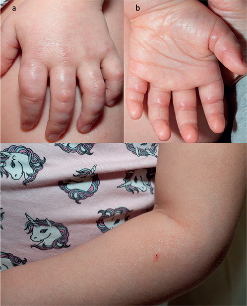
Fig. 1. Clinical presentation. (a–b) Numerous hard painful subcutaneous nodules on the hands. (c) Grouped itchy flesh-coloured papules on the left arm, some with subtle sheen.
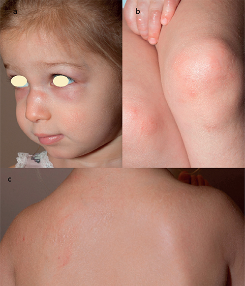
Fig. 2. Clinical presentation. (a) Periorbital swelling and hard subcutaneous nodules on the nose. (b–c) Grouped and itchy flesh-coloured papules, some with subtle sheen, on the knees, neck, and shoulders.
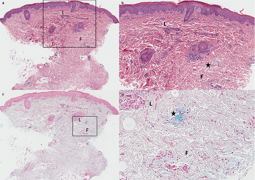
Fig. 3. Histology: subcutaneous nodule. (a) Haematoxylin and eosin (H&E, 20×) staining shows an unremarkable epidermis. Subtle foci of increased fibroblast density (F) and mucin deposits are observed in superficial and deep dermal layers. A diffuse, superficial, perivascular, and periadnexal lymphocytic infiltrate is noted (L). (b) At higher magnification (H&E, 40×), mucin deposits appear as pale, ill-defined areas within the dermis (star). (c–d) Alcian blue (AB) staining confirms dermal mucin as noticeable at lower (c, 20×) and higher (d, 100×) magnification (star).
DISCUSSION
SHJCM is an extremely rare and underrecognized entity with a striking clinical presentation, which may lead to misdiagnosis and unnecessary interventions (4, 6). The condition is characterized by a sudden onset of pruritic papules and painful subcutaneous nodules, most commonly affecting the face, dorsum of the hands, and periarticular regions. Although children are mostly affected a few adult cases have been described (3). Unlike cutaneous mucinoses seen in adults, SHJCM lacks any known association with systemic diseases (4, 6–8). The presence of painful nodules, particularly on pressure sites, may prompt clinicians to suspect a connective tissue disorder or inflammatory conditions. Histopathological examination is essential in distinguishing SHJCM from other inflammatory skin disorders. The histological hallmark is the presence of well-demarcated mucin deposits within the dermis as well as subcutis, accompanied by a sparse perivascular lymphocytic infiltrate (5, 9). According to the literature, areas of fibroblast proliferation are also a common feature in SHJCM (10). A main challenge in managing SHJCM is reassurance of the affected patients and their families concerning its benign and self-limiting nature. Due to its rarity, many physicians may not be familiar with this entity and initiate unnecessary treatments such as immunosuppressants or systemic corticosteroids. The exact aetiology of SHJCM remains unclear, though infections have been suggested as a potential trigger.
In conclusion, SHJCM presents as a unique diagnostic challenge due to its rapid onset and striking clinical features. Timely and accurate diagnosis through clinical assessment and histopathological confirmation is essential for recognizing and managing SHJCM as a distinct yet benign paediatric condition. Due to its self-limited course, reassurance of the patient and a conservative approach focused on relief of symptoms should be chosen.
ACKNOWLEDGEMENT
IRB approval status: Written informed consent to publication of this case report was obtained from the parents of the reported child.
REFERENCES
- Colomb D, Racouchot J, Vittori F. Mucinose d’évolution spontanément régressive sans paraprotéinémie chez une jeune fille. Lyon Med 1973; 230: 474–475.
- Rongioletti F, Rebora A. Updated classification of papular mucinosis, lichen myxedematosus, and scleromyxedema. J Am Acad Dermatol 2001; 44: 273–281. https://doi.org/10.1067/mjd.2001.111630
- Jang KA, Han MH, Choi JH, Sung KJ, Moon KC, Koh JK. Recurrent self-healing cutaneous mucinosis in an adult. Br J Dermatol 2000; 143: 650–651. https://doi.org/10.1111/j.1365-2133.2000.03731.x
- Kofler H, Lipsker D, Maurer H, et al. Self-healing juvenile cutaneous mucinosis: challenging diagnosis and management. J Dtsch Dermatol Ges 2014; 12: 815–817. https://doi.org/10.1111/ddg.12379
- Luchsinger I, Coulombe J, Rongioletti F, Haspeslagh M, Dompmartin A, Melki I, et al. Self-healing juvenile cutaneous mucinosis: clinical and histopathologic findings of 9 patients: the relevance of long-term follow-up. J Am Acad Dermatol 2018; 78: 1164–1170. https://doi.org/10.1016/j.jaad.2017.10.023
- Barreau M, Dompmartin-Blanchère A, Jamous R, Chababi M, Soutou B, Reynier-Rezzi J, et al. Nodular lesions of self-healing juvenile cutaneous mucinosis: a pitfall! Am J Dermatopathol 2012; 34: 699–705. https://doi.org/10.1097/DAD.0b013e3182459345
- Carder KR, Fitzpatrick JE, Weston WL, Morelli JG. Self-healing juvenile cutaneous mucinosis. Pediatr Dermatol 2003; 20: 35–39. https://doi.org/10.1046/j.1525-1470.2003.03008.x
- Hershko K, Sagi E, Ingber A. Self-healing juvenile cutaneous mucinosis in an infant. Acta Derm Venereol 2002; 82: 145–146. https://doi.org/10.1080/00015550252948266
- Bonerandi JJ, Andrac L, Follana J, Ortonne JP, Cetran JP, Piacentino G. Self-healing juvenile cutaneous mucinosis: clinical, histological and ultrastructural study (in French; author’s translation). Ann Dermatol Venereol 1980; 107: 51–57.
- Cowen EW, Scott GA, Mercurio MG. Self-healing juvenile cutaneous mucinosis. J Am Acad Dermatol 2004; 50: S97–100. https://doi.org/10.1016/j.jaad.2003.07.019