QUIZ SECTION
Solitary Facial Nodule in a Young Female Patient: A Quiz
Diogo DE SOUSA1, Claúdia BRAZÃO1, Pedro VASCONCELOS1 and Paulo FILIPE1–3
1Dermatology Department, Centro Hospitalar Universitário Lisboa Norte, Lisbon, 2Dermatology Universitary Clinic, Faculdade de Medicina da Universidade de Lisboa, Lisbon, and 3Dermatology Research Unit, Instituto de Medicina Molecular, University of Lisbon, Lisbon, Portugal.
E-mail: diogo.sousa@chln.min-saude.pt
Citation: Acta Derm Venereol 2025; 105: adv44549. DOI: https://doi.org/10.2340/actadv.v105.44549.
Copyright: © 2025 The Author(s). Published by MJS Publishing, on behalf of the Society for Publication of Acta Dermato-Venereologica. This is an Open Access article distributed under the terms of the Creative Commons Attribution-NonCommercial 4.0 International License (https://creativecommons.org/licenses/by-nc/4.0/).
Published: Sep 4, 2025.
Competing interests and funding: The authors have no conflicts of interest to declare.
A 36-year-old woman was referred to the dermatology clinic with a solitary red, itchy papule on her left inferior palpebra for the previous 2 months. She denied all other review of systems symptoms with the exception of fatigue. She did not have any other medical conditions. Physical examination revealed an erythematous and indurated 1×1.5-cm papule on the periocular region (Fig. 1A). Dermoscopy showed arborizing well defined vessels and brownish follicular openings, on a yellow and milky background (Fig. 1B). A punch biopsy was taken for histopathological examination. PET/CT examination of the neck, chest, abdomen, and pelvis revealed no abnormalities.
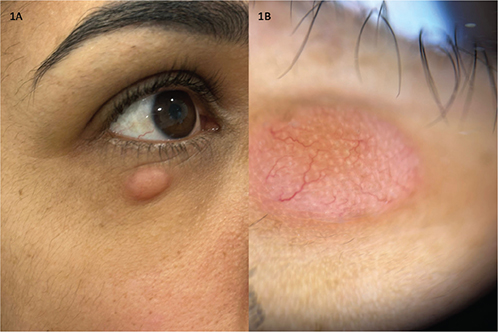
Fig. 1. Clinical presentation. (A) erythematous and indurated 1×1.5-cm papule on the periocular region. (B) Dermoscopy revealing arborizing vessels and brownish follicular openings, on a yellow-milky background.
Histopathology revealed a dense dermal and hypodermal infiltrate of small- to-medium-sized lymphocytes arranged in a nodular pattern, with pleomorphic nuclei, without epidermotropism (Fig. 2). Immunohistochemistry demonstrated intense and diffuse CD3 and CD4 staining, moderate staining for CD8 and CD20, and scarce staining for PD1 and BCL-6, with negative CD10 and CD30 stain. Monoclonal TCR-gamma rearrangement was present in the skin biopsy.
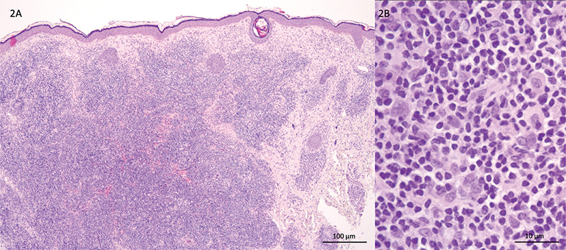
Fig. 2. Skin biopsy histology. (A) Lymphocytic infiltration involving deep portion of the dermis (haematoxylin and eosin, x40). (B) Higher magnification shows mixed infiltrate of small and medium-sized lymphocytes with scattered plasma cells and macrophages (haematoxylin and eosin, x400).
What is your diagnosis?
1: Cutaneous pseudolymphoma
2: Basal cell carcinoma
3: Primary cutaneous CD4+ small-/medium- sized T-cell lymphoproliferative disorder
4: Lymphomatoid papulosis
See next page for answer.
ANSWERS TO QUIZ
Solitary Facial Nodule in a Young Female Patient: A Commentary
Diagnosis: Primary cutaneous CD4+ small-/medium-sized T-cell lymphoproliferative disorder
Primary cutaneous CD4+ small/medium- sized T-cell lymphoproliferative disorder (PCSM-TCLPD) is a rare and heterogeneous entity, with suggested derivation from follicular T-helper lymphocytes. In 2016, the World Health Organization (WHO) working group reclassified this entity as a lymphoproliferative disorder, rather than a lymphoma, due to accumulating evidence of its benign prognosis (2).
In this case a PCSM-TCLPD diagnosis was made, based on clinical and histopathological features. Interestingly, the lesion completely regressed following the diagnostic biopsy. At 1-year follow-up, there were no signs of local recurrence or new lesions.
Classically PCSM-TCLPD manifests as singular asymptomatic lesion with an anatomic predilection for the head, neck, and upper extremities, although multifocal lesions have also been described (2). It commonly presents as a slow-growing erythematous or violaceous papule, plaque, nodule, or tumour (3). Ulceration is rarely present (3). The clinical course is widely variable: lesions can appear abruptly, enlarging over several months, slowly grow over years, persist with minimal change, diminish over the course of weeks, and spontaneously resolve (3, 4). Atypical presentations include acneiform, alopecia, leprosy-like disease, drug-induced (methotrexate–etanercept combination, vemurafenib, interleukin (IL)-2 therapy, triple-agent immunosuppressive therapy with cyclosporine, prednisolone, and azathioprine) (4).
Pileri et al. described a dermoscopy common vascular pattern in patients with PCSMP-TLPD, which included an orange-pink background with overlying short, fine, curvy or serpiginous blood vessels, and, when located on the face, yellow dots regarded as pilosebaceous gland openings (5).
By definition PCSMP-TLPD shows dense, diffuse, or nodular infiltrates within the dermis with a tendency to infiltrate the subcutis, with a predominance of small/medium-sized pleomorphic T cells (6). These lymphomas have a CD3+, CD4+, CD8-, CD30- phenotype, sometimes with loss of pan-T-cell markers (6). A low Ki-67 proliferation index is found in most cases (4). Clonal T cell receptor β and/or γ chain rearrangement has been detected in most cases, and comprise useful criteria to differentiate these small/medium-sized pleomorphic cutaneous T cell lymphomas from pseudo-T-cell lymphomas (2, 6).
The optimal treatment for PCSMP-TLPD has not yet been defined (6). Several therapeutic options have been used for solitary lesions, including surgical excision, radiotherapy, or a combination of both, as well as topical, intralesional, or oral steroids, doxycycline, amoxicillin, cryotherapy, bexarotene, cyclophosphamide either as monotherapy or in association with prednisolone, multi-agent chemotherapy (cyclophosphamide, hydroxydaunomycin, oncovin, and prednisone [CHOP] therapy) and systemic interferon-alfa (2, 4, 6). There are also cases of spontaneous remission following biopsy (2, 4).
PCSMP-TLPD generally has a favourable prognosis and indolent behaviour, especially with solitary lesions, with 5-year survival rates of 98.4 % (4). The minimal group of patients whose outcomes are unfavourable may present with an aggressive course, and multiple, large and rapidly growing, recurrent, treatment-resistant lesions (4).
ACKNOWLEDGEMENTS
IRB approval status: Written informed consent was obtained from the patient for the publication of this case report and accompanying images.
REFERENCES
- Surmanowicz P, Doherty S, Sivanand A, Parvinnejad N, Deschenes J, Schneider M, et al. The clinical spectrum of primary cutaneous CD4+ small/medium-sized pleomorphic T-cell lymphoproliferative disorder: an updated systematic literature review and case series. Dermatology 2021; 237: 618–628. https://doi.org/10.1159/000511473
- Swerdlow SH, Campo E, Pileri SA, Harris NL, Stein H, Siebert R, et al. The 2016 revision of the World Health Organization classification of lymphoid neoplasms. Blood 2016; 127: 2375–2390. https://doi.org/10.1182/BLOOD-2016-01-643569
- Gru AA, Wick MR, Eid M. Primary cutaneous CD4+ small/medium T-cell lymphoproliferative disorder: clinical and histopathologic features, differential diagnosis, and treatment. Semin Cutan Med Surg 2018; 37: 39–48. https://doi.org/10.12788/J.SDER.2018.006
- Salah E. Primary cutaneous CD4+ small/medium pleomorphic T-cell lymphoproliferative disorder: where do we stand? A systematic review. J Ger Soc Dermatology 2019; 17: 123–136. https://doi.org/10.1111/DDG.13691
- Pileri A, Agostinelli C, Sabattini E, Tabanelli M, Gaspari V, Leuzzi M, et al. Primary cutaneous small/medium-sized pleomorphic T-cell lymphoproliferative disorder shows a common vascular pattern at dermoscopy. J Eur Acad Dermatol Venereol 2018; 32: e318–e321. https://doi.org/10.1111/JDV.14872
- Willemze R, Jaffe ES, Burg G, Cerroni L, Berti E, Swerdlow SH, et al. WHO-EORTC classification for cutaneous lymphomas. Blood 2005; 105: 3768–3785. https://doi.org/10.1182/BLOOD-2004-09-3502