QUIZ SECTION
Giant Plaques on the Back with Gyrus-like Alteration: A Quiz
Mingxuan MA, Shuli GU, Jiaao FANG and Guangwen YIN
The First Affiliated Hospital of Zhengzhou University in China, No.1 East Road, ZhengZhou, Henan, China. E-mail: gwyin67@126.com
Citation: Acta Derm Venereol 2025; 105: adv44562. DOI: https://doi.org/10.2340/actadv.v105.44562.
Copyright: © 2025 The Author(s). Published by MJS Publishing, on behalf of the Society for Publication of Acta Dermato-Venereologica. This is an Open Access article distributed under the terms of the Creative Commons Attribution-NonCommercial 4.0 International License (https://creativecommons.org/licenses/by-nc/4.0/).
Published: September 1, 2025
A 21-year-old man presented with skin lesions. Six years ago, scattered skin-coloured papules were observed on the upper back with no obvious cause or significant subjective symptoms. The number and size of the papules gradually increased and became fused. Physical examination revealed 2 large skin-coloured plaques on the back and waist, measuring approximately 55 × 50 × 0.5 cm and 16 × 8 × 0.2 cm, respectively. The surface exhibited gyrus-like alteration with a soft texture and clear boundaries. Scattered skin-coloured papules with a tendency to fuse were observed on the left shoulder, sacrum, and buttocks (Fig. 1). Histopathological examination revealed a slightly pigmented basal epidermal layer with a small amount of mature adipose tissue in the superficial dermis (Fig. 2).
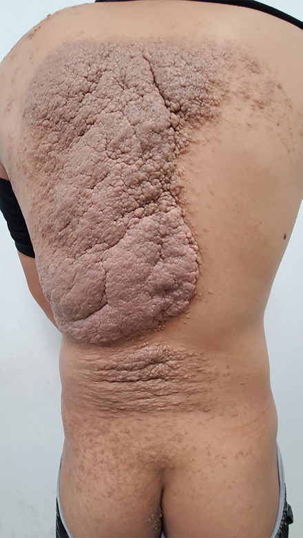
Fig. 1. Skin-coloured plaques on the back and waist, whose surface exhibited gyrus-like alterations with clear boundaries. Scattered skin-coloured papules with a tendency to fuse were observed on the left shoulder, sacrum, and buttocks.
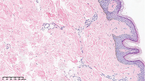
Fig. 2. Histopathological examination revealed a slightly pigmented basal epidermal layer with a small amount of mature adipose tissue in the superficial dermis (HE ×100).
What is your diagnosis?
1. Giant skin tag
2. Lipofibroma
3. Nevus lipomatosus cutaneous superficialis
4. Focal dermal hypoplasia
See next page for answer.
Giant Plaques on the Back with Gyrus-like Alteration: A Commentary
Diagnosis: Nevus lipomatosus cutaneous superficialis
Nevus lipomatosus cutaneous superficialis (NLCS) is a rare cutaneous hamartoma characterized by the presence of mature ectopic adipose tissue within the dermis. The pathogenesis of this disease remains unclear, with the main theories including: (i) lipomatous metaplasia caused by degenerative changes in dermal connective tissue; (ii) a true nevus formed by developmental displacement of adipose tissue; (iii) differentiation of perivascular adipoblasts into adipocytes (1).
In 1921, Hoffmann and Schuhelle were the first to report this disease and classified it into 2 clinical types: classical and solitary. The classical type mostly manifests at birth or before the age of 30, preferentially involving the lower back and buttocks. The skin lesions present as multiple soft papules or nodules with skin-coloured or light-yellow hues; the overlying skin is smooth or wrinkled, occasionally exhibiting a cerebriform appearance. Meanwhile, the solitary type is characterized by a single skin-coloured papule or nodule, with no obvious age predilection or anatomical site preference. Most cases are asymptomatic, although a few might show abnormal growth or morphological changes, including giant NLCS, comedo-like alterations, foul-smelling secretions, and ulcerative lesions (2).
The characteristic histopathological features of NLCS include the proliferation of mature ectopic adipose tissue within the dermis, accounting for 10–50% of the lesion. Adipocytes are often distributed in clusters around blood vessels or eccrine glands; they may also appear as single cells between collagen bundles. Increased density of collagen fibres and fibroblasts, along with perivascular infiltration of mononuclear cells and spindle cells, is occasionally observed. Additionally, the epidermis shows acanthosis, basket-weave hyperkeratosis, and increased basal pigmentation, as well as focal elongation and fusion of epidermal ridges (3).
This disease should be differentiated from disorders such as giant skin tag, lipofibroma, and focal dermal hypoplasia. While giant skin tag is clinically similar to the solitary type of NLCS, histopathological examination can confirm the absence of adipocytes in the dermis of giant skin tag cases. Meanwhile, lipofibroma typically presents as a pedunculated mass, characterized by a mixture of adipose tissue and dense collagen fibres around skin appendages. Focal dermal hypoplasia (Goltz syndrome) shares similar histopathological features with NLCS; however, in addition to ectopic adipose tissue proliferation in the dermis, it exhibits widespread collagen fibre rarefaction and absence of skin appendages. In cases where adipocyte components in NLCS lesions contain numerous spindle cells (representing immature adipocytes), they should be differentiated from other dermal spindle cell lesions such as neurofibroma, leiomyoma, and dermatofibroma (4).
Treatment is typically not required for NLCS except for cosmetic purposes. Surgical excision is curative for this condition, and no recurrence has been reported to date. Other treatment modalities include electrocautery, cryotherapy, phosphatidylcholine injection, and CO2 laser therapy (5). Notably, the patient in this case had giant skin lesions, and reconstructing tissue defects after lesion excision was challenging. Thus, early surgical excision is necessary for such cases to achieve a favourable prognosis. Furthermore, potential abnormal morphological changes should be vigilantly monitored.
ACKNOWLEDGEMENT
Funding sources: The authors sincerely thank the Henan Zhongyuan Talents Program (Yucai Series).
REFERENCES
- Kim YJ, Choi JH, Kim H, Nam SH, Choi YW. Recurrence of nevus lipomatosus cutaneous superficialis after CO(2) laser treatment. Arch Plast Surg 2012; 39: 671–673. https://doi.org/10.5999/aps.2012.39.6.671
- Alotaibi H, Alsaif F, Alali A, Almashali M, Al-Dabeeb D, Altaweel AA. Nevus lipomatosis cutaneous superficialis: a single-center case series of 5 patients. Case Rep Dermatol 2018; 10: 138–144. https://doi.org/10.1159/000488900
- Buch AC, Panicker NK, Karve PP. Solitary nevus lipomatosus cutaneous superficialis. J Postgrad Med 2005; 51: 47–48.
- Dhamija A, Meherda A, D’Souza P, Meena RS. Nevus lipomatosus cutaneous superficialis: an unusual presentation. Indian Dermatol Online J 2012; 3: 196–198. https://doi.org/10.4103/2229-5178.101819
- Pakhan A, Singh S, Dudhe M, Patil S. A giant nevus lipomatosus cutaneous superficialis treated with serial surgical excision: a case report. Cureus 2024; 16: e75054. https://doi.org/10.7759/cureus.75054