RESEARCH LETTER
A Series of Halo Congenital Nevi
Christina STEFANAKI, Eythymia SOURRA, Christina CHLORIDOU and Alexander STRATIGOS
First Dermatology Clinic, Andreas Sygros University Skin Hospital, Athens, Greece. E-mail: cstefana@otenet.gr
Citation: Acta Derm Venereol 2025; 105: adv44608. DOI: https://doi.org/10.2340/actadv.v105.44608.
Copyright: © 2025 The Author(s). Published by MJS Publishing, on behalf of the Society for Publication of Acta Dermato-Venereologica. This is an Open Access article distributed under the terms of the Creative Commons Attribution-NonCommercial 4.0 International License (https://creativecommons.org/licenses/by-nc/4.0/).
Submitted: Aug 12, 2025. Accepted after revision: Oct 20, 2025. Published: Oct 30, 2025.
To the Editor,
Halo nevus (HN) is a benign melanocytic nevus surrounded by an achromic rim that simulates a halo, resulting in regression of the nevus (1). The estimated incidence of HN in the population is around 1% and there is no predilection for sex or race (1, 2). Children and young adults are predominantly affected, with an average age of onset of 15 years (1, 2).
Halo formation may also rarely be observed with small and medium-sized congenital melanocytic nevi (CMN) and may result in subsequent involution of the nevus (3).
In a cohort of 524 patients with CMN we identified 13 CMN with halo phenomenon, representing 2.4% of the cohort. Six CMN were small, 6 were medium-sized CMN measuring 1.5–10 cm, and 1 CMN was large (> 20 cm). No patient had a family history of melanoma or immunosuppression. Only 2 patients suffered from vitiligo and thyroiditis, while the rest were healthy. Patients’ age at presentation and disease characteristics are presented in Table I.
Dermoscopic evaluation was performed in all CMN and digital dermoscopic follow-up was available for 10 patients ranging between 3 and 10 years. No vascular structures or blue-white veil were identified. No patient developed melanoma in the follow-up period. Two CMN disappeared completely, whereas in the rest the colour regressed by 10–90% (Fig. 1). Dermoscopic characteristics of halo CMN are presented in Table II.
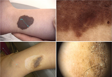
Fig. 1. Macroscopic image of medium-sized halo congenital nevus before and after 9 years of follow-up and dermoscopic pictures of the same nevus before and after the halo phenomenon.
Halo phenomenon in CMN has been reported previously and in particular large CMN have been associated with intralesional and perilesional depigmentation and extralesional vitiligo (3–7).
Halo phenomenon has not been fully understood but it is tempting to speculate that the abundance of melanocytes and nevus cells in CMN triggers an immunological response leading to the destruction of melanocytes and pigment loss (4, 5).
There is a strong relationship between halo nevi and vitiligo (3, 6); however, we observed vitiligo in only 1 of our patients. The relationship between CMN and vitiligo has been investigated before and it has been found that patients with vitiligo with CMN had an earlier age of onset, which was even more pronounced in the case of halo CMN (7). In a previously reported series of 8 children with halo large CMN, 3 had extensive vitiligo and 1 had vitiligo adjacent to the nevus (4).
The question remains whether patients with CMN develop a halo phenomenon more frequently than other individuals. Our series of 13 patients represented 2.4% of a larger cohort of 524 patients with CMN, suggesting a much higher prevalence than the usual 1% prevalence of halo nevi in the general population.
The most important question remains whether halo phenomenon is protective against melanoma. Cutaneous malignant melanoma has been reported in 1 patient with a halo large CMN (8). On the other hand, 2 large series support the protective role of vitiligo against the development of malignant melanoma (9, 10). It is unknown whether patients in our series will develop melanoma in the future but we did not detect any worrisome features in a follow-up period of 3–10 years.
Dermoscopically, halo CMN in our series demonstrated a globular or homogeneous pattern and typical features of CMN like target network, target and haloed globules, blotches, milia-like cysts, perifollicular hypopigmentation, and skin furrow hypopigmentation in the pigmented part and those features gradually faded during the hypopigmentation process (see Fig. 1). We did not detect a cerebriform appearance and a blue-white veil in any of the nevi in our series as has been observed by other authors (11).
Further studies should clarify the halo phenomenon in CMN, which in the case of medium and large CMN may in part improve the patient’s cosmetic appearance.
ACKNOWLEDGEMENTS
The authors would like to thank the nurses of the Pediatric Dermatology Department Ioanna Katsirea and Kyriakoula Kalentzi for their valuable contribution.
REFERENCES
- Aouthmany M, Weinstein M, Zirwas MJ, Brodell RT. The natural history of halo nevi: a retrospective case series. J Am Acad Dermatol 2012; 67: 582–586. https://doi.org/10.1016/j.jaad.2011.11.937
- Weyant G, Chung C, Helm K. Halo nevus: review of the literature and clinicopathologic findings. Int. J Dermatol 2015; 54: 30–447. https://doi.org/10.1111/ijd.12843
- Stierman SC, Tierney EP, Shwayder TA. Halo congenital nevocellular nevi associated with extralesional vitiligo: a case series with review of the literature. Pediatr Dermatol 2009; 26: 414–424. https://doi.org/10.1111/j.1525-1470.2009.00938.x
- Polat Ekinci A, Kiliç S, Baykal C. Pigment loss in patients with large congenital melanocytic nevi: various clinical presentations documented in a large series. Pediatr Dermatol 2016; 33: 307–310. https://doi.org/10.1111/pde.12823
- Cui J, Bystryn JC. Melanoma and vitiligo are associated with antibody responses to similar antigens on pigment cells. Arch Dermatol 1995; 131: 314–318. https://doi.org/10.1001/archderm.1995.01690150078015
- Kim HS, Goh BK. Vitiligo after halo formation around congenital melanocytic nevi. Pediatr Dermatol 2009; 26: 755–756. https://doi.org/10.1111/j.1525-1470.2009.01030.x
- Van Geel N, Van Poucke L, Van de Maele B, Speeckaert R. Relevance of congenital melanocytic naevi in vitiligo Br J Dermatol 2015; 172: 1052–1057. https://doi.org/10.1111/bjd.13444
- Bouffard D, Barnhill RL, Mihm MC, Sober AJ. Very late metastasis (27 years) of cutaneous malignant melanoma arising in a halo giant congenital nevus. Dermatology 1994; 189: 162–166. https://doi.org/10.1159/000246823
- Paradisi A, Tabolli S, Didona B, Sobrino L, Russo N, Abeni D. Markedly reduced incidence of melanoma and nonmelanoma skin cancer in a nonconcurrent cohort of 10,040 patients with vitiligo. J Am Acad Dermatol 2014; 71: 1110–1116. https://doi.org/10.1016/j.jaad.2014.07.050
- Teulings HE, Overkamp M, Ceylan E, Nieuweboer-Krobotova L, Bos JD, Nijsten T, et al. Decreased risk of melanoma and nonmelanoma skin cancer in patients with vitiligo: a survey among 1307 patients and their partners. Br J Dermatol 2013; 168: 162–171. https://doi.org/10.1111/bjd.12111
- Nath AK, Thappa DM, Rajesh NG. Spontaneous regression of a congenital melanocytic nevus. Indian J Dermatol Venereol Leprol 2011; 77: 507–510. https://doi.org/10.4103/0378-6323.82418