QUIZ SECTION
Unilateral Blaschkolinear Agminated Pigmented Lesion in an 11-year-old Girl: A Quiz
Tubanur ÇETINARSLAN1*, Şule Yıldız SAĞCAN TERCAN1, Peyker TEMIZ2, Mustafa Turhan ŞAHIN1 and Aylin TÜREL ERMERTCAN1
1Department of Dermatology and Venereology, Manisa Celal Bayar University, Manisa, Turkey, and 2Department of Pathology, Manisa Celal Bayar University, Manisa, Turkey. *Email: t_sarmis@windowslive.com
Citation: Acta Derm Venereol 2026; 106: adv-2025-0099. DOI: https://doi.org/10.2340/actadv.v106.adv-2025-0099.
Copyright: © 2026 The Author(s). Published by MJS Publishing, on behalf of the Society for Publication of Acta Dermato-Venereologica. This is an Open Access article distributed under the terms of the Creative Commons Attribution-NonCommercial 4.0 International License (https://creativecommons.org/licenses/by-nc/4.0/).
Submitted: Accepted after revision:
Published: Jan 21, 2026.
Competing interests and funding:
An 11-year-old female patient was referred to our dermatology department for numerous pigmented papules on her abdomen. The patient stated that lesions had been present since birth. On dermatological examination, there were multiple well-defined clustered pigmented papules with terminal hairs (Fig. 1). Dermoscopic examination revealed a homogeneous pattern with diffuse brownish areas and symmetrically distributed peripherally globules (Fig. 2). We performed a punch biopsy, and histopathological examination revealed nevoid cell nests arranged in the dermis and nevoid cell groups containing melanin located superficially in the dermis (Fig. 3).
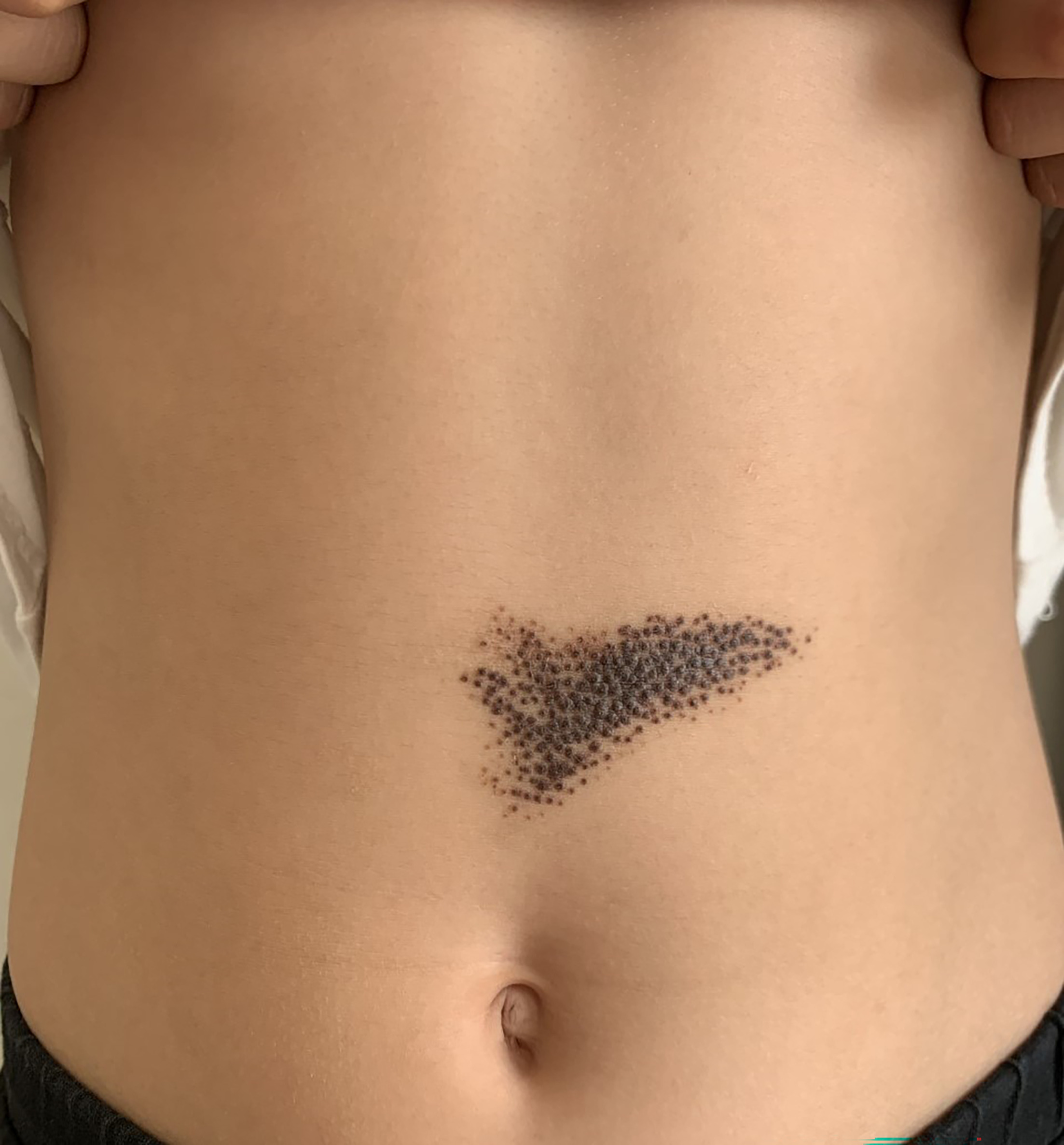
Fig. 1. Numerous well-defined clustered pigmented papules with terminal hairs.

Fig. 2. Homogeneous pattern with diffuse brownish areas and symmetrically distributed peripherally globules.

Fig. 3. Nevoid cell groups containing melanin located superficially in the dermis (haematoxylin-eosin stain, original magnification x100).
What is your diagnosis?
1: Partial unilateral lentiginosis
2: Nevus spilus
3: Agminated melanocytic nevus
4: Becker nevus
See next page for answer.
ANSWERS TO QUIZ
Unilateral Blaschkolinear Agminated Pigmented Lesion in an 11-Year-Old Girl: A Commentary
Diagnosis: Agminated melanocytic nevus
Agminated melanocytic nevi are very rare and occur as a grouped or clustered melanocytic lesions following a block-like pattern or Blaschko lines, on normal-appearing background skin (1). This type of nevi may show histopathological features of congenital melanocytic compound nevi (2), junctional nevi (3) and congenital melanocytic dermal nevi (4).
It is unclear whether agminated melanocytic nevi are congenital or acquired. Bragg et al. (1). reported four cases with acquired agminated melanocytic naevi and suggested that agminated melanocytic naevi are acquired and commonly observed after puberty. Contrary to this view, there are reported congenital agminated nevi cases in the literature (2, 3), as in our case. We suggest that agminated melanocytic nevi may have acquired or congenital forms, just like other melanocytic nevi.
The relationship between the agminated melanocytic nevi and mosaicism is another issue that needs to be clarified. Congenital type of agminated melanocytic nevi are often dermatomal distribution or follow Blaschko's lines (2, 3, 4, 5). It has been suggested that the distribution pattern in these patients may be due to somatic mutation that occurs in the early stages of embryogenesis (2, 3, 4). The term agminated refers to scattered, disorganized lesions, but the term of blaschkolinear is a pattern of lines on the skin that represent the developmental growth pattern during epidermal cell migration (5). Therefore, it is debatable whether the use of the term agminated in all patients is correct.
The occurrence of Langerhans cell histiocytosis (LCH) in some of these patients raises the question of whether there is any association between agminated melanocytic naevi and LCH (6, 7). Agminated melanocytic nevi reported in the literature in association with LCH have been reported as acquired melanocytic nevi (6, 7). The first case with agminated melanocytic nevus and LCH was reported in 2010 by Berk and Lane (8). In this case, systemic chemotherapy was thought to be the responsible factor because of the occurrence of agminated melanocytic nevus after several years of the remission of LCH (8). In contrast with this, it has been suggested that regulatory T cells or pro-inflammatory cytokines from LCH cells may induce the nevus in cases with agminated melanocytic nevus and LCH (7). Furthermore, Carreno-Gayosso et al. (9) reported a somatic mutation in BRAFV600E that leads to the overexpression and activation of the mitogen-activated protein kinase–Extracellular signal-regulated kinase (ERK) signal pathway in a patient with LCH and agminated atypical melanocytic nevus. This may lead to the occurrence of numerous nevi in the skin areas affected by LCH (9). On the other hand, there are other reported malignancies, including neuroendocrine tumour and breast cancer (1). There was no associated disease or malignancy in our patient. Data regarding the association of agminated melanocytic nevus with malignancy or LCH remain unclear, and further studies are needed.
It is not clear whether agminated melanocytic nevi are premalignant. There are reported cases of melanoma developed on the basis of agminated melanocytic nevus (10) and dysplastic nevus associated with agminated melanocytic nevus (9). Routine follow-up should be recommended in patients with agminated melanocytic nevus, whether acquired or congenital.
Agminated melanocytic nevi are rare lesions that need to be distinguished from other agminated lesions, including nevus spilus, partial unilateral lentiginosis, Becker nevus, speckled lentiginous nevi and segmental neurofibromatosis. Further genetic and clinical studies are needed to elucidate the pathogenesis of agminated melanocytic nevi. Our personal opinion is that patients with agminated melanocytic nevus should be monitored routinely.
REFERENCES
- Bragg JW, Swindle L, Halpern AC, Marghoob AA. Agminated acquired melanocytic nevi of the common and dysplastic type. J Am Acad Dermatol 2005; 52: 67–73. https://doi.org/10.1016/j.jaad.2004.05.033
- Effendy I, Happle R. Linear arrangement of multiple congenital melanocytic nevi. J Am Acad Dermatol 1992; 27: 853–854. https://doi.org/10.1016/0190-9622(92)70265-h
- Nachbar F, Merkle T, Ruzicka T, Plewig G. Congenital junctional nevi following Blaschko’s lines. Eur J Dermatol 1993; 3: 478–479.
- Brunner M, Vardarman E, Megahed M, Ruzicka T. Congenital agminated segmental naevi. Br J Dermatol 1995; 133: 315–316. https://doi.org/10.1111/j.1365-2133.1995.tb02638.x
- Moss C, Larkins S, Stacey M, Blight A, Farndon PA, Davison EV. Epidermal mosaicism and Blaschko’s lines. J Med Genet 1993; 30: 752–755. https://doi.org/10.1136/jmg.30.9.752
- Makino T, Matsui Y, Torai R, Mizawa M, Shimizu T. Agminated flexural melanocytic nevus associated with Langerhans cell histiocytosis. J Dermatol 2020; 47: e275–e276. https://doi.org/10.1111/1346-8138.15391
- Feldstein S, Funk T, Smith JC, Bruckner AL. Agminated flexural melanocytic nevi: a late sequela of Langerhans cell histiocytosis? Pediatr Dermatol 2015; 32: e89–91. https://doi.org/10.1111/pde.12563
- Berk DR, Lane AT. Acquired bilateral agminated Spitz nevi in a child with Langerhans cell histiocytosis. Pediatr Dermatol 2010; 27: 282–284. https://doi.org/10.1111/j.1525-1470.2010.01139.x
- Carreño-Gayosso EA, Hernández-Peralta SL, Guevara-Gutiérrez E, Solís-Ledezma G. Agminated atypical melanocytic nevus associated with Langerhans cell histiocytosis. An Bras Dermatol 2019; 94: 455–457. https://doi.org/10.1590/abd1806-4841.20198620
- Corradin MT, Alaibac M, Fortina AB. A case of malignant melanoma arising from an acquired agminated melanocytic naevus. Acta Derm Venereol 2007; 87: 432–433. https://doi.org/10.2340/00015555-0256