QUIZ SECTION
An Elderly Man with Fever and a Pustular Eruption: A Quiz
Jakob Lillemoen DRIVENES1*, Dag Sollesnes HOLSEN1 and Ingeborg Margrethe BACHMANN1,2
1Department of Dermatology, Haukeland University Hospital, Bergen, Norway, and 2Institute of Medical Science, University of Bergen, Bergen, Norway. *Email: jakob.lillemoen.drivenes@helse-bergen.no
Citation: Acta Derm Venereol 2026; 106: adv-2025-0192. DOI: https://doi.org/10.2340/actadv.v106.adv-2025-0192.
Copyright: © 2026 The Author(s). Published by MJS Publishing, on behalf of the Society for Publication of Acta Dermato-Venereologica. This is an Open Access article distributed under the terms of the Creative Commons Attribution-NonCommercial 4.0 International License (https://creativecommons.org/licenses/by-nc/4.0/).
Submitted: Accepted after revision:
Published: Jan 21, 2026.
Competing interests and funding:
A 73-year-old man presented to our institution’s department of dermatology with acute-onset fever and a generalized skin eruption. His medical comorbidities included hypercholesterolaemia treated with rosuvastatin 10 mg daily, hypertension treated with amlodipine 10 mg daily, polymyalgia rheumatica treated with prednisolone 3.75 mg daily and chronic plaque psoriasis for >15 years. Over the preceding year, his psoriasis had evolved from stable plaques into a more guttate morphology. A few days prior to the consultation, he developed a fever (maximum 38.7°C) followed by rapid and widespread worsening of his psoriasis. On examination, he exhibited a diffuse erythematous and guttate eruption over the trunk and extremities, many of which were studded with deep-seated pustules and areas of exudative scaling (Figs 1 and 2). Mucosal and palmoplantar involvement were absent. Laboratory analysis revealed a C-reactive protein level of 128 mg/L (<1) and lymphopenia of 0.4×10⁹/L (1.2–3.1).
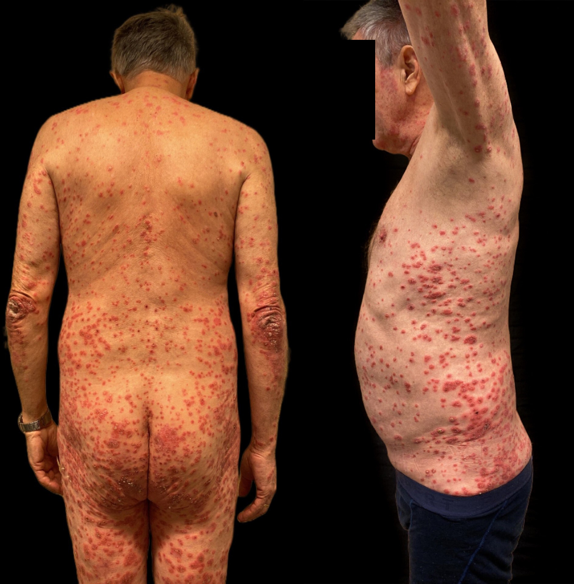
Fig. 1. Diffuse guttate and pustular psoriasis lesions with erythema and areas of exudative scaling on the trunk.
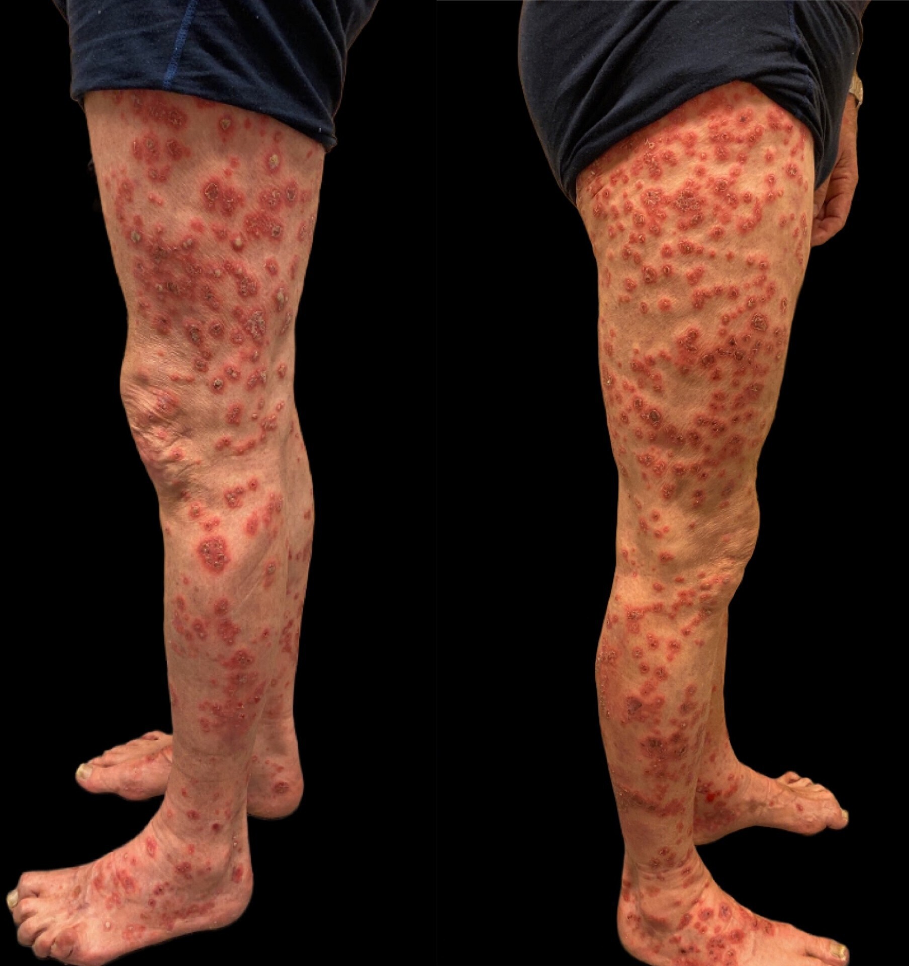
Fig. 2. Diffuse guttate and pustular psoriasis lesions with erythema and areas of exudative scaling on the legs.
What is your diagnosis?
1: Impetiginized psoriasis
2: Acute generalized pustular psoriasis
3: Hand, foot and mouth disease
4: Acute generalized exanthematous pustulosis
See next page for answer.
ANSWERS TO QUIZ
An Elderly Man with Fever and a Pustular Eruption: A Commentary
Diagnosis: Hand, foot and mouth disease (psoriasis coxsackium)
The patient reported that he had been in close contact with his grandchildren approximately 10 days before his fever and rash appeared, and the grandchildren had recently been diagnosed with hand, foot and mouth disease (HFMD). He was admitted to the dermatology inpatient ward for evaluation and treatment and was placed in contact isolation as a hygiene precaution. Polymerase chain reaction (PCR) testing confirmed Coxsackievirus A6 from nasopharyngeal swabs, pustular lesions and faeces. Bacterial cultures were negative. The condition was diagnosed as psoriasis coxsackium – a rare Koebner phenomenon triggered by enteroviral infection in patients with psoriasis. The patient was treated with oral acitretin (25 mg daily), clobetasol propionate ointment once daily, and potassium permanganate baths. He showed marked improvement over 2 weeks, with resolution of pustules, scaling and systemic symptoms. He remained in remission at the 2-month follow-up.
Psoriasis coxsackium is a rare clinical entity in which enteroviral infection triggers a psoriatic flare through a Koebner phenomenon. While eczema coxsackium has previously been described, its psoriatic counterpart is exceedingly rare (1, 2). Psoriasis is a chronic, immune-driven skin disorder that impacts an estimated 125 million people globally. The most prevalent subtype is plaque psoriasis, which represents over 80% of all cases. Its development is linked to a self-perpetuating inflammatory process, largely mediated through the T-helper 17 pathway. Genetic susceptibility constitutes a key determinant in disease development, while environmental triggers can worsen its clinical expression (3). In this patient, we hypothesize that Coxsackievirus A6 acted as an immunologic trigger, transforming his chronic psoriasis into an acute pustular and guttate flare. Coxsackievirus A6 has emerged as a major cause of HFMD outbreaks in the United States and globally. Compared to classical strains like A16, A6 is associated with more widespread and atypical cutaneous presentations and has been increasingly linked to adult-onset HFMD (4). Histologically, Coxsackievirus A6 has been shown to induce keratinocyte necrosis and spongiosis, disrupting skin barrier function and promoting local inflammation (5). In genetically predisposed individuals with psoriasis, this epidermal insult may act as a potent Koebnerizing stimulus. The Koebner phenomenon, a hallmark of psoriasis, refers to the development of psoriatic lesions at sites of cutaneous injury or inflammation (3). This can be elicited by physical trauma, infection or even certain dermatoses. In this context, both the cutaneous manifestations of viral replication and the ensuing systemic immune response may synergistically provoke a flare of underlying disease.
Clinically, psoriasis coxsackium presents a diagnostic challenge. The sudden onset of pustular lesions, particularly in areas affected by HFMD, may mimic acute generalized pustular psoriasis or impetiginized psoriasis. Distinguishing among these entities is crucial, as management strategies differ substantially. PCR testing for enteroviruses from skin or mucosal lesions offers a valuable diagnostic tool and can help confirm the presence of viral involvement. In this case, PCR positivity for Coxsackievirus A6 supported the diagnosis and enabled appropriate treatment planning. Management is supportive, given the self-limited nature of HFMD, and should address both the psoriatic component and potential viral complications. However, in patients with significant psoriatic exacerbation, intervention may be warranted to control inflammation and prevent progression. In this case, systemic retinoid therapy was selected due to its efficacy in pustular variants of psoriasis and its non-immunosuppressive profile – an important consideration in the context of active viral infection (6). Adjunctive topical corticosteroids and antiseptic soaks facilitated symptom control and reduced the risk of secondary bacterial colonization. The patient’s rapid clinical improvement without complications suggests that early recognition and targeted therapy can lead to favourable outcomes.
This case underscores the importance of recognizing viral triggers in patients with skin disorders, particularly during periods of increased enteroviral circulation or intergenerational contact. Awareness of psoriasis coxsackium may prevent diagnostic delay and guide appropriate treatment.
REFERENCES
- Cole DW, Wang B, Fullen DR, Helfrich YR. Psoriasis coxsackium. JAAD Case Rep 2022; 25: 22–24. https://doi.org/10.1016/j.jdcr.2022.05.004
- Wu CY, Lin FL. Hand-foot-and-mouth-disease-induced Koebner phenomenon in psoriasis. J Dtsch Dermatol Ges 2019; 17: 549–551. https://doi.org/10.1111/ddg.13824
- Armstrong AW, Read C. Pathophysiology, clinical presentation, and treatment of psoriasis: A review. JAMA 2020; 323: 1945. https://doi.org/10.1001/jama.2020.4006
- Kimmis BD, Downing C, Tyring S. Hand-foot-and-mouth disease caused by coxsackievirus A6 on the rise. Cutis 2018; 102: 353–356.
- Horsten HH, Kemp M, Fischer TK, Lindahl KH, Bygum A. Atypical hand, foot, and mouth disease caused by Coxsackievirus A6 in Denmark: A diagnostic mimicker. Acta Derm Venereol 2018; 98: 350–354. https://doi.org/10.2340/00015555-2853
- Hoegler KM, John AM, Handler MZ, Schwartz RA. Generalized pustular psoriasis: A review and update on treatment. J Eur Acad Dermatol Venereol 2018; 32: 1645–1651. https://doi.org/10.1111/jdv.14949