SHORT COMMUNICATION
Dermoscopic Findings of a Rare Variant of Elastolytic Giant Cell Granuloma: Papular Form
Muhammed Ali MERGEN1, Tubanur ÇETINARSLAN1*, Peyker TEMIZ2, Mustafa Turhan ŞAHIN1 and Aylin TÜREL ERMERTCAN1
1Department of Dermatology and Venereology, Manisa Celal Bayar University, Manisa, Turkey, and 2Department of Pathology, Manisa Celal Bayar University, Manisa, Turkey. *Email: t_sarmis@windowslive.com
Key words: elastolytic giant cell granuloma; dermoscopy; papular form; familial mediterranean fever; O'Brien granuloma.
Citation: Acta Derm Venereol 2026; 106: adv-2025-0076. DOI: https://doi.org/10.2340/actadv.v106.adv-2025-0076.
Copyright: © 2026 The Author(s). Published by MJS Publishing, on behalf of the Society for Publication of Acta Dermato-Venereologica. This is an Open Access article distributed under the terms of the Creative Commons Attribution-NonCommercial 4.0 International License (https://creativecommons.org/licenses/by-nc/4.0/).
Submitted: Oct 6, 2025. Accepted after revision: Nov 18, 2025.
Published: Feb 09, 2026.
Competing interests and funding: The authors have no conflicts of interest to declare.
Elastolytic giant cell granuloma (EGCG), also called actinic granuloma or O'Brien granuloma, is a rare, idiopathic, chronic inflammatory skin disease that typically occurs in sun-exposed areas in adults (1, 2). EGCG usually presents with well-defined annular erythematous plaques with slightly elevated borders, atrophic and/or hypopigmented centres. The papular variant of EGCG is quite rare and may pose a diagnostic challenge due to its deviation from the classic annular morphology (3, 4). Herein, we report an EGCG case accompanied by Familial Mediterranean Fever (FMF) and describe the dermoscopic findings of the papular type of EGCG.
CASE
A 50-year-old female patient presented with itchy swellings on hands for 3 years. She had a diagnosis of FMF for 23 years. She is under follow-up by the Rheumatology Department and is being treated with colchicine 0.5 mg 3 times daily. On dermatological examination, a plaque composed of numerous papules measuring approximately 2 mm × 2 mm on the dorsum of the right hand was observed. The plaque displayed central atrophy and hypopigmentation, with a raised, hyperpigmented and mildly erythematous to violaceous border (Fig. 1A). Dermoscopy revealed irregularly distributed polymorphic vessels (linear and spermatozoa-like) on a pale pink background, along with white and yellow-orange structureless areas. In the dermoscopic evaluation of the papules, linear vessels extending from the periphery towards the centre were prominent. Fine white scales were noted at the periphery of the papules (Fig. 1B, C).

Fig. 1. (A) A plaque composed of numerous papules measuring approximately 2 mm × 2 mm on the dorsum of the right hand. (B) Polymorphous vessels irregularly distributed on a pale pink background (black arrow), white structureless areas (blue star) and yellow-orange structureless areas (black circle). (C) Linear vessels extending from the periphery to the centre of the papule (purple arrow) and fine white scales at the edge of the papule (orange circle) (Dermlite DL4, 10× magnification, polarized mode).
On laboratory examination, no abnormalities were detected except dyslipidaemia and elevated creatinine (1.19 mg/dL). The Purified protein derivative (PPD) test measured 12 mm (Bacillus Calmette-Guerin (BCG)-vaccinated). No pathological finding was observed in abdominal ultrasonography and chest X-ray. Tuberculosis, sarcoidosis and malignancy were ruled out.
A skin biopsy was obtained from the dorsum of the right hand. On histopathological examination, numerous granulomatous structures were observed below the normal epidermis. Within the dermis, dense eosinophilic areas between these granulomas, corresponding to degeneration of collagen and elastic fibres and granulomas containing multinucleated giant cells with cytoplasmic fragments of elastic fibres, were seen (Fig. 2A, B). The patient was diagnosed with EGCG considering the clinical and histopathological findings. Regular sunscreen was advised and clobetasol propionate cream was started for 15 days, followed by topical tacrolimus ointment. Annual follow-up by the pulmonology and internal medicine departments was recommended. By the fourth month of treatment, the lesions had regressed (Fig. 3).

Fig. 2. (A) A dermal granulomatous structure with a central area of degeneration and peripheral palisading histiocytes (Hematoxylin & Eosin, ×40). (B) A multinucleated giant cell containing phagocytosed elastic fibre fragments within its cytoplasm (Hematoxylin & Eosin, ×400).
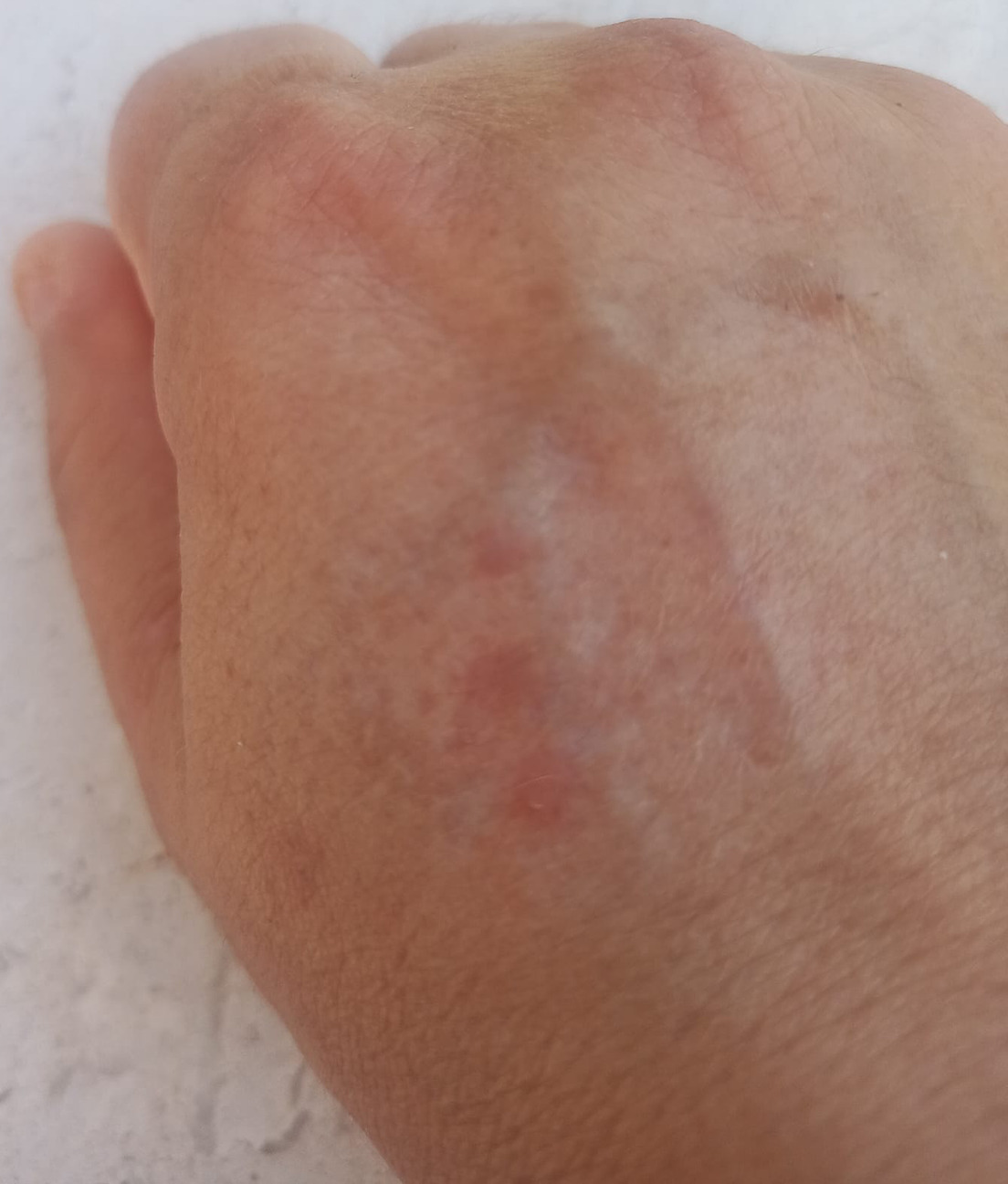
Fig. 3. Clinical improvement after treatment.
DISCUSSION
Although EGCG was initially described as actinic granuloma, it was later observed to occur in the absence of actinic damage as well, leading to the adoption of a more general term: annular EGCG (1, 2). While the disease classically presents with annular plaques, the identification of less common morphologies – such as papular, reticular, giant, generalized and mixed forms – has made the broader designation of EGCG more appropriate. The papular form typically presents as numerous firm, erythematous or skin-coloured papules, with diameters ranging between 2 and 12 mm (3).
Dermoscopy of the annular type of EGCG typically reveals yellowish-orange, structureless areas at the active edge in the peripheral border and homogeneous, reticular, well-focused vessels and white areas on a pale pink background in the centre (4, 5). We showed a similar pale pink background, white structureless areas and irregularly distributed polymorphic vessels (linear and spermatozoa-like). The presence of a yellowish-orange background, white areas and linear to reticular vessels may be useful dermoscopic clues suggestive of granulomatous dermatitis (4, 5). However, distinguishing EGCG from other granulomatous dermatoses, particularly sarcoidosis, based solely on dermoscopic features can be challenging, and histopathological examination is required.
Although the exact aetiopathogenesis of EGCG remains unclear, several hypotheses have been proposed in the literature. According to O'Brien’s actinic hypothesis, a granulomatous inflammatory response is triggered by a cell-mediated autoimmune reaction secondary to sun-induced damage of dermal elastic tissue. In contrast, the inflammation theory posits that the granulomatous reaction arises independently of actinic radiation and instead results from a primary inflammatory process that ultimately leads to elastic fibre degradation (1). EGCG has also been reported in association with various systemic conditions, including diabetes mellitus, hypertension, temporal arteritis, thyroiditis, vitiligo and malignancies (3). Therefore, it is hypothesized that alterations in elastin antigenicity – arising from both environmental and host-related factors – may trigger a CD4-mediated autoinflammatory response, ultimately leading to elastin degradation. Our case had a 23-year history of FMF, which is an autoinflammatory disorder caused by mutations in the Mediterranean Fever (MEFV) gene, which regulates the inflammatory response and has occasionally been reported in association with various dermatological conditions (6, 7).
To the best of our knowledge, the coexistence of EGCG and FMF has not been previously reported in the literature. Although it remains unclear whether the coexistence of EGCG and FMF in this case is incidental or indicative of an underlying autoimmune/autoinflammatory mechanism in EGCG development, it raises the possibility of a role for autoinflammatory processes in EGCG pathogenesis. This observation highlights the importance of further investigation into this potential relationship and contributes to the existing literature.
REFERENCES
- Mistry AM, Patel R, Mistry M, Menon V. Annular elastolytic giant cell gran. Cureus 2020; 12: e11456. https://doi.org/10.7759/cureus.11456
- O’Brien JP. Actinic granuloma. An annular connective tissue disorder affecting sun- and heat-damaged (elastotic) skin. Arch Dermatol 1975; 111: 460–466. https://doi.org/10.1001/archderm.1975.01630160050003
- Qian YT, Liu JW, Liu W, Chen T, Tan Y, Ma DL. A retrospective study of 105 patients with elastolytic giant cell granuloma and a proposal for a new clinical classification. Acta Derm Venereol 2022; 102: adv00684. https://doi.org/10.2340/actadv.v102.1985
- Chauhan P, Jindal R, Shirazi N. Dermoscopy of annular elastolytic giant cell granuloma in Fitzpatrick skin type V. Indian Dermatol Online J 2022; 13: 820–821. https://doi.org/10.4103/idoj.IDOJ_849_20
- Errichetti E, Cataldi P, Stinco G. Dermoscopy in annular elastolytic giant cell granuloma. J Dermatol 2019; 46: e66–e67. https://doi.org/10.1111/1346-8138.14539
- Kanazawa N. Designation of autoinflammatory skin manifestations with specific genetic backgrounds. Front Immunol 2020; 11: 475. https://doi.org/10.3389/fimmu.2020.00475
- Kishida D, Nakamura A, Yazaki M, Tsuchiya-Suzuki A, Matsuda M, Ikeda S ichi. Genotype–phenotype correlation in Japanese patients with familial Mediterranean fever: differences in genotype and clinical features between Japanese and Mediterranean populations. Arthritis Res Ther 2014; 16: 439. https://doi.org/10.1186/s13075-014-0439-7