ORIGINAL REPORT
Cross-sectional Study of Dermatitis Herpetiformis with Focus on Neurological Comorbidities
Tamás MALKOVICS1,2*, Zoltán CSÁK1,2, Enkhbilguun SODBUYAN2, Iván LAMBERTUS3, Sarolta KÁRPÁTI1 and Miklós SÁRDY1,4
1Department of Dermatology, Venereology, and Dermatooncology, Semmelweis University, Budapest, Hungary, 2Károly Rácz Conservative Medicine Division, Doctoral College, Semmelweis University, Semmelweis University, Budapest, Hungary, 3Department of Neurology, Semmelweis University, Budapest, Hungary, and 4Department of Dermatology and Allergy, University Hospital, LMU Munich, Munich, Germany.
Corr:Tamás Malkovics, Department of Dermatology, Venereology, and Dermatooncology, Semmelweis University, 41 Mária Street, 1085 Budapest, Hungary. Email: malkovics.tamas@semmelweis.hu
Key words: celiac disease; dermatitis herpetiformis; gluten-sensitivity; transglutaminase.
Citation: Acta Derm Venereol 2026; 106: adv-2025-0004. DOI: https://doi.org/10.2340/actadv.v106.adv-2025-0004.
Copyright: © 2026 The Author(s). Published by MJS Publishing, on behalf of the Society for Publication of Acta Dermato-Venereologica. This is an Open Access article distributed under the terms of the Creative Commons Attribution-NonCommercial 4.0 International License (https://creativecommons.org/licenses/by-nc/4.0/).
Submitted: Sept 3, 2025. Accepted after revision: Mar 17, 2026.
Published: May 11, 2026.
Competing interests and funding: The authors have no conflicts of interest to declare.
This study was supported by the National Research, Development and Innovation Office (NKFIH), Hungary, under grant numbers 138420 and 2024-1.2.3-HURIZONT-2024-00055.
The study was approved by the Regional and Institutional Research Ethics Committee of Semmelweis University (No. 9/2021).
Dermatitis herpetiformis (DH) is a chronic autoimmune blistering skin disease strongly associated with coeliac disease. This cross-sectional study investigated the clinical, serological and neurological characteristics of 114 DH patients (70 males, 44 females; mean age: 50.2 years; 13 newly, 101 previously diagnosed) at Semmelweis University, Budapest, between 2020 and 2023. Diagnosis was confirmed by direct immunofluorescence microscopy. Serum autoantibodies against transglutaminase 2 (TG2), 3 (TG3) and 6 (TG6) were quantified using immunoassays. While at the time of evaluation, 65.8% of patients adhered to a strict gluten-free diet (GFD), 34.2% followed an incomplete or no GFD. Autoantibodies against TG2 and TG3 were each detected in 24.6% of patients, while TG6 autoantibodies were detected in 53.5%. Neurological comorbidities were reported in 43.9%, with headache and radiculopathy being the most prevalent. Hashimoto’s thyroiditis was present in 19.3%, and anaemia in 32.5% of patients. These findings, derived from a globally relevant patient cohort, underscore the systemic nature of DH and its strong association with transglutaminase autoimmunity, neurological and autoimmune comorbidities, emphasizing the need for early diagnosis, strict adherence to a GFD and a multidisciplinary approach to optimize patient outcomes.
SIGNIFICANCE
This cross-sectional study of 114 dermatitis herpetiformis (DH) patients at Semmelweis University (2020–2023) highlights DH’s systemic nature and strong link to coeliac disease. TG2, TG3 and TG6 autoantibodies were detected in 24.6%, 24.6% and 53.5% of patients, respectively. Despite the diagnosis, 34.2% did not follow a strict gluten-free diet. Neurological issues affected 43.9% (notably headache and radiculopathy), while Hashimoto’s thyroiditis and anaemia were present in 19.3% and 32.5%, respectively. Findings stress the importance of early diagnosis, strict dietary adherence and multidisciplinary care to manage DH effectively.
Dermatitis herpetiformis (DH) is a rare autoimmune blistering disorder characterized by intense itching and subepidermal vesicles. It is the extraintestinal manifestation of coeliac disease (CD), with both conditions involving immunological, environmental and identical genetic factors (1, 2). While in CD, tissue transglutaminase 2 (TG2) is the primary autoantigen, in DH, transglutaminase 3 (TG3) serves as the main autoantigen. The precipitation of TG3-IgA immune complexes in the dermal papillae and blood vessels leads to the typical skin lesions on predilection sites as elbows, knees and buttocks (3, 4, 5).
This autoimmune condition is reversible in both CD and DH with adherence to a strict gluten-free diet (GFD). Epitope spreading, due to epitope homology between transglutaminases, can lead to other gluten-induced autoimmune conditions, such as gluten ataxia and gluten neuropathies (4).
In this study, we evaluated the general and neurological condition of a subset of our DH patients alongside their laboratory parameters. By collecting epidemiological data, we aimed to highlight challenges in patient care, particularly regarding the frequently occurring comorbidities. We paid particular attention to various neurological symptoms and disorders, which may remain under-recognised in the context of gluten-induced systemic autoimmunity in everyday clinical practice.
PATIENTS, MATERIALS AND METHODS
Between 2020 and 2023, 114 DH patients (13 newly diagnosed; 101 diagnosed between 1979–2019) were evaluated at the Department of Dermatology, Venereology and Dermatooncology, Semmelweis University, Budapest. Inclusion required a diagnosis of DH, while exclusion criteria were lack of informed consent and age under 18 years. Laboratory and serology parameters, skin and neurologic status, and comorbidities were evaluated. DH diagnosis was confirmed by skin biopsy using direct immunofluorescence microscopy at the time of diagnosis. Sera were obtained from all patients and stored at −78 °C.
The mean age was 50.2 years; gender distribution: 70/114 (61.4%) males and 44/114 (38.6%) females. During the study period, 13/114 (11.4%) DH patients were newly diagnosed. While 65.8% (75/114) of patients adhered to a strict GFD, 34.2% showed partial or no adherence due to economic and/or lifestyle factors. Specifically, 17.5% (20/114) followed an incomplete GFD, and 15.8% (18/114) did not follow a GFD at all. A total of 11.4% (13/114) of patients were treated with dapsone to manage itchy and painful skin lesions, including 7 on a strict GFD.
GFD strictness was assessed based on patients' self-reported adherence and the clinician’s evaluation of dietary habits. A strict GFD excluded both intentional and unintentional gluten consumption.
Autoantibodies against transglutaminase 2 (TG2), 3 (TG3) and 6 (TG6) were measured using commercially available assays.
The reference ranges for all laboratory examinations, information on ethics approval and statistical analyses, are available in Appendix S1.
RESULTS
Onset
Mean age at diagnosis was 34.26±18.53, while the mean age at skin symptom onset was 29.26±18.78 years. The average diagnostic delay, defined as the time between symptom onset and diagnosis, was 5.00±8.06 years overall (males 6.10±9.0, females 3.3±5.8), with median diagnostic delay of 1.0 year (males 1.5, females 1.0). Mean duration was 20.95±13.45 years from symptom onset and 15.95±11.61 years from diagnosis.
Transglutaminase autoimmunity
Newly diagnosed DH patients showed a higher frequency of transglutaminase seropositivity (Fig. 1A, B).

Fig. 1. Autoimmunity in newly diagnosed dermatitis herpetiformis. (A) Anti-TG2 and anti-DGP IgG/IgA. (B) Anti-TG6 IgA (red, equivocal range 2.6–4.0 AU/mL) and anti-TG6 IgG (green, equivocal range 3.3–5.1 AU/mL). AU: arbitrary unit; DGP: deamidated gliadin peptides; GFD: gluten-free diet; TG: transglutaminase.
All patients were tested for circulating transglutaminase antibodies (including anti-TG2 IgA/IgG, anti-TG3 IgA and anti-TG6 IgA/IgG). TG2 autoantibody positivity was observed in 24.6% (28/114) of patients, with 24.6% (28/114) testing positive for anti-TG2 IgA (Table I, Fig. 2A) and 0.9% (1/114) for anti-TG2 IgG (Fig. 2B). Among patients positive for anti-TG2 IgA/IgG, 53.6% (15/28) were also positive for anti-TG3 IgA. Lack of a strict GFD was significantly linked to higher IgA TG2 autoantibody levels (p<0.001). IgA TG2 autoantibody levels differed significantly between the incomplete vs no GFD (p<0.01) and GFD vs no GFD (p<0.001) groups, whereas the incomplete vs GFD groups showed no significant difference (p>0.05).
Table I. A. Anti-TG2 and anti-TG3 IgA circulating autoantibodies in DH patients. B. Anti-TG6 IgA and IgG circulating autoantibodies in DH patients. C. Anti-TG6 IgA and IgG circulating antibodies in DH patients with diet status. D . Anti-DGP IgA and IgG circulating antibodies in DH patients
| A. Anti-TG2 and anti-TG3 IgA circulating autoantibodies in DH patients | |||||||
|---|---|---|---|---|---|---|---|
| Diet type | Number of patients (%) | Positive anti-TG2 IgA | Negative anti-TG2 IgA | Positive anti-TG3 IgA | Negative anti-TG3 IgA | ||
| Gluten-free | 75 (65.8%) | 12 (10.5%) | 63 (55.3%) | 12 (10.5%) | 63 (55.3%) | ||
| Incomplete | 21 (18.4%) | 5 (4.4%) | 16 (14.0%) | 1 (0.9%) | 20 (17.5%) | ||
| No diet | 18 (15.8%) | 11 (9.7%) | 7 (6.1%) | 15 (13.2%) | 3 (2.6%) | ||
| B. Anti-TG6 IgA and IgG circulating autoantibodies in DH patients | |||||||
| anti- TG6 IgG positive | anti-TG6 IgG equivocal | anti-TG6 IgG negative | |||||
| Anti-TG6 IgA | anti-TG6 IgA positive | 18 | 12 | 25 | |||
| anti-TG6 IgA equivocal | 1 | 11 | 16 | ||||
| anti-TG6 IgA negative | 5 | 3 | 23 | ||||
| C. Anti-TG6 IgA and IgG circulating antibodies in DH patients with diet status | |||||||
| Diet | Number of patients (%) | Positive (IgA) | Equivocal range (IgA) | Negative (IgA) | Positive (IgG) | Equivocal range (IgG) | Negative (IgG) |
| Gluten-free | 75 (65.8%) | 34 (29.8%) | 18 (15.8%) | 23 (20.2%) | 12 (10.5%) | 16 (14.0%) | 47 (41.2%) |
| Incomplete | 21 (18.4%) | 11 (9.6%) | 5 (4.4%) | 5 (4.4%) | 6 (5.3%) | 8 (7.0%) | 7 (6.1%) |
| No diet | 18 (15.8%) | 11 (9.6%) | 4 (3.5%) | 3 (2.6%) | 6 (5.3%) | 2 (1.8%) | 10 (8.8%) |
| D. Anti-DGP IgA and IgG circulating antibodies in DH patients | |||||||
| Diet | Number of patients (%) | Positive anti-DGP IgA | Negative anti-DGP IgA | Positive anti-DGP IgG | Negative anti-DGP IgG | ||
| Gluten-free | 75 (65.8%) | 7 (6.1%) | 68 (59.6%) | 6 (5.3%) | 69 (60.5%) | ||
| Incomplete | 21 (18.4%) | 2 (1.8%) | 19 (16.7%) | 4 (3.5%) | 17 (14.9%) | ||
| No diet | 18 (15.8%) | 8 (7.0%) | 10 (8.8%) | 9 (7.9%) | 9 (7.9%) | ||
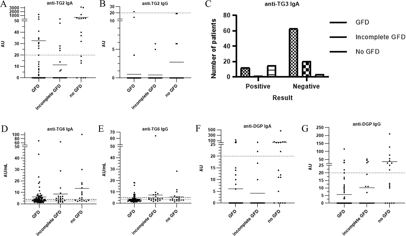
Fig. 2. Circulating (auto)antibodies in dermatitis herpetiformis. (A) Anti-TG2 IgA titres. (B) Anti-TG2 IgG titres. (C) Anti-TG3 IgA positivity (according to the manufacturer’s specifications, this ELISA assay did not report antibody titres but instead provided qualitative outcomes, i.e., only positive or negative results; this is why no dot diagram was possible). (D) Anti-TG6 IgA titres. (E) Anti-TG6 IgG titres. (F) Anti-DGP IgA titres. (G) Anti-DGP IgG titres. AU: arbitrary unit; GFD: gluten-free diet; TG: transglutaminase.
Circulating anti-TG3 IgA antibodies were detected in 24.6% (28/114) of patients (Table IA, Fig. 2C), half of them (14/28) were also TG2-seropositive. Among TG3 seropositive patients, 12/28 (42.9%) reported strict adherence to a GFD, including 5/28 (17.9%) who had elevated autoantibody levels exclusively against TG3. As with TG2, a significant correlation was found between dietary adherence and TG3 autoantibody levels (p<0.001). TG3 autoantibody levels differed significantly between the incomplete vs no GFD (p<0.01) and GFD vs no GFD (p<0.001) groups. On the other hand, the incomplete vs strict GFD groups showed no significant difference (p>0.05).
Circulating IgA and/or IgG antibodies against TG6 were detected in 53.5% (61/114) of patients, while 26.3% (30/114) exhibited at least one isotype of autoantibodies within the equivocal range. No antibodies against TG6 were detected in 20.2% (23/114) of patients. The association between adherence to a GFD and anti-TG6 IgG and IgA levels is presented in Table IB and visually illustrated in Fig. 2D, E. For further details on correlation between anti-TG6 IgG and IgA autoantibody levels, see Table IC. A significant correlation was found between adherence to a GFD and anti-TG6 IgG levels (p=0.0095) when incomplete adherence was classified as non-adherence. Specifically, a significant difference was observed between the incomplete and the fully adherent GFD group (p=0.0149). In contrast, anti-TG6 IgA antibody levels did not show a correlation with GFD adherence.
An elevation of anti-TG6 antibody titres was observed in some cases after several years on a GFD.
As TG2 IgA increases, both TG3 IgA (ρ=0.504, p<0.0001) and TG2 IgG (ρ=0.422, p<0.0001) tend to increase in a consistent (monotonic) pattern across patients and show a statistically significant and meaningful correlation (ρ≥0.4 and p<0.05). No statistically significant correlation was found between other isotypes of TG autoantibodies.
The occurrence and overlap of anti-TG autoantibodies are illustrated in Fig. 3.

Fig. 3. The occurrence and overlap of autoantibodies against TG2, TG3 and TG6 in dermatitis herpetiformis patients, demonstrating their co-occurrence and potential immunological associations. TG: transglutaminase.
Anti-deamidated gliadin peptides (DGP) antibodies (IgG/IgA) were elevated in 20.2% (23/114) of DH patients. Specifically, elevation of anti-DGP IgA, IgG or IgA+IgG antibodies was observed in 3.5% (4/114), 5.3% (6/114) or 11.4% (13/114) of patients, respectively (Table ID, Fig. 2F, G). Both anti-DGP antibody levels showed a significant correlation with adherence to a GFD when incomplete adherence was classified as non-adherence (p=0.0011 and p=0.062, respectively). However, no statistically significant difference was observed between the fully adherent and incomplete GFD groups for either isotype.
Immunodeficiency
An increased prevalence of partial IgA deficiency has been reported in CD; limited data are available regarding its frequency in DH (6). Partial IgA deficiency was observed in 2/114 patients (1.8%) (40 and 42 years old), with 2 additional patients having childhood records of partial IgA deficiency.
One patient (1/114, 0.9%) was diagnosed with hypogammaglobulinaemia, accompanied by pernicious anaemia, including anti-parietal cell antibody positivity and elevated MCV.
Thyroid disorders
Thyroid function and organ-specific autoantibodies were assessed in all participants. Thyroid disorders were identified in 31/114 patients (27.2%) – 14/31 males (45.2%) and 17/31 females (54.8%) – with 24/31 patients (77.4%) diagnosed with Hashimoto’s thyroiditis, 16/24 (66.7%) of which were previously undiagnosed. Anti-thyroid peroxidase autoantibodies were positive in 21/111 (18.9%), and antithyroglobulin autoantibodies in 18/109 patients (16.5%), respectively.
Notably, 3/16 (18.8%) previously undiagnosed cases of Hashimoto’s thyroiditis exhibited abnormal serum hormone values. Subclinical hypothyroidism with elevated TSH levels was observed in 2 of 3, while hyperthyroidism was present in 1 of 3 cases. In addition, Basedow’s disease was previously diagnosed in 1/114 patients (0.9%). Nontoxic goitre was identified in 2 (1.75%), hypothyroidism without serologic evidence of thyroid autoimmunity was present in 3 patients (2.6%).
Interestingly, TSH levels were significantly elevated in the cohort of patients on an incomplete GFD or no GFD (median: 1.835) compared to those adhering to a strict GFD (median: 1.444, p=0.0317). This difference was no longer observed, excluding patients receiving L-thyroxine. Serum thyroid hormone levels showed no significant changes in our cohorts.
Anaemia
Anaemia was observed in 37/114 patients (32.5%) with DH (14/37, 37.8% males and 23/37, 62.2% females). Among them, 32 (86.5%) were characterized by decreased RBC counts, accompanied by reduced haemoglobin levels in 20 (62.5%) or normal haemoglobin levels in 12 cases (37.5%). In 5 patients (13.5%), only haemoglobin levels were reduced. Serum iron deficiency was detected in 8/37 anemic patients (21.6%), with a higher occurrence in females (75.0%, 6/8) compared to males (25.0%, 2/8). In 3/114 cases (2.6%), decreased iron levels were present without manifesting as anaemia. Pernicious anaemia was confirmed in 2 females. Of the 114 DH patients, 13 (11.4%) were receiving dapsone therapy, and 8 of these patients (61.5%) exhibited haemolytic anaemia (5/8, 62.5% male and 3/8, 37.5% female), accounting for 21% of all anemic cases. Additionally, 45/114 patients (39.5%) reported a history of previously diagnosed anaemia, most commonly associated with underlying iron deficiency.
Other immune-mediated disorders
Additional autoimmune conditions were present in 9/114 patients (7.9%), see Table II for details.
Table II. Autoimmune diseases associated with dermatitis herpetiformis in our study
| Disease | Patient no. | Percentage of 114 patients |
|---|---|---|
| Hashimoto’s thyroiditis | 24 | 21.0% |
| Raynaud’s syndrome | 3 | 2.6% |
| Vitiligo | 2 | 1.8% |
| Type 1 diabetes mellitus | 2 | 1.8% |
| Pernicious anaemia | 2 | 1.8% |
| Alopecia Areata | 1 | 0.9% |
| Progressive systemic sclerosis | 1 | 0.9% |
| Basedow’s disease | 1 | 0.9% |
Neurologic involvement
Neurological complaints (Table III) were reported by 50/114 patients (43.9%), most frequently headache (21.9%, 25/114) and radiculopathy (12.3%, 14/114). Neuropathy-related complaints occurred in 5 patients (4.4%), with one histology confirmed small fibre neuropathy. Ataxia was observed in 2 patients (1.8%).
Table III. Neurological manifestations in dermatitis herpetiformis patients in our study
| Manifestation | Patient no. | Percentage of 114 patients |
|---|---|---|
| Headache | 25 | 21.9% |
| Radiculopathy | 14 | 12.3% |
| Neuropathy | 5 | 4.4% |
| Essential tremor | 3 | 2.6% |
| Ataxia | 2 | 1.8% |
| Epileptic seizure | 2 | 1.8% |
| Restless leg syndrome | 2 | 1.8% |
| Attention deficit hyperactivity disorder | 2 | 1.8% |
| Schwannoma | 1 | 0.9% |
| Parkinson disease | 1 | 0.9% |
| Vestibular neuritis | 1 | 0.9% |
A woman with a childhood CD developed neurological symptoms at the age of 26, several years after discontinuing a GFD. Initial manifestations included facial tingling and numbness, which progressively spread to the limbs and finally to the entire body. Ten years later, she developed cerebellar ataxia and DH, with serologic and histological findings confirming gluten-related disease. A GFD improved both the ataxia and skin lesions, but peripheral neuropathy persisted. Subsequent histological analysis confirmed small-fibre neuropathy. In the later course of the disease, she was additionally diagnosed with a schwannoma.
The clinical symptoms of lower limb ataxia in a man who had only recently started a GFD were not pronounced.
In one patient, severe headaches were associated with gluten consumption, and brain MRI revealed white matter lesions. Several other patients reported previously frequent headaches that resolved or markedly improved after initiation of a GFD. Epileptic seizures occurred in 2/114 patients (1.8%), both TG6 antibody-positive. One of them also had a history of severe alcohol dependence. Restless legs syndrome had been previously diagnosed in 2, while essential tremor was diagnosed in 3 patients.
Attention deficit hyperactivity disorder (ADHD) was diagnosed in 2 patients; one had hallucinations that resolved with a GFD. Other neurological findings included Parkinson’s disease and vestibular neuritis in one patient each.
In cases where the effect of a GFD was not reported, patients did not report any improvement in neurologic symptoms, or the effect was not evaluable – for example, due to the presence of comorbidities.
Additionally, 10/114 patients (8.8%) reported a history of mood disorders, 4 of whom were anti-TG6 IgA seropositive (Table III).
Among patients with neurological complaints, TG6 antibody testing revealed that 26/50 individuals (52.0%) tested positive for anti-TG6 autoantibodies (using the cut-offs recommended by the manufacturer), with no significant differences observed (p>0.05) based on dietary status (Table IV, Fig. 4A, B).

Fig. 4. (A) Anti-TG6 IgA titres, (B) anti-TG6 IgG titres, (C) anti-TG2 IgA titres and (D) anti-TG2 IgG titres of circulating autoantibodies in patients with or without neurological symptoms, analysed in relation to their gluten-free diet status. AU: arbitrary unit; GFD: gluten-free diet; NS: neurological symptoms; TG: transglutaminase.
Table IV. Anti-TG6 IgA and IgG serology results in patients with neurologic manifestations
| Anti-TG6 IgG | ||||
|---|---|---|---|---|
| anti- TG6 IgG positive | anti-TG6 IgG equivocal | anti-TG6 IgG negative | ||
| Anti-TG6 IgA | anti-TG6 IgA Positive | 8 (16%) | 6 (12%) | 10 (20%) |
| anti-TG6 IgA equivocal | 0 (0%) | 4 (8%) | 7 (14%) | |
| anti-TG6 IgA negative | 2 (4%) | 2 (4%) | 11 (22%) | |
Elevated serum anti-TG2 IgA was accompanied by neurological manifestations in 7.9% (9/114) of cases. Anti-TG2 IgA levels were significantly different between patients with neurological symptoms on a strict or an incomplete GFD and those without neurological symptoms who were not following a GFD (p<0.001) (Fig. 4C, D). Anti-TG3 IgA positivity was observed in 30.0% (15/50) of DH patients with neurological symptoms, accounting for 53.6% (15/28) of all anti-TG3 IgA–positive patients.
Of the patients with neurological complaints, 64.0% (32/50) adhered strictly to a GFD, while 16.0% (8/50) reported dietary lapses, and 20.0% (10/50) did not follow a GFD (Fig. 4).
Abdominal complaints
The majority of patients (69/114, 60.5%) reported abdominal complaints. Among them, 12 patients (17.4%) followed an incomplete GFD, and 11 (15.9%) did not adhere to a GFD.
In 15/69 cases (21.7%), gastrointestinal symptoms were present before starting a GFD and improved after the introduction of the diet.
The most common abdominal complaint was bloating (22/69, 31.9%) and abdominal pain (17/69, 24.6%), with 5 of them (29.4%) describing the pain as cramping. Nausea (3/69, 4.4%), bowel hypermotility (2/69, 2.9%), and vomiting (1/69, 1.4%) were less frequent.
The underlying diagnoses of the symptoms were not clarified in all cases, thus the abdominal complaints were not necessarily related to DH.
Miscarriage
Among 44 female DH patients, 35 (79.5%) had been pregnant, totalling 91 pregnancies. Eight patients (22.9%) had elective abortions (13 procedures), and 8 (22.9%) experienced altogether 13 miscarriages. Overall, 14.3% (13/91) of pregnancies ended in miscarriage.
Other diseases
The most common allergic condition was allergic rhinitis, affecting 25/114 patients (21.9%). Bronchial asthma was observed in 9/114 patients (7.9%). Osteopenia was documented in a single patient and osteoporosis was proven in 12/114 (10.5%) cases.
DISCUSSION
DH is a rare disease, with its highest reported prevalence in Finland. A long-term Finnish study (1970–2014) revealed a nearly equal gender ratio (51% (229/446) males and 49% (217/446) females) and a median diagnostic delay of 10 months, which improved over time from 12 to 8 months (7).
In contrast, our cohort demonstrated a slight male predominance (56.1% of 492 patients diagnosed between 1,994 and the study’s conclusion), aligning with Finnish data.
In our study, strict adherence to a GFD was considered as complete diet adherence, while partial or absent adherence was categorized as failure to follow a diet. Although clinical manifestations may vary with intermittent gluten exposure, gluten consumption could still provoke or sustain the autoimmune response against transglutaminases, potentially leading to elevated antibody levels and complications as shown on Figs 1 and 2.
Our data supports that a strict GFD can significantly reduce serum levels of anti-TG2 IgA, anti-TG3 IgA, anti-TG6 IgG and anti-DGP IgA/IgG. No correlation was found between a GFD and anti-TG6 IgA or anti-TG2 IgG levels, reinforcing the limited diagnostic and monitoring value of anti-TG2 IgG. Interestingly, anti-TG6 IgA levels did not correlate with GFD adherence, while anti-TG6 IgG levels showed a significant correlation (p=0.0095) when patients on an incomplete and no GFD were merged. If these groups were not merged, a significant difference was observed only between the GFD and incomplete GFD groups. In some cases, elevated TG6 autoantibody levels persisted even after a long-term strict GFD.
Previous studies exploring TG6 autoantibody positivity and gluten-related neurological deficits predominantly focused on CD (8, 9), thus limited data are available in DH patients. According to a prospective cohort, 40% of CD patients had circulating IgA and/or IgG autoantibodies against TG6, and seropositive individuals showed reduced brain volume of specific brain regions on MRI. It supports a link between autoimmunity to TG6 and atrophy of central nervous system in patients with CD (8). After 7 years, 50% of the cohort remained seropositive for one or more gluten-related antibodies, including 7 patients with newly diagnosed coordination problems (9). In Finland, 39% of DH patients were TG6 seropositive (13/33, IgA in 11 patients, IgG in 3, and both in 1) at diagnosis, decreasing to 26% after 1 year on a GFD. Our findings supported that a strict GFD results in reduced IgA autoantibody titres against TG2 and TG3, though not for TG6. We could only confirm this effect for anti-TG6 IgG autoantibodies (10, 11). Screening for multiple TG isoenzymes and DGP may enhance diagnostic sensitivity and follow-up examinations.
Neurological involvement is better documented in CD. In a study, 228 CD patients with neurologic involvement (67% IgG and/or IgA anti-TG6 positive) were evaluated. The most common findings were cerebellar ataxia (41%), peripheral neuropathy (30%) and encephalopathy (21%) (12). Neurological dysfunction was reported in 10–22% of CD patients in other studies (13). In our study, 43.9% reported neurological symptoms, and 52% of them tested positive for at least one isotype of TG6 antibodies. The frequency of ataxia and neuropathy was increased compared to other conditions, supporting routine neurological evaluation in DH management.
Headaches were reported by 21.9% of patients, lower than the global prevalence of approximately 50%, yet gluten-related headaches may raise suspicion of underlying neurological disease, as demonstrated by our patient who presented with headaches and white matter abnormalities on MRI.
Autoimmune thyroid diseases are prevalent in both CD and DH (14, 15, 16). In our study, significantly higher serum TSH levels were found between patients adhering to a strict GFD and those not following a GFD. This has not been found in other studies, highlighting the need for further investigation into the underlying mechanisms. Particularly in subgroups of DH patients with concurrent thyroid diseases.
In Finland, the frequency of anaemia in 250 DH patients was 12% at diagnosis, rising to 19% after 1 year, mainly due to dapsone treatment (17). In India, 23% (15/65) of untreated DH patients had anaemia, with iron deficiency in 20% (13/65) of patients and pernicious anaemia in 2 (3%) (18). Consistent with existing data, 25% of our patients had anaemia, excluding those on dapsone.
Additionally, autoimmune diseases were reported in 23% of Indian DH patients, including thyroid abnormalities, pernicious anaemia, vitiligo, alopecia areata, chronic immune thrombocytopenic purpura and discoid lupus erythematosus (18). This shows a similar pattern of associated diseases as in a previous study from Finland (14) and our own study, supporting the notion that DH is often accompanied by other immune-mediated conditions (3). However, the underlying causality remains unclear.
Miscarriage risk in our study was consistent with reported population-level rates (15.3%) (19).
Limitations
The limitations of this study include the relatively modest sample size (though 114 DH patients are considerable for a monocentric study of a rare disease) and the potential selection bias, as elderly patients with multiple comorbidities, mobility issues or patients from remote areas were less likely to participate. Due to the cross-sectional design and variable timing of patient evaluation, longitudinal relationships between gluten exposure, autoantibody dynamics and neurological manifestations could not be assessed, while reliance on patient-reported data may introduce recall and reporting bias. Another limitation is the challenge in interpreting the cutoff values of the manufacturers for the TG assays which have not been validated in a DH patient population.
Conclusions
Our study provided insights into the multifaceted nature of DH, its associations with other conditions and into the long-term effects of gluten sensitivity and the GFD. By deepening our understanding of this patient population, we can better tailor disease management.
Screening for comorbidities is recommended when clinical signs are present. Given the high frequency of thyroid disorders, thyroid function should be tested at diagnosis and monitored during follow-up. Although neurological evaluation is not yet routine, it should be considered in all patients with gluten sensitivity, as early detection and treatment may improve outcomes.
REFERENCES
- Kárpáti S, Sárdy M, Németh K, Mayer B, Smyth N, Paulsson M, et al. Transglutaminases in autoimmune and inherited skin diseases: the phenomena of epitope spreading and functional compensation. Exp Dermatol 2018; 27: 807–814. https://doi.org/10.1111/exd.13449
- Görög A, Antiga E, Caproni M, Cianchini G, De D, Dmochowski M, et al. S2k guidelines (consensus statement) for diagnosis and therapy of dermatitis herpetiformis initiated by the European Academy of Dermatology and Venereology (EADV). J Eur Acad Dermatol Venereol 2021; 35: 1251–1277. https://doi.org/10.1111/jdv.17183
- Malkovics T, Joura MI, Koszorú K, Sárdy M. Dermatitis herpetiformis und sonstige Formen der Weizensensitivität. Dermatologie 2023; 74: 955–960. https://doi.org/10.1007/s00105-023-05243-1
- Sárdy M, Kárpáti S, Merkl B, Paulsson M, Smyth N. Epidermal transglutaminase (TGase 3) is the autoantigen of dermatitis herpetiformis. J Exp Med 2002; 195: 747–757. https://doi.org/10.1084/jem.20011299
- van Beek N, Holtsche MM, Atefi I, Olbrich H, Schmitz MJ, Pruessmann J, et al. State-of-the-art diagnosis of autoimmune blistering diseases. Front Immuno l2024; 15: 1363032. https://doi.org/10.3389/fimmu.2024.1363032
- Samolitis NJ, Hull CM, Leiferman KM, Zone JJ. Dermatitis herpetiformis and partial IgA deficiency. J Am Acad Dermato l2006; 54: S206–9. https://doi.org/10.1016/j.jaad.2005.06.033
- Mansikka E, Salmi T, Kaukinen K, Collin P, Huhtala H, Reunala T, et al. Diagnostic delay in dermatitis herpetiformis in a high-prevalence area. Acta Derm Venereol 2018; 98: 195–199. https://doi.org/10.2340/00015555-2818
- Hadjivassiliou M, Croall ID, Zis P, Sarrigiannis PG, Sanders DS, Aeschlimann P, et al. Neurologic deficits in patients with newly diagnosed celiac disease are frequent and linked with autoantibodies to transglutaminase 6. Clin Gastroenterol Hepatol 2019; 17: 2678–2686. https://doi.org/10.1016/j.cgh.2019.03.014
- Hadjivassiliou M, Croall ID, Grünewald RA, Trott N, Sanders DS, Hoggard N. Neurological evaluation of patients with newly diagnosed coeliac disease presenting to gastroenterologists: a 7-year follow-up study. Nutrients 2021; 13: 1846. https://doi.org/10.3390/nu13061846
- Reunala T, Salmi TT, Hervonen K, Laurila K, Kautiainen H, Collin P, et al. IgA antiepidermal transglutaminase antibodies in dermatitis herpetiformis: a significant but not complete response to a gluten-free diet treatment. Br J Dermatol 2015; 172: 1139–1141. https://doi.org/10.1111/bjd.13387
- Hietikko M, Hervonen K, Ilus T, Salmi T, Huhtala H, Laurila K, et al. Ex vivo culture of duodenal biopsies from patients with dermatitis herpetiformis indicates that transglutaminase 3 antibody production occurs in the gut. Acta Derm Venereol 2018; 98: 366–372. https://doi.org/10.2340/00015555-2849
- Hadjivassiliou M, Rao DG, Grìnewald RA, Aeschlimann DP, Sarrigiannis PG, Hoggard N, et al. Neurological dysfunction in coeliac disease and non-coeliac gluten sensitivity. Am J Gastroenterol 2016; 111: 561–567. https://doi.org/10.1038/ajg.2015.434
- Al-Toma A, Volta U, Auricchio R, Castillejo G, Sanders DS, Cellier C, et al. European Society for the Study of Coeliac Disease (ESsCD) guideline for coeliac disease and other gluten-related disorders. United European Gastroenterol J 2019; 7: 583–613. https://doi.org/10.1177/2050640619844125
- Reunala T, Collin P. Diseases associated with dermatitis herpetiformis. Br J Dermatol 1997; 136: 315–318.
- Cunningham MJ, Zone JJ. Thyroid abnormalities in dermatitis herpetiformis. Prevalence of clinical thyroid disease and thyroid autoantibodies. Ann Intern Med 1985; 102: 194–196. https://doi.org/10.7326/0003-4819-102-2-194
- Malandrini S, Trimboli P, Guzzaloni G, Virili C, Lucchini B. What about TSH and anti-thyroid antibodies in patients with autoimmune thyroiditis and celiac disease using a gluten-free diet? A systematic review. Nutrients 2022; 14: 1681. https://doi.org/10.3390/nu14081681
- Alakoski A, Pasternack C, Reunala T, Kaukinen K, Huhtala H, Mansikka E, et al. Anaemia in dermatitis herpetiformis: prevalence and associated factors at diagnosis and one-year follow-up. Acta Derm Venereol 2021; 101: adv00443. https://doi.org/10.2340/00015555-3795
- Handa S, Dabas G, De D, Mahajan R, Chatterjee D, Saika UN, et al. A retrospective study of dermatitis herpetiformis from an immunobullous disease clinic in north India. Int J Dermatol 2018; 57: 959–964. https://doi.org/10.1111/ijd.14029
- Quenby S, Gallos ID, Dhillon-Smith RK, Podesek M, Stephenson MD, Fisher J, et al. Miscarriage matters: the epidemiological, physical, psychological, and economic costs of early pregnancy loss. Lancet 2021; 397: 1658–1667. https://doi.org/10.1016/S0140-6736(21)00682-6