SHORT COMMUNICATION
The Candle Spot: An Unreported Sign of SULT2B1-nEDD
Sarah MILESI1* , Christine CHIAVERINI2, Laura CHÊNE2, Nathalie JONCA3,4 and Juliette MAZEREEUW-HAUTIER1
1Departement of Dermatology, CHU Toulouse, Toulouse, France, 2Departement of Dermatology, CHU Nice, Nice, France, 3Toulouse Institute for Infectious and Inflammatory Diseases (INFINITy), Toulouse University, CNRS, Inserm, Toulouse, France, and 4Department of Cell Biology and Cytology, Federative Institute of Biology, Purpan University Hospital, Toulouse, France. *Email: milesi.sa@chu-toulouse.fr
Citation: Acta Derm Venereol 2026; 106: adv-2025-0264. DOI: https://doi.org/10.2340/actadv.v106.adv-2025-0264.
Copyright: © 2026 The Author(s). Published by MJS Publishing, on behalf of the Society for Publication of Acta Dermato-Venereologica. This is an Open Access article distributed under the terms of the Creative Commons Attribution-NonCommercial 4.0 International License (https://creativecommons.org/licenses/by-nc/4.0/).
Submitted: Dec 12, 2025. Accepted after revision: Apr 7, 2026.
Published: Jun 1, 2026.
Competing interests and funding: The authors have no conflicts of interest to declare.
Congenital ichthyosis (CI), characterized by abnormal scaling often associated with erythroderma, is now classified within the group of epidermal differentiation disorders (EDD). This group comprises palmo-plantar keratoderma (pEDD), syndromic (sEDD) and non-syndromic (nEDD) forms of ichthyosis. EDDs are named based on the gene and grouped based on the function of the gene product (e.g. cholesterol metabolism enzyme-associated disorders including SULT2B1-nEDD) (1).
SULT2B1 encodes a sulfotransferase that converts cholesterol into cholesterol sulfate. Loss of function of this enzyme results in the accumulation of toxic cholesterol metabolites and disruption of normal cornification (2). Only a few patients with SULT2B1-nEDD have been reported in the literature (2, 3) and the clinical description remains lacking. Here, we report a new case involving a novel variant associated with a distinctive phenotype of “candle spot like” lesions.
CASE REPORT
A young boy originally from Tunisia was referred to our department for evaluation of skin anomalies. He had no personal or family medical history (three siblings, non-consanguineous parents). He presented at birth with scaling, which gradually worsened with age. By the age of 11, he exhibited generalized skin thickening and scaling (sparing the skin folds) with more pronounced involvement on the extensor surfaces of legs and the dorsal aspects of hands. In these areas, small (5 to 10 mm in diameter) round white patches with an appearance of “candle-spots” were observed (Fig. 1), which, according to the parents, were due to skin fragility. Erythema was very subtle, and no pruritus was reported. Additional findings included pectus excavatum, recurrent onychomycosis (with otherwise normal-appearing nails) and mild learning disabilities.
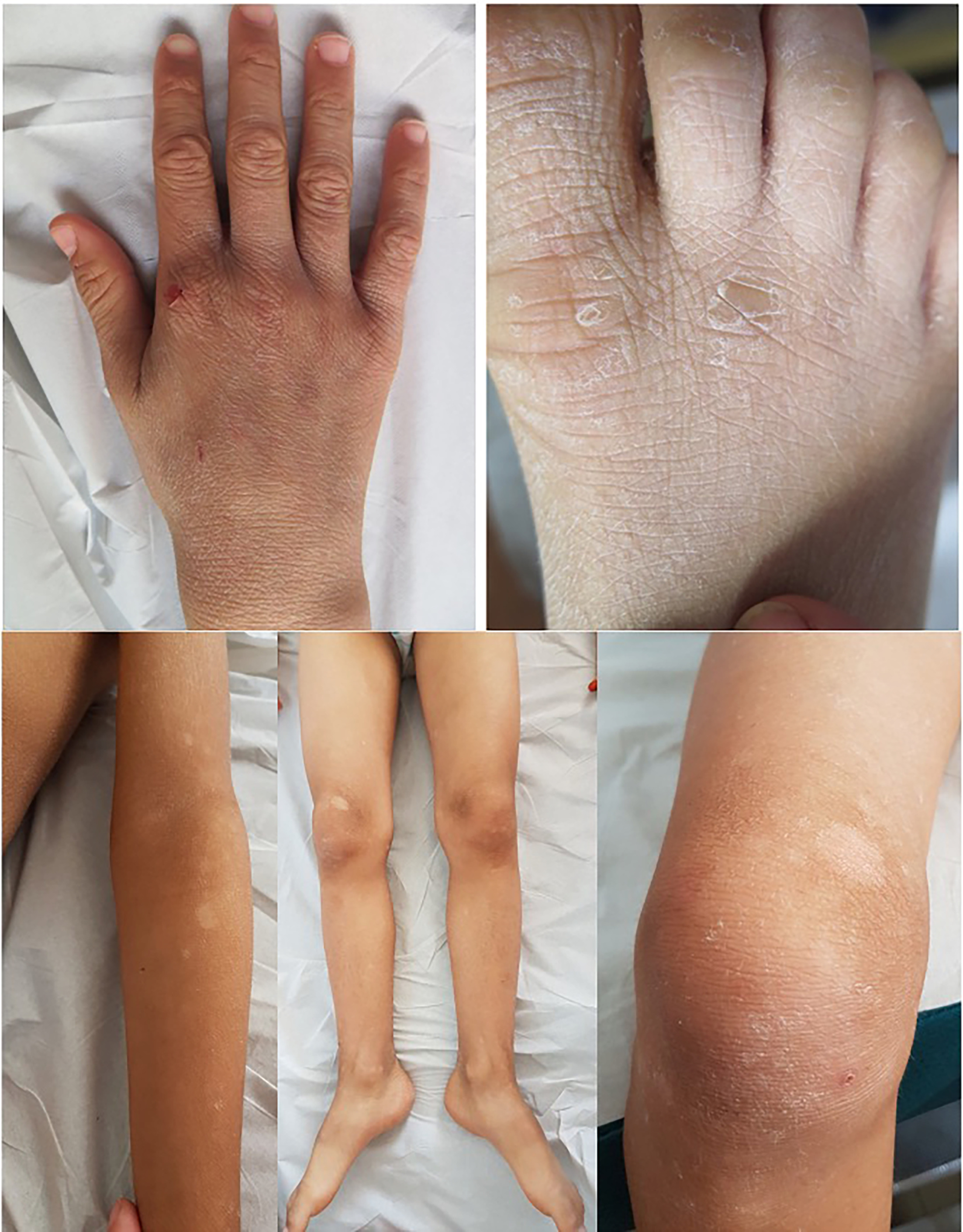
Fig. 1. Clinical presentation of the patient. Marked skin thickening and scaling, predominantly on the extensor surfaces of the legs and dorsal aspects of the hands. Small (5–10 mm) round white patches with a “candle-spot” appearance are visible in these areas.
Using a next-generation sequencing (NGS) panel of 108 genes involved in EDD, we identified the c.102C>G (p.(Tyr34Ter)) variant in exon 2 of SULT2B1 gene (NM_177973.2) in a homozygous (or possibly hemizygous) state. Immunofluorescence analysis of SULT2B1 expression in a skin biopsy revealed a complete loss of the intense staining observed in the granular keratinocytes, in contrast to healthy control skin (Fig. 2).

Fig. 2. Analysis of SULT2B1 expression by immunofluorescence in the patient’s skin. Immunofluorescence staining of a skin section from a healthy control (A) and the patient (B) using an antibody specific to SULT2B1 (red), showing the complete absence of SULT2B1 protein in the stratum granulosum of the patient’s epidermis. Nuclei are countersained with DAPI (blue).
Based on previous evidence of toxic cholesterol metabolites accumulation in SULT2B1-nsEDD, a combination therapy consisting of 2% topical lovastatin and 2% cholesterol alongside regular emollients was applied to one side of the body over a period of more than two months. No skin clinical improvement of the skin was observed.
DISCUSSION
To date, nine patients with nEDD carrying SULT2B1 variants have been reported; their characteristics are summarized in Table I. Seven of them were born as collodion babies, while the remaining two presented at birth with scaling and erythema. During childhood, ichthyosis was only briefly described by the authors and/or inconstantly documented through photographs. The condition ranged in severity and was characterized by skin thickening and variable scaling patterns, including white scales or brown thick scales of lamellar appearance with occasional erythroderma. Lesions were diffuse, often sparing the face and soles. None of the reported cases exhibited the particular “candle spots” appearance seen in our patient, which may reflect a certain degree of skin fragility, similar to that observed in epidermolytic ichthyosis caused by KRT2 variants (KRT2-nEDD).
Table I. SULT2B1 cases in literature
| Family, Patient, Origin | Skin examination | Extracutaneous anomalies | SULT2B1a nucleotide variant | SULT2B1b protein variant | ClinVar reference | Pathogenicity | Publication | |
|---|---|---|---|---|---|---|---|---|
| At birth | During infancy | |||||||
| Family1, P1, Tunisian | Collodion baby | Moderate to severe lesions made of skin thickening and scaling (lamellar aspect) sparing face and folds | None | c.[446C>T];[446C>T] | p.[Pro149Leu];[Pro149Leu] | VCV000426107.5 | 5 (pathogenic) | (2) |
| Family1, P2, Tunisian | Collodion baby | Not described | None | c.[446C>T];[446C>T] | p.[Pro149Leu];[Pro149Leu] | VCV000426107.5 | 5 (pathogenic) | (2) |
| Family1, P3, Tunisian | c.[446C>T];[446C>T] | p.[Pro149Leu];[Pro149Leu] | VCV000426107.5 | 5 (pathogenic) | (2) | |||
| Family2, P4, Turkish | Collodion baby | Skin thickening and erythema | None | c.[364dupA]; [821G>] |
p.[Met122AsnfsTer73];[Arg274Gln] | [VCV000426109.5];[VCV000426108.5] | 5 (pathogenic) (bot alleles) | (2) |
| Family3, P5, Kurdish | Scaling and erythema | Skin thickening and scaling sparing face and folds | None | c.[71+2T>A];[71+2T>A] | p.[?];[?] | VCV000426110.5 | 5 (pathogenic) | (2) |
| Family3, P6, Kurdish | Skin thickening and scaling sparing face and folds, mild erythroderma | c.[71+2T>A];[71+2T>A] | p.[?];[?] | VCV000426110.5 | 5 (pathogenic) | (2) | ||
| Family4, P7, Pakistani | Collodion baby | Skin thickening and scaling (lamellar type) sparing face and folds. Eclabion Ectropion |
Allergic rhinoconjunctivitis | c.[419C>T];[419C>T] | p.[Ala140Val];[Ala140Val] | Absent | 3 (uncertain significance) | (3) |
| Family4, P8, Pakistani | c.[419C>T];[419C>T] | p.[Ala140Val];[Ala140Val] | Absent | 3 (uncertain significance) | (3) | |||
| Family4, P9, Pakistani | c.[419C>T];[419C>T] | p.[Ala140Val];[Ala140Val] | Absent | 3 (uncertain significance) | (3) | |||
| Our patient | Dry skin | Generalized skin thickening and scaling (sparing folds) with more pronounced involvement on the extensor surfaces of legs and the dorsal aspects of hands. | Pectus excavatum, Recurrent onychomycosis, Mild learning disabilities. | c.[102C>G];[102C>G] | p.[Tyr34Ter];[Tyr34Ter] | SCV006310947 | 4 (likely pathogenic) | This report |
|
a Reference sequence SULT2B1 NM_177973.2. b Reference sequence SULT2B1 NP_814444.1 |
||||||||
The nonsense variant identified in our patient has not been previously reported in the literature or in genomic databases (dbSNP, GnomAD). Immunostaining confirmed the absence of SULT2B1 expression in the patient’s skin, likely due to nonsense-mediated mRNA decay. Given that previously reported loss-of-function variants in SULT2B1 have been established as causative of ns-EDD (OMIM#617571) (2), we classified this novel variant as likely pathogenic.
In conclusion, we reported here a case of SULT2B1-nEDD caused by a novel variant, presenting with a previously unreported distinctive “candle-spot” phenotype. This clinical feature may aid in the suspicion of SULT2B1 involvement in cases of EDD and could support the interpretation of variants of uncertain significance.
REFERENCES
- Hernández-Martín Á, Paller AS, Sprecher E, Akiyama M, Granier Tournier C, Aldwin-Easton M, et al. A proposal for a new pathogenesis-guided classification for inherited epidermal differentiation disorders. Br J Dermatol 2025; 193: 544–548. https://doi.org/10.1093/bjd/ljaf065
- Heinz L, Kim GJ, Marrakchi S, Christiansen J, Turki H, Rauschendorf MA, et al. Mutations in SULT2B1 cause autosomal-recessive congenital ichthyosis in humans. Am J Hum Genet 2017; 100: 926–939. https://doi.org/10.1016/j.ajhg.2017.05.007
- Fozia F, Nazli R, Alam Khan S, Bari A, Nasir A, Ullah R, et al. Novel homozygous mutations in the genes TGM1, SULT2B1, SPINK5 and FLG in four families underlying congenital ichthyosis. Genes 2013; 12: 373. https://doi.org/10.3390/genes12030373