SHORT COMMUNICATION
Detection of Circulating Tumour DNA in Plasma during the Treatment of In-transit Melanoma Metastases with Topical Imiquimod
Miguel MANSILLA-POLO1,2 , Blanca de UNAMUNO-BUSTOS1,2*, Fernando NAVARRO-BLANCO1,2, Gema PÉREZ-SIMÓ3, Sarai PALANCA-SUELA3 and Rafael BOTELLA-ESTRADA1,2,4
1Dermatology Department, Hospital Universitario y Politécnico La Fe, Valencia, Spain, 2Instituto de Investigación Sanitaria La Fe (IIS La Fe), Valencia, Spain, 3Molecular Biology Laboratory, Service of Clinical Analysis, Hospital Universitario y Politécnico La Fe, Valencia, Spain, and 4Faculty of Medicine, Universidad de Valencia, Valencia, Spain. *Email: deunamuno_bla@gva.es
Citation: Acta Derm Venereol 2026; 106: adv-2025-0229. DOI: https://doi.org/10.2340/actadv.v106.adv-2025-0229.
Copyright: 2026 ©Author(s). Published by MJS Publishing, on behalf of the Society for Publication of Acta Dermato-Venereologica. This is an Open Access article distributed under the terms of the Creative Commons Attribution-NonCommercial 4.0 International License (https://creativecommons.org/licenses/by-nc/4.0/).
Submitted: Nov 26, 2025. Accepted after revision: Apr 21, 2026.
Published: Jun 16, 2026.
Competing interests and funding: The authors have no conflicts of interest to declare.
Oral and written consent of patients were obtained to publish the images.
Liquid biopsy has emerged as a minimally invasive technique that allows real-time monitoring of tumour dynamics through the detection of circulating tumour DNA (ctDNA) (1). It is increasingly being used in oncology for cancer screening, molecular profiling and longitudinal treatment monitoring. By analysing cell-free DNA fragments released into the circulation – primarily through tumour cell apoptosis or necrosis – liquid biopsy provides valuable insights into tumour burden, minimal residual disease, treatment response and the emergence of resistance mechanisms (1, 2). The development of highly sensitive techniques such as digital droplet PCR (ddPCR) and next-generation sequencing (NGS) has greatly improved the detection of low-frequency mutations, extending the clinical utility of ctDNA to a wide range of solid tumours, including melanoma (1). In melanoma, ctDNA is particularly useful for monitoring response to therapy and identifying resistance mutations, especially in patients receiving immune checkpoint blockade or targeted therapies (1, 2, 3). However, its detection remains limited in certain clinical scenarios, particularly in patients with isolated in-transit metastases (ITMs), where ctDNA levels are often undetectable despite clear clinical evidence of disease. In this article, we report two cases of stage IIIC melanoma treated with topical imiquimod for ITMs in which ctDNA became detectable exclusively during treatment. These findings suggest that imiquimod may facilitate ctDNA release, possibly through tumour cell destruction and subsequent release of genetic material into the circulation.
RESULTS
Patient 1: An 84-year-old woman was diagnosed with a 3.8-mm-thick, ulcerated melanoma of the left arm with lymphovascular invasion. Sentinel lymph node biopsy failed to identify a sentinel lymph node (Fig. 1). Three years later, the patient developed ITMs, at which time the disease was classified as stage IIIC (T3bN1cM0). Tumour genotyping revealed an NRAS mutation (c.182A>G), with wild-type BRAF. Three years after initial excision, the patient developed ITMs, which were surgically resected. Adjuvant nivolumab was initiated but was discontinued after 3 months due to disease progression, with the development of additional ITMs. Over the next 3 years, the patient underwent repeated cycles of topical treatment with imiquimod 5% cream, applied Monday to Friday for 8 weeks per cycle. A total of 4 treatment cycles were administered over the follow-up period, whenever new lesions appeared. A significant clinical response was observed. Notably, ctDNA analysis during treatment cycles consistently detected NRAS mutant DNA in plasma, whereas no ctDNA was detectable during off-treatment periods, even in the presence of visible lesions. Similar ctDNA elevations were observed during each treatment cycle. No systemic disease progression was observed during three years of follow-up after initiation of imiquimod therapy, including more than 12 months after the final treatment cycle. Disease monitoring consisted of regular dermatological examination every 3–6 months and annual whole-body PET-CT imaging, which showed no evidence of distant metastases.
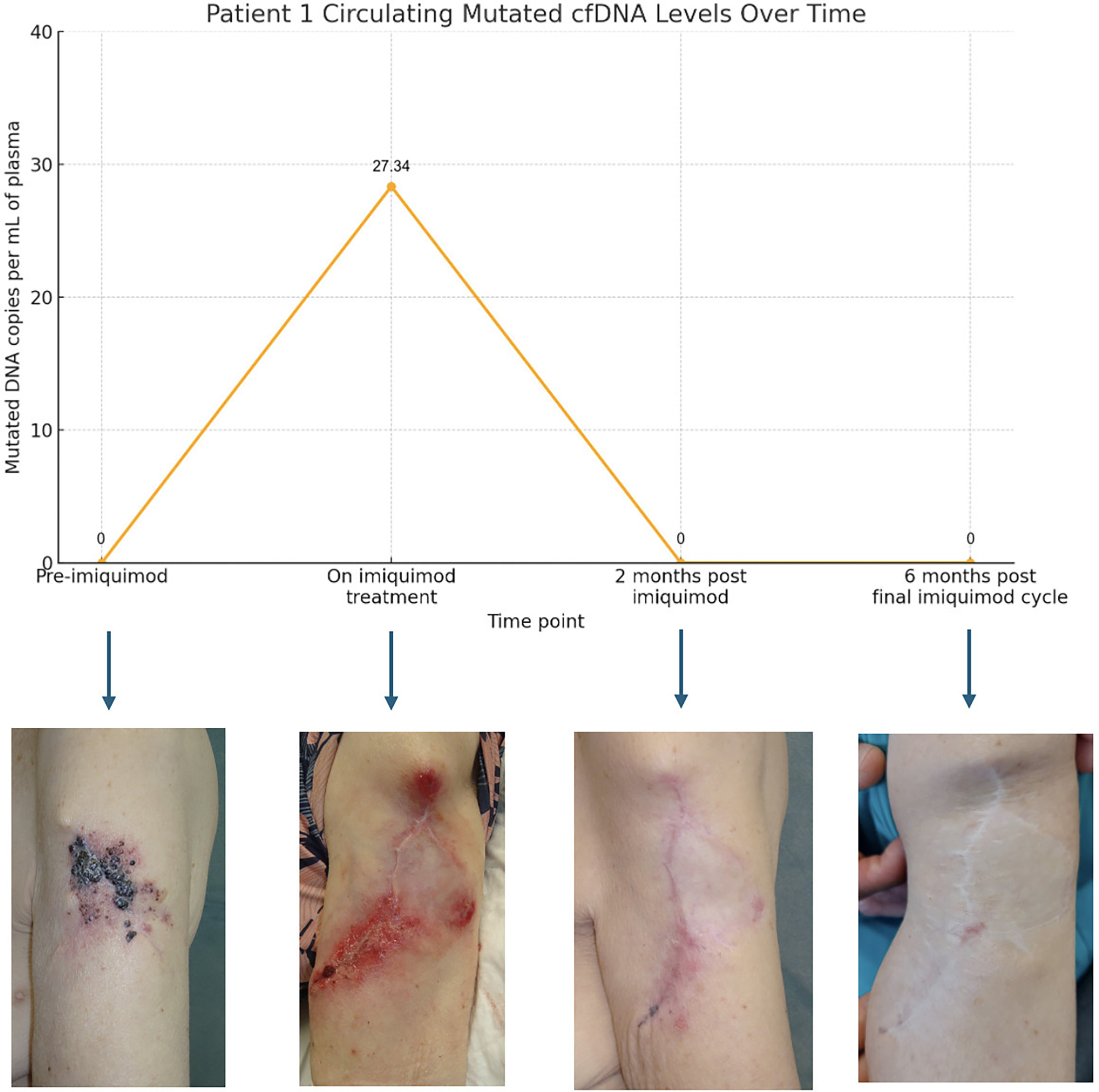
Fig. 1. Clinical and circulating tumour DNA (ctDNA) kinetics in Patient 1 during topical imiquimod treatment. This figure illustrates ctDNA levels measured at representative time points during one imiquimod treatment cycle in Patient 1 with in-transit melanoma metastases (ITMs), alongside corresponding clinical images. ctDNA was undetectable prior to treatment, became transiently detectable during active imiquimod therapy (27.34 copies/mL) and returned to undetectable levels at subsequent post-treatment assessments. Similar transient ctDNA elevations were consistently observed during other imiquimod treatment cycles, whereas ctDNA remained undetectable during off-treatment periods, even in the presence of clinically visible lesions. Clinical images show lesion appearance before treatment, inflammatory changes during therapy, and progressive regression following treatment.
Patient 2: A 91-year-old woman with a 2.5 mm ulcerated melanoma of the left leg, classified as stage IIIC due to the presence of ITMs detected at the time of wide local excision (Fig. 2). SLNB was negative and the patient declined adjuvant therapy. Tumour genotyping revealed an NRAS mutation (c.181C>A), with wild-type BRAF. For recurrent ITMs, she was treated with imiquimod 5% cream applied Monday to Friday in repeated cycles. As in Patient 1, ctDNA corresponding to her known tumour mutations became detectable in plasma exclusively during imiquimod treatment periods, while remaining undetectable before treatment and during drug-free intervals, even when cutaneous disease was clinically evident. The patient received 2 imiquimod treatment cycles in total. There was no systemic progression during 2 years of follow-up, including more than 9 months after completion of the final imiquimod cycle. Follow-up included regular dermatological examination and imaging surveillance with PET-CT scans, which confirmed absence of systemic disease.
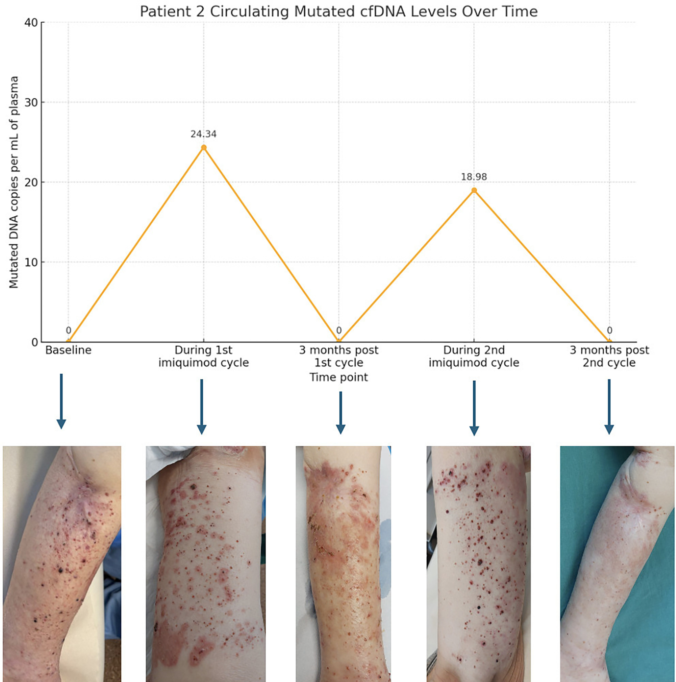
Fig. 2. Clinical and ctDNA kinetics in Patient 2 during 2 separate imiquimod treatment cycles. This figure shows ctDNA levels measured at representative time points before, during, and after 2 separate imiquimod treatment cycles in Patient 2 with recurrent ITMs, together with corresponding clinical images. ctDNA was undetectable at baseline and during treatment-free intervals but became transiently detectable during both imiquimod treatment cycles (24.34 and 18.98 copies/mL, respectively), before returning to undetectable levels after treatment discontinuation. This pattern was reproducible across treatment cycles. Clinical images demonstrate the presence of metastatic lesions before treatment, inflammatory response during therapy, and marked clinical improvement after treatment.
DISCUSSION
We report two patients with stage IIIC melanoma and isolated in-transit metastases in whom ctDNA became transiently detectable during treatment with topical imiquimod. To our knowledge, this pattern has not previously been described in melanoma ITMs managed with imiquimod.
In melanoma, ctDNA detection is generally associated with tumour burden, particularly in patients with nodal or visceral metastatic disease (2, 3, 4, 5). However, patients with disease confined to cutaneous or subcutaneous in-transit metastases may have undetectable ctDNA despite clinically apparent lesions (2, 3, 6). In the two cases presented here, ctDNA remained negative during treatment-free intervals, including periods in which ITMs were present, and became detectable only during active imiquimod treatment. It subsequently returned to undetectable levels after treatment. This repeated temporal relationship argues against systemic progression as the most likely explanation and suggests that ctDNA release may have been linked to the local therapeutic effect of imiquimod.
Imiquimod is a TLR7/8 agonist with recognised local antitumour activity, driven by immune activation and, at least in part, by direct cytotoxic effects on tumour cells (7). Although serial biopsies were not obtained and tumour apoptosis could not be histologically confirmed during treatment, imiquimod-induced tumour cell death offers a plausible biological explanation for the transient increase in ctDNA observed in both patients (8, 9). This interpretation is also consistent with the absence of distant metastatic disease on clinical assessment and imaging follow-up, despite detectable ctDNA during treatment cycles.
These observations are relevant for the interpretation of ctDNA in patients receiving locoregional therapies. In this context, new ctDNA detection or a transient increase in ctDNA should not necessarily be regarded as evidence of systemic progression. Rather, ctDNA kinetics should be interpreted in relation to treatment timing, clinical response and radiological findings, particularly when the intervention is expected to induce local tumour cell death (10).
This report has several limitations, including the small number of patients, the use of targeted ddPCR assays restricted to known NRAS mutations, the lack of histological confirmation of apoptosis during treatment and the limited follow-up expected in a short clinical report. Nevertheless, the consistency of the pattern across repeated treatment cycles supports the hypothesis that ctDNA may, in selected patients with melanoma ITMs treated with imiquimod, reflect local tumour cell death rather than occult systemic progression. Larger studies are needed to determine whether this finding is reproducible and whether ctDNA kinetics could contribute to response-adapted management in locoregional melanoma.
To our knowledge, no previous reports have documented ctDNA release associated with topical imiquimod for ITMs in melanoma. Our findings open new avenues for exploring ctDNA as a real-time indicator of treatment-induced tumour cell death in cutaneous metastases and may inform response-adaptive strategies in patients with limited disease burden.
REFERENCES
- Martínez-Vila C, Teixido C, Aya F, Martín R, González-Navarro EA, Alos L, et al. Detection of circulating tumor DNA in liquid biopsy: current techniques and potential applications in melanoma. Int J Mol Sci 2025; 26: 861. https://doi.org/10.3390/ijms26020861
- Genta S, Araujo DV, Hueniken K, Pipinikas C, Ventura R, Rojas P, et al. Bespoke ctDNA for longitudinal detection of molecular residual disease in high-risk melanoma patients. ESMO Open 2024; 9: 103978. https://doi.org/10.1016/j.esmoop.2024.103978
- Slusher N, Jones N, Nonaka T. Liquid biopsy for diagnostic and prognostic evaluation of melanoma. Front Cell Dev Biol 2024; 12: 1420360. https://doi.org/10.3389/fcell.2024.1420360
- Lucci A, Addanki S, Chiang YJ, Meas S, Sarli VN, Upshaw JR, et al. Presence of circulating tumor cells predates imaging detection of relapse in patients with stage III melanoma. Cancers 2023; 15: 3630. https://doi.org/10.3390/cancers15143630
- Marchisio S, Ricci AA, Roccuzzo G, Bongiovanni E, Ortolan E, Bertero L, et al. Monitoring circulating tumor DNA liquid biopsy in stage III BRAF-mutant melanoma patients undergoing adjuvant treatment. J Transl Med 2024; 22: 1074. https://doi.org/10.1186/s12967-024-05783-7
- Palacios-Diaz RD, de Unamuno-Bustos B, Carratalá-García A, Pérez-Simó G, Moreno-Ramírez D, Ferrándiz L, et al. Circulating tumor DNA predicts tumor progression and poor survival in patients with stage III melanoma. Melanoma Res 2025; 35: 259–267. https://doi.org/10.1097/CMR.0000000000001041
- Scarfì F, Patrizi A, Veronesi G, Lambertini M, Tartari F, Mussi M, et al. The role of topical imiquimod in melanoma cutaneous metastases: a critical review of the literature. Dermatol Ther 2020; 33: e14165. https://doi.org/10.1111/dth.14165
- Kumar AB, Jakub JW, Lester SC, Markovic SN, Baum CL. In-transit melanoma metastases: evaluation and management for the dermatologist. J Am Acad Dermatol 2025; 93: 188–195. https://doi.org/10.1016/j.jaad.2025.03.005
- El-Khattouti A, Selimovic D, Hannig M, Taylor EB, Abd Elmageed ZY, Hassan SY, et al. Imiquimod-induced apoptosis of melanoma cells is mediated by ER stress-dependent Noxa induction and enhanced by NF-κB inhibition. J Cell Mol Med 2016; 20: 266–286. https://doi.org/10.1111/jcmm.12718
- Di Nardo L, Del Regno L, Di Stefani A, Mannino M, Fossati B, Catapano S, et al. The dynamics of circulating tumour DNA (ctDNA) during treatment reflects tumour response in advanced melanoma patients. Exp Dermatol 2023; 32: 1785–1793. https://doi.org/10.1111/exd.14901