QUIZ SECTION
Widespread Erythematous Vesiculobullea: A Quiz
Xiaoting SONG1, Dong LAN2 and Fang LIU1*
1Department of Dermatology, Beijing Chaoyang Hospital, Capital Medical University, Beijing, China, and 2Department of Dermatology, Jingxi Campus, Beijing Chaoyang Hospital, Capital Medical University, Beijing, China. *Email: roseliufang@qq.com
Citation: Acta Derm Venereol 2026; 106: adv-2025-0240. DOI: https://doi.org/10.2340/actadv.v106.adv-2025-0240.
Copyright: 2026 ©Author(s). Published by MJS Publishing, on behalf of the Society for Publication of Acta Dermato-Venereologica. This is an Open Access article distributed under the terms of the Creative Commons Attribution-NonCommercial 4.0 International License (https://creativecommons.org/licenses/by-nc/4.0/).
Submitted: Accepted after revision:
Published: Jun 29, 2026.
Competing interests and funding:
A previously healthy woman in her 50s presented with 4 days of widespread erythematous vesiculobullea with burning, without fever or arthralgias. On the first day of the rash, she received a 7 mg intramuscular injection of betamethasone, while the skin lesions continued to spread out. At the current presentation, she had normal vital signs, multiple erythematous papules that coalesced into plaques with a transparent, vesicle-like appearance on the trunk and limbs (Fig. 1). Blood count, C-reactive protein, erythrocyte sedimentation rate and immunoglobulin on day 1 and the test of EBV on day 7 are unremarkable. Punch biopsy of the skin lesion was performed for histopathological examination with haematoxylin-eosin (H&E) staining (Fig. 2).
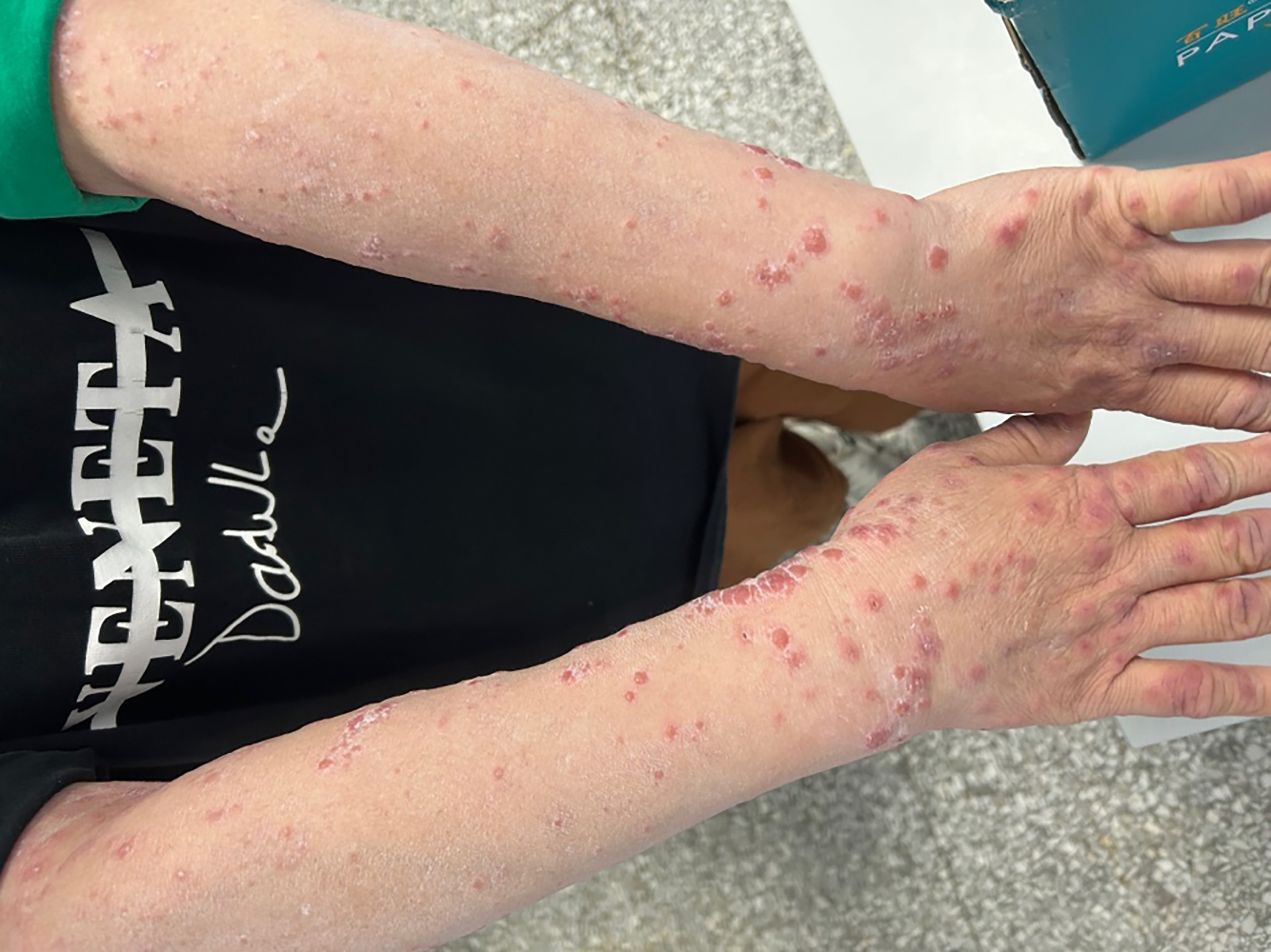
Fig. 1. Clinical presentation. Multiple erythematous papules with a vesicle-like appearance on the upper limbs.

Fig. 2. Histopathological finding. Significant papillary oedema and diffuse dermal neutrophilic and eosinophilic infiltrate.
Histopathological examination revealed massive papillary dermal oedema and a diffuse nodular and perivascular neutrophilic and eosinophilic infiltration, with no fibrinoid change in vessel walls (Fig. 2).
What is your diagnosis?
A. Bullous pemphigoid
B. Gianotti-Crosti syndrome
C. Varicella
D. Sweet’s syndrome
See next page for answer.
ANSWERS TO QUIZ
Widespread Erythematous Vesiculobullea: A Commentary
Diagnosis: Sweet’s syndrome
Given the pseudovesicular appearance and classic histological findings, she was diagnosed with Sweet’s syndrome and treated with oral prednisone at 30 mg daily, showing a rapid resolution after 3 days. After gradually tapering the prednisone, she was left with only hyperpigmentation and did not relapse during the follow-up period.
Sweet’s syndrome is an inflammatory, noninfectious skin reaction. It is clinically characterized by rapid onset tender, burning, erythematous plaques or papules, with an uneven mammillated surface, most commonly affecting the face and extremities, with less frequent involvement of the trunk. Skin lesions usually heal without scarring, while there may be residual pigmentation attributed to hemosiderin. It is commonly accompanied by fever (50–80%), malaise, preceding upper respiratory infections or flu-like symptoms, arthralgias and ocular involvement. Despite the alternative name of acute febrile neutrophilic dermatosis, a fever is not always present. Laboratory evaluation typically shows elevated inflammatory markers, although normal values do not exclude the diagnosis. The clinical variants include bullous, cellulitis-like, necrotizing, and neutrophilic dermatosis of the dorsal hands, etc. (1). The characteristic histologic presentation is significant papillary oedema and diffuse dermal neutrophilic infiltrate. Marked oedema of the papillary dermis may lead to the appearance of subepidermal vesiculation. Leukocytoclasis with nuclear dust formation is typically present, while vasculitis and fibrinoid extravasation are generally absent. Histological variants, including histiocytoid, lymphocytic, eosinophilic and subcutaneous types, have been reported.
The case should be differentiated from clinically similar conditions. Bullous pemphigoid (BP) is the most common autoimmune subepidermal blistering disease, typically presents as generalized pruritic bullous in older adults. In the early stage of disease, or some rare variants, it can be nonspecific, with pruritic eczematous, papular or urticarial lesions. Subepidermal blister in histopathology, positive result of direct immunofluorescence (DIF) test and/or circulating autoantibodies (anti-BP180, anti-BP230) can help in diagnosis (2). Gianotti-Crosti syndrome (GCS) is a self-limited dermatosis primarily affecting children under 6 years of age. It presents as a symmetrical eruption of flat-topped maculopapular or papulovesicular exanthema, which can coalesce and form plaques, predominantly on the extremities, gluteal region and extensor surfaces. It is often associated with viral infections (3). Varicella (chickenpox), caused by varicella-zoster virus, is a highly contagious disease with an average incubation period of 2 weeks and peak incidence during childhood. It presents as an acute vesicular eruption, which evolves in successive crops, resulting in simultaneous papules, vesicles, pustules, crusted lesions and healing lesions (4). In this case, the typical histological features lead to the diagnosis of Sweet’s syndrome.
Sweet’s syndrome is usually idiopathic but can be associated with infections, malignancies, autoimmune diseases, medications and pregnancy, thus is thought to be an immune-mediated hypersensitivity reaction triggered by a preceding event. Therefore, individuals presenting with features of Sweet’s syndrome should be carefully evaluated to identify any underlying conditions (5).
Without treatment, lesions may persist for weeks or months. High-quality treatment studies for Sweet’s syndrome remain limited. Corticosteroids are considered first-line therapy due to the rapid clinical response observed in most patients. The most effective therapy is prednisone 0.5–1.0 mg/kg daily for 4–6 weeks. Indomethacin is supported by the strongest evidence and may be used as a second-line agent for steroid-refractory cases. Retrospective studies support the use of acitretin, colchicine and dapsone, while isolated case reports have demonstrated efficacy for biologic agents. Ultimately, treatment selection should be individualized based on patient comorbidities, medical history and preferences (1).
ACKNOWLEDGMENTS
We thank the patients for granting permission to publish this information.
REFERENCES
- Joshi TP, Friske SK, Hsiou DA, Duvic M. New practical aspects of sweet syndrome. Am J Clin Dermatol 2022; 23: 301–318. https://doi.org/10.1007/s40257-022-00673-4
- Terra JB, Meijer JM, Jonkman MF, Diercks GFH. The n- vs. u-serration is a learnable criterion to differentiate pemphigoid from epidermolysis bullosa acquisita in direct immunofluorescence serration pattern analysis. Br J Dermatol 2013; 169: 100–105. https://doi.org/10.1111/bjd.12308
- Afonso A, Cachão J, Pinto Junior VL, Gouveia T. Gianotti-Crosti syndrome: a challenging exanthema. BMJ Case Rep 2021; 14: 14. https://doi.org/10.1136/bcr-2020-240747
- Gershon AA, Breuer J, Cohen JI, Cohrs RJ, Gershon MD, Gilden D, et al. Varicella zoster virus infection. Nat Rev Dis Primers 2015; 1: 15016. https://doi.org/10.1038/nrdp.2015.16
- Villarreal-Villarreal CD, Ocampo-Candiani J, Villarreal-Martínez A. Sweet syndrome: a review and update. Actas Dermosifiliogr 2016; 107: 369–378. https://doi.org/10.1016/j.ad.2015.12.001