ORIGINAL REPORT
Distinctive Reactivity to the C-terminal Epitope of BP180 Characterizes Immune Checkpoint Inhibitor–associated Bullous Pemphigoid, and an ELISA Based on the BP180 Ectodomain Enables Prompt Diagnosis in a Subset of Patients
Anna PIRA1† , Martina MERLI2,3†, Feliciana MARIOTTI1*, Marco ANDREANI4, Angelo Valerio MARZANO5,6, Clara DE SIMONE7,8, Rosa FALCONE9, Giuseppe TESTA4, Mariarosa BATTARRA4, Maurizio ROMAGNUOLO5,10, Pietro SOLLENA8,11, Federica DE GALITIIS9, Emiliano ANTIGA12, Roberto MAGLIE12, Dario DIDONA13,14, Jo Linda Maria SINAGRA13, Marzia CAPRONI15, Michael HERTL16, Emanuele COZZANI17, Pamela VEZZOLI18, Laura CALABRESE8,19, Giovanni PAOLINO20, Sofia VERKHOVSKAIA9, Luca FANIA14,21, Giulia GASPARINI17, Anna Rita GIAMPETRUZZI14, Biagio DIDONA13, Pietro QUAGLINO2 and Giovanni DI ZENZO1
1Molecular and Cell Biology Laboratory, Istituto Dermopatico dell’Immacolata (IDI)-IRCCS, Rome, Italy, 2Section of Dermatology, Department of Medical Sciences, University of Turin, Turin, Italy, 3Dermatology Clinic, Department of Medical Sciences, A.O. Ordine Mauriziano, Turin, Italy, 4Laboratorio di Immunogenetica dei Trapianti, IRCCS Ospedale Pediatrico Bambino Gesù, Rome, Italy, 5Dermatology Unit, Fondazione IRCCS Ca’ Granda Ospedale Maggiore Policlinico, Milan, Italy, 6Department of Pathophysiology and Transplantation, Università Degli Studi di Milano, Milan, Italy, 7Dermatology, Department of Medical and Surgery Sciences, Fondazione Policlinico Universitario A. Gemelli IRCCS, Rome, Italy, 8Dermatology, Department of Medicine and Translational Surgery, Università Cattolica del Sacro Cuore, Rome, Italy, 9Department of Oncology and Dermatological Oncology, Istituto Dermopatico dell’Immacolata (IDI)-IRCCS, Rome, Italy, 10Department of Medical Biotechnologies, University of Siena, Siena, Italy, 11Dermatology, Gemelli Isola - Ospedale Isola Tiberina, Rome, Italy, 12Section of Dermatology, Department of Health Sciences, University of Florence, Florence, Italy, 13Rare Diseases Unit, Istituto Dermopatico dell’Immacolata (IDI)-IRCCS, Rome, Italy, 14Dermatology Clinic, Istituto Dermopatico dell’Immacolata (IDI)-IRCCS, Rome, Italy, 15Immunopathology and Rare Skin Diseases Unit, Section of Dermatology, Department of Health Sciences, Azienda Unità Sanitaria Locale Toscana Centro, University of Florence, Florence, Italy, 16Department of Dermatology and Allergology, Philipps-Universität Marburg, Marburg, Germany, 17Section of Dermatology, Department of Health Sciences (DISSAL), IRCCS San Martino Polyclinic Hospital, University of Genoa, Genoa, Italy, 18Dermatology Unit, ASST Papa Giovanni XXIII Hospital, Bergamo, Italy, 19Department of Medical, Surgical and Neurological Sciences, Dermatology Section, University of Siena, Siena, Italy, 20Unit of Dermatology, IRCCS Ospedale San Raffaele, Milan, Italy, and 21Department of Life Science, Health, and Health Professions, Link University of Rome, Rome, Italy.
†These authors contributed equally to this work.
Corr: Feliciana Mariotti, Molecular and Cell Biology Laboratory, Istituto Dermopatico dell’Immacolata (IDI)-IRCCS, Via Monti di Creta 104, 00167 Rome, Italy. *Email: f.mariotti@idi.it
Key words: autoantibodies; BP180; bullous pemphigoid; epitope profile; humoral response; immune checkpoint inhibitors.
Citation: Acta Derm Venereol 2026; 106: adv-2026-0336. DOI: https://doi.org/10.2340/actadv.v106.adv-2026-0336.
Copyright: © The Authors 2026. Published by MJS Publishing, on behalf of the Society for Publication of Acta Dermato-Venereologica. This is an Open Access article distributed under the terms of the Creative Commons Attribution-NonCommercial 4.0 International License (https://creativecommons.org/licenses/by-nc/4.0/).
Submitted: Jan 9, 2026. Accepted after revision: May 25, 2026.
Published: Jun 29, 2026.
Competing interests and funding: This study was supported by the “Progetto Ricerca Corrente” of the Italian Ministry of Health, Rome, Italy.
The data that support the findings of this study are available from the corresponding author upon reasonable request.
The study was conducted in accordance with the Ethic Committees of the participating institutions. Ethical Approval: EC 440/2022.
Anna Pira, Feliciana Mariotti, Rosa Falcone, Giuseppe Testa, Mariarosa Battarra, Maurizio Romagnuolo, Roberto Maglie, Dario Didona, Jo Linda Maria Sinagra, Marzia Caproni, Emanuele Cozzani, Giovanni Paolino, Luca Fania, Anna Rita Giampetruzzi, Giulia Gasparini, Biagio Didona, Pietro Quaglino, and Giovanni Di Zenzo have no conflicts of interest to declare. Martina Merli received payments for data collection in studies sponsored by Almirall and has been a speaker for Abbvie, Novartis and Giuliani. Marco Andreani is President of European Federation for Immunogenetics (EFI) (unpaid position). Angelo Valerio Marzano has been an advisor, speaker and/or consultant for AbbVie, Amgen, Boehringer-Ingelheim, Bristol Myers Squibb, Incyte, Leopharma, Novartis, Pfizer, Sanofi, and UCB. Clara De Simone received payments for lectures/presentations for Abbvie, Almirall, Biogen, Eli Lilly, Sanofi, UCB Pharma and participated on a Data Safety Monitoring Board or Advisory Board for Abbvie, Almirall, and Bristol-Myers Squibb. Pietro Sollena received payments for lectures/presentations and received support for attending meetings and/or travel by for Almirall, L’Oreal and Pierre Fabre; and participated on a Data Safety Monitoring Board or Advisory Board for Almirall and L’Oreal. Federica De Galitiis has been a consultant/speaker for Novartis, Bristol, Pierre Fabre and Merk. Emiliano Antiga: has been an advisor, speaker and/or consultant for AbbVie, Almirall, Incyte, Leopharma, Lilly, Pfizer, Sanofi. Michael Hertl: has been a consultant for Topas Therapeutics and Argenx; has been a speaker for Novartis, Janssen Cilag, Almiral, Sanofi; received support for attending meetings and/or travel by Janssen Cilag, and participated on a Data Safety Monitoring Board or Advisory Board for Argenx. Pamela Vezzoli: received support for attending meetings and/or travel by Abbvie, Almirall, Novartis, Sanofi. Laura Calabrese: received payments for lectures/presentations for Abbvie and Almirall and received support for attending meetings and/or travel by Abbvie, Almirall, Novartis, Sanofi. Sofia Verkhovskaia has been a speaker for Novartis.
Growing evidence has linked bullous pemphigoid (BP) to immune checkpoint inhibitor (ICI) therapy in cancer treatment. However, the immunological features of ICI-associated BP (ICI-BP) are not yet fully elucidated. In order to characterize the humoral response in ICI-BP patients and investigate whether their epitope profile differs from idiopathic BP (IBP), 53 ICI-BP patients were enrolled, immunologically characterized and compared with 59 IBP patients. ICI-BP had a distinctive IgG humoral profile, with reduced reactivity toward BP230 and recognition of multiple BP180 epitopes beyond the immunodominant extracellular noncollagenous 16A domain (NC16A). Specifically, reactivity to BP180 ectodomain was present in 94% of ICI-BP and 78% of IBP (p=0.044). Moreover, BP180 C-terminal epitope was more frequently targeted in ICI-BP than IBP (72% vs 41%, p=0.002). Notably, the combined use of an in-house BP180 ectodomain ELISA and the commercial BP180 test increased diagnostic sensitivity from 83% to 100%. Enhanced IgG reactivity toward nonimmunodominant epitopes, and especially C-terminal epitope recognition, characterize the humoral immune response in ICI-BP. Our data suggest that combining NC16A and full-length BP180 ectodomain ELISAs may help reduce diagnostic delay in ICI-BP patients, in whom a timely diagnosis is crucial to appropriately manage the disease and ultimately avoid discontinuation of cancer therapy.
SIGNIFICANCE
Managing immunotherapy–associated bullous pemphigoid (ICI-BP) requires balancing autoimmune disease control and oncologic therapy continuation. This study offers an integrated clinical, immunological and genetic characterization of a large ICI-BP cohort. Compared to idiopathic BP, ICI-BP shows a distinctive immunological signature that could be leveraged for earlier BP diagnosis. The joint use of a commercial and in-house assay to detect autoantibodies to different regions of BP180, the main target in BP, allowed diagnosis in 100% of patients. This approach may enable prompt BP diagnosis and its timely management, avoiding unnecessary immunotherapy discontinuation and improving outcomes for patients receiving life-prolonging cancer immunotherapy.
INTRODUCTION
Bullous pemphigoid (BP) is an autoimmune skin disorder characterized by subepidermal blistering, resulting from autoantibodies (autoAbs) directed against hemidesmosomal components: BP180 and BP230 (1, 2). Evidence from case-control studies has linked a broad range of medications to BP (3). Additionally, there is growing interest in BP as an immune-related adverse event (irAE) associated with immune checkpoint inhibitors (ICIs), whose use is steadily increasing in oncology (4). ICIs are monoclonal antibodies targeting cytotoxic T-lymphocyte antigen 4 and programmed cell death protein 1 or its ligand (PD-1/PD-L1), thereby enhancing the T-cell-mediated immune response against tumour cells. While ICIs have revolutionized cancer treatment, their mechanism of action also causes nonspecific immune activation leading to adverse events affecting multiple organ systems, including the skin (4). Specifically, ICIs block inhibitory T-cell pathways, enhancing their activity and, in some cases, trigger B-cell-mediated production of autoAbs against BP180 and BP230, potentially leading to the onset of BP. Although ICI-associated BP (ICI-BP) is an uncommon irAE, with reported incidence rates ranging from 0.2% to 0.8% (5, 6, 7), it can lead to oncological treatment discontinuation.
The association between BP and specific HLA alleles in the Italian population has been demonstrated (8); however, HLA allele typing has been performed in few ICI-BP patients and data on genetic susceptibility in this population remain scarce (9, 10).
BP180 is the primary BP autoantigen, with its immunodominant region, the noncollagenous 16A domain (NC16A), accounting for approximately 80–90% of IgG, 65% of IgA, and 80% of IgE autoAb reactivity (1, 11, 12). Nevertheless, additional epitopes within the extracellular domain of BP180 (ECD-BP180) have also been identified (13, 14) (Fig. 1).
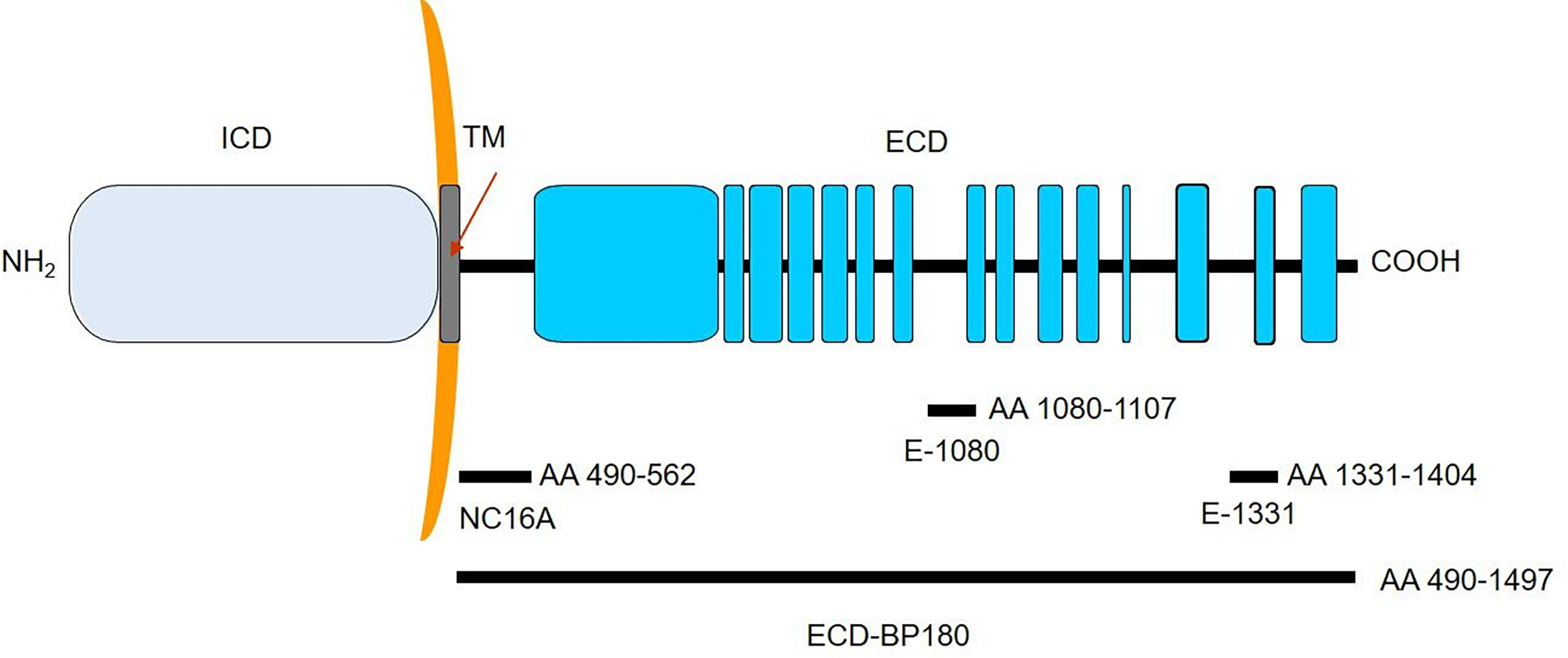
Fig. 1. Schematic representation of the BP180 antigen. NC16A immunodominant domain, E-1080 and E-1331 encompass residues 490–562, 1080–1107 and 1331–1404, respectively. The entire extracellular portion of BP180 (ECD-BP180) spans amino acids 490 to 1497. Black and light blue boxes refer to non-collagenous and collagenous protein domains, respectively
In ICI-BP, the humoral response is predominantly directed against BP180 rather than BP230, suggesting a selective immune recognition of more exposed antigens at the dermoepidermal junction (4).
Although some studies described the clinical features of ICI-BP (4, 15), its immunological characteristics remain insufficiently explored. Characterization of the ICI-BP humoral response could enable the identification of a specific diagnostic approach for timely diagnosis, allowing appropriate disease management and ultimately preventing discontinuation of cancer therapy.
MATERIALS AND METHODS
Study population
Fifty-six patients were retrospectively and prospectively enrolled in the Dermatology Units of 10 Italian hospitals between January 2020 and September 2024. Three patients were excluded, as one was affected with mucous membrane pemphigoid and 2 developed BP more than 12 months after therapy interruption. Therefore, this study includes 53 patients who developed BP during cancer treatment with ICIs or up to 12 months after discontinuation. Fifty-nine idiopathic BP (IBP) and 30 gliptin-associated BP (GABP) were also retrospectively enrolled as controls. The diagnosis of BP was established according to the current European Guidelines for BP (16). This study was approved by the local Ethics Committee and was conducted in accordance with the Declaration of Helsinki guidelines. All patients gave written informed consent.
Typing of human leucocyte antigens
Thirty-two ICI-BP patients were typed for the HLA-A, B, C, DRB1, DRB3/4/5, DQA1, DQB1, DPA1 and DPB1 loci at high resolution using an NGS method on the S5 Ion Torrent platform with the FastPlex Kit (both from Thermo Fisher, Canoga Park, CA, USA). Occasionally, typing was performed using the SSO method with LABType XR and analysed on the Luminex LABScan3D™ (both from Thermo Fisher, Canoga Park, CA, USA). A group of Italian healthy donors (HD) and IBP patients, which had been previously characterized (8), were used as comparisons.
Enzyme-linked immunosorbent assays
IgG autoAbs targeting BP180-NC16A and BP230 were detected with commercial ELISA tests (MBL International, Woburn, MA, USA), with a cut-off value ≥9 U/mL. The immunological profiles of patients with active lesions at the time of blood draw, regardless of time from disease onset or initiation of BP-directed therapy, were further characterized. IBP and GABP controls were selected according to the same criterion and IgG reactivity against other epitopes of BP180 (Fig. 1) was assessed. Specifically, autoAbs directed against ECD-BP180, covering amino acid residues 490–1497, were detected with a recently described in-house ELISA (cut-off value ≥10.02 pemphigoid index value, PIV) (11), and IgG reactivity to the mid-portion (E-1080) and COOH-terminus (E-1331) regions of BP180 ectodomain (spanning amino acids 1080–1107 and 1331–1404, respectively) was also assessed by using ELISAs with GST-1080 and GST-1331 (cut-off ≥14.9 PIV and ≥4.5 PIV, respectively) (13) (Fig. 1). PIV represents the standardization of extinction data on the positive and negative internal standards, which were set to 1 and 0 OD units (17). Moreover, IgE and IgA reactivity against BP180-NC16A and BP230 was also measured by slightly modifying MBL’s protocol for its commercial tests as previously reported (IgE cut-offs: BP180 ≥3.2 PIV, BP230 ≥4.5 PIV; IgA cut-offs: BP180 >13.8 PIV, BP230 >14.5 PIV) (12).
Direct and indirect immunofluorescence
Direct and indirect immunofluorescence (DIF and IIF) were performed as previously described (1, 18).
Statistical analysis
Differences on immunological profiles were assessed through the Fisher exact (probability) test and the Mann–Whitney U test using GraphPad Prism software version 9.4.1. Differences in allele and phenotype frequencies for HLA alleles across the analysed groups were assessed through the χ2 test. Bonferroni correction was also applied. Differences were considered significant when the p-value was ≤0.05.
RESULTS
Demographic and clinical features of 53 patients with immune checkpoint inhibitor-associated bullous pemphigoid
Fifty-three patients with ICI-associated BP had a mean age of 74.9, and the majority (88.7%) were males (Table I). BP presented with a median delay of 41 weeks from therapy initiation and the median time to diagnosis was 49 weeks (Table I). ICI-BP with mucosal involvement were 16.3% (Table I). The majority of patients was treated for lung cancer (34.0%), followed by melanoma (30.2%) and renal/urothelial cancer (18.9%), while the remaining 16.9% of patients were treated for multiple basal cell carcinomas (n=1), squamous cell carcinoma (n=2), tracheal cancer (n=1), colorectal cancer (n=3), hepatocellular carcinoma (n=1) and parotid cancer (n=1) (Table I). Anti-PD-1 monoclonal antibodies pembrolizumab and nivolumab were equally employed, while a minority of patients received other anti-PD-(L)1 agents (Table I). In more than half of patients, oncologic therapy was discontinued after BP onset, whereas 17.5% experienced only a temporary interruption. A total of 17.5% of patients continued oncologic treatment despite BP onset. In the remaining 10.0% of patients, discontinuation occurred: (i) before BP onset, (ii) after complete tumour response or (iii) due to tumour progression (Table I).
Table I. Demographic and clinical characteristics of patients with idiopathic, immune checkpoint inhibitor-associated and gliptin-associated bullous pemphigoid
| 59 IBP | 53 ICI-BP | 30 GABP | |
|---|---|---|---|
| Mean age - years (±SD) | 77.8 (±9.6) | 74.9 (±8.3) | 79.3 (±7.4) |
| Males - n/N (%) | 30/58 (51.7) | 47/53 (88.7) | 15/30 (50.0) |
| Mucosal involvementa – n/N (%) | 13/59 (22.0) | 8/49 (16.3) | 7/30 (23.3) |
| Median delay – weeks (range) | / | 41 (5–353) | 35 (1–511) |
| Median time to diagnosis – weeks (range) | / | 49 (8–357) | 43 (1–546) |
| DIF (linear IgG and/or C3)b – n/N (%) | 52/56 (93.0) | 37/40 (92.5) | 20/23 (87.0) |
| IIF (epidermal side, IgG)c – n/N (%) | 57/58 (98.3) | 32/37 (80.0) | 25/29 (86.2) |
| Mode of oncologic therapy administrationd – n/N (%) | |||
| Discontinued because of BP onset | 22/40 (55.0) | ||
| Discontinued after BP onset with CR | 2/40 (5.0) | ||
| Discontinued after BP onset with PD | 1/40 (2.5) | ||
| Temporarily interrupted because of BP onset | 7/40 (17.5) | ||
| Uninterrupted | 7/40 (17.5) | ||
| BP onset after discontinuation of cancer therapy | 1/40 (2.5) | ||
| Tumour type – n/N (%) | |||
| Lung cancer | 18/53 (34.0) | ||
| Melanoma | 16/53 (30.2) | ||
| Renal/Urothelial cancer | 10/53 (18.9) | ||
| Other | 9/53 (17.0) | ||
| Immune checkpoint inhibitor – n/N (%) | |||
| Nivolumab | 24/53 (45.3) | ||
| Pembrolizumab | 24/53 (45.3) | ||
| Atezolizumab, cemiplimab, spartalizumab | 5/53 (9.4) | ||
|
BP: bullous pemphigoid; CR:complete tumor response; DIF:direct immunofluorescence; GABP:gliptin-associated bullous pemphigoid; IBP: idiopathic bullous pemphigoid; ICI-BP: immune checkpoint inhibitor-associated bullous pemphigoid; IIF: indirect immunofluorescence; PD: tumor progressive disease. |
|||
HLA allele frequencies were similar in immune checkpoint inhibitor-associated bullous pemphigoid and idiopathic bullous pemphigoid
To identify HLA alleles that could reflect a higher susceptibility to develop BP in ICI-treated patients in the Italian population, we analysed 32 ICI-BP Italian patients, comparing them with 1,017 HD and 28 IBP patients.
As previously described in IBP (8), ICI-BP patients of this study showed a higher frequency of HLA alleles DQB1*03 : 01 (51.6% vs 32.0%, p=0.001 and Bonferroni-corrected p [pc]=0.020) and DQA1*05 : 01P (56.3% vs 39.5%, p=0.009, pc=not statistically significant) in the comparison with HD (Table II). The HLA-DQB1*03 : 01 phenotype frequency was also higher in ICI-BP (81.3% vs 54.5%, p=0.003, pc=0.050). Both allele and phenotype frequencies of DRB1*11 : 04 were higher in ICI-BP (25.0% vs 14.1%, p=0.030 and 43.8% vs 23.0%, p=0.007) (Table II). Of note, ICI-BP patients also had significantly higher allele and phenotype frequencies for the locus A*31 : 01 (allele: 9.4% vs 1.6, p<0.001, pc=0.040; phenotype: 18.8% vs 3.0%, p<0.00001, pc<0.001) (Table II).
Table II. Comparison of HLA profile between 32 ICI-BP patients, 28 IBP and 1,017 healthy donors
Immune checkpoint inhibitor-associated bullous pemphigoid patients show a peculiar IgG humoral response against the BP autoantigens in comparison with idiopathic bullous pemphigoid patients
Sera from 50 ICI-BP patients with active lesions were tested and compared with IBP immunological profiles. Furthermore, as GABP has previously been reported to have a typical immunological response (12), the ICI-BP group was also compared with gliptin users.
ICI-BP patients showed a peculiar IgG response: although no major differences were found in regard to BP180-NC16A, both reactivity and mean titres to the other BP180 epitopes were significantly higher in ICI-BP than in IBP, while the opposite was observed for the response to BP230 (Table SI, Fig. 2). Specifically, ECD-BP180 reactivity was more frequent in ICI-BP than IBP patients (93.6% vs 78.0%, p=0.044), with higher mean titres (104.1 vs 77.0 PIV, p=0.037) (Table SI, Fig. 2). Notably, COOH-terminal epitope of BP180 (E-1331) was more frequently targeted in ICI-BP than IBP (72.3% vs 40.7%, p=0.002), with higher IgG titres (PIV: 95.1 vs 36.8, p=0.006) (Table SI, Fig. 2). ICI-BP also showed increased reactivity to the BP180 mid-portion epitope, E-1080, (55.3% vs 32.2% in IBP (p=0.028); however, autoAb titres did not differ significantly (Table SI,Fig. 2). As for BP230, both reactivity (24.0%) and titres (25.8 U/ml) were heavily lower in ICI-BP than IBP (55.9% and 68.2 U/ml; p=0.001 and p<0.001, respectively) (Table SI,Fig. 2).

Fig. 2. IgG, IgE and IgA autoantibody reactivity to BP180/BP230 in idiopathic and drug-associated bullous pemphigoid patients. Bar plot representation of IgG, IgE and IgA positivity to BP230 and BP180 (NC16A, ectodomain [ECD], E-1080, E-1331, spanning amino acids 490–562, 490–1497, 1080–1107 and 1331–1404, respectively) epitopes in idiopathic, immune checkpoint inhibitor-associated and gliptin-associated bullous pemphigoid patients (IBP, ICI-BP and GABP respectively).
Compared with IBP, ICI-BP and GABP showed similarly high IgG reactivity to ECD-BP180 and low positivity to BP230. Of note, reactivity to NC16A was not reduced in ICI-BP in contrast to GABP. Another interesting difference concerned anti-E-1331 mean titres, which were higher in ICI-BP (p=0.007) (Table SI).
In addition, ICI-BP patients showed higher IgE titres to BP180-NC16A than IBP (PIV: 53.7 vs 15.5, p=0.030) (Table SI). Regarding IgA, reactivity against BP180-NC16A was also higher in ICI-BP than in IBP, although not statistically significant (Table SI). The comparison with GABP also showed some differences in IgE and IgA responses: ICI-BP patients had higher IgE reactivity to BP180-NC16A (70.6% vs 26.7%, p<0.001) and higher IgA reactivity to both BP180-NC16A (76.5% vs 36.7%, p=0.003) and BP230 (58.8% vs 26.7%, p=0.019) (Table SI). ICI-BP also seemed to have higher IgE mean titres to NC16A (p=0.052) (Table SI).
Longer delays in BP onset are associated with an increased IgG reactivity to the immunodominant epitope of BP
Stratification by age, tumour type, drug employed, and time to onset revealed that only time to BP onset (≤ or > 40 weeks from therapy initiation) significantly affected serum IgG profiles (Table SII, Table SIII). Specifically, higher autoAb titres in patients with delays longer than 40 weeks for ECD-BP180 (PIV: 135.0 vs 70.5, p<0.001) and E-1331 (PIV: 129.8 vs 53.4, p=0.013) (Table SII) was observed. Of note, patients with longer delays were more reactive to BP180-NC16A than those with BP onset within 40 weeks from therapy initiation (95.7% vs 68.0%, p=0.032) (Table SII).
An ELISA based on ECD-BP180 increases diagnostic sensitivity of BP180 and BP230 commercial ELISAs
Commercial ELISAs failed to detect BP autoAbs in 7 patients who were double negative on both BP180-NC16A and BP230, and 1 patient showed reactivity only to BP230. Interestingly, all 8 patients showed IgG reactivity against other BP180 epitopes (Fig. 3). Specifically, 3 (37.5%), 7 (87.5%) and all 8 patients reacted to E-1331, E-1080 and ECD-BP180, respectively (Fig. 3). Thus, our in-house ECD-BP180 ELISA increased the sensitivity of commercial BP180-NC16A test, showing a role in reducing diagnostic delay in ICI-BP patients.

Fig. 3. ELISA reactivity to BP180 and BP230 epitopes in 47 immune checkpoint inhibitor-associated bullous pemphigoid (ICI-BP) patients. Scatter plot representations of IgG reactivity to: (a) BP180-NC16A and BP230 in commercial MBL tests; (b) BP180 ectodomain (ECD-BP180); (c) midportion epitope of BP180 (E-1080); (d) COOH-terminal epitope of BP180 (E-1331). Red dots represent the 7 patients whose results for both BP180-NC16A and BP230 were negative. Green dots represent 1 patient who was negative for BP180-NC16A and positive for BP230. The dotted lines represent the cut-offs of the assays.
Among the 8 NC16A-negative patients, DIF was positive in 4, negative in one, and unavailable in 3. In the latter cases, IIF showed dermo-epidermal junction staining in one, was negative in one and unavailable in the third. The DIF negative patient was positive on IIF. Thus, in 2/8 NC16A-negative patients (25.0%), DIF and IIF were negative or unavailable, highlighting the diagnostic value of ECD-BP180 ELISA. While DIF remains the gold standard for BP diagnosis, this assay offers a sensitive, less invasive and time-efficient alternative, particularly useful in fragile patients such as those with ICI-BP.
DISCUSSION
Managing ICI-BP is clinically challenging, requiring a comprehensive approach that addresses both the control of autoimmune disease and the continuation of oncological therapy. In this context, reducing diagnostic delay is therefore crucial, as a timely diagnosis of BP and prompt treatment initiation can improve patient outcomes and enable prompt resumption or prevent the interruption of immunotherapy.
In the present study, the immunological characterization of ICI-BP provides a foundation for enhancing current diagnostic methodologies and reducing delays in its diagnosis.
Consistent with previous studies (4, 15, 19, 20), our ICI-BP patients were mainly elderly males. While the age distribution aligns with the typical onset of BP (21), the observed male predominance may reflect the higher incidence in males of certain malignancies commonly treated with ICIs (22, 23). Most patients were treated for lung cancer (n=18, 34.0%), followed by melanoma (n=16, 30.2%) and renal/urothelial cancers (n=10, 18.9%). However, due to the design of this study, the prevalence of different tumour types does not indicate higher risk of BP development.
Consistent with data reported in an Italian population, ICI-BP was significantly linked to the HLA allele DQB1*03 : 01, indicating a predisposition profile comparable to that observed in IBP (8). Similarly, alleles DRB1*11 : 04 and DQA1*05 : 01P are also associated with BP onset. Notably, the rare allele HLA-A*31 : 01 showed a significant association with ICI-BP compared to HD, suggesting a role for MHC class I molecules in the occurrence of anti-BP180 immune response in at least a subset of patients. In fact, in these cases, autoimmunity targeting BP180 may originate from a cytotoxic T lymphocyte-mediated reaction induced by immunotherapy, potentially involving specific MHC class I alleles, such as HLA-A*31 : 01. Given these findings, BP onset in ICI-treated patients has a multifactorial aetiology that likely includes ageing and HLA genotype.
To the best of our knowledge, this is the largest cohort of ICI-BP patients immunologically and genetically characterized to date. Regarding BP180-NC16A reactivity, consistent with a recent investigation (15) reporting that 9/11 ICI-BP patients were positive by ELISA, we found reactivity in 41/50 patients in our cohort. However, we observed a distinct immunological profile compared with both IBP and GABP. IgG reactivity to BP180-NC16A is similar to IBP, but differs from GABP, where reactivity is typically reduced (12), highlighting mechanistic differences in BP induction between GABP and ICI-BP. Both subgroups exhibited IgG reactivity against additional BP180 epitopes, with a tendency toward higher reactivity than IBP. Notably, ICI-BP patients recognize the COOH-terminal region of BP180 more frequently than IBP and GABP, a finding that may reflect the underlying pathogenic mechanism. The immune-stimulatory activity of ICIs could promote recognition of tumour-expressed BP180 (24, 25, 26), subsequently leading to immune recognition of the antigen in the skin and resulting in dermo-epidermal separation. The most exposed BP180 portion in tumour tissue is likely the extracellular COOH-terminal domain, especially if the tumour cells lose contact with their molecular counterpart on the basement membrane. Consequently, it is plausible that the immune system more readily targets the COOH-terminal region, facilitating autoAb production and BP development. In this context, a longer latency period before BP onset is associated with prolonged ICIs stimulation, leading to higher serum autoAb concentrations against BP180-NC16A, ECD-BP180, and E-1331. Longer delays in BP onset were associated with an increased IgG reactivity to BP180-NC16A and not to other epitopes, potentially indicating an epitope spreading phenomenon from other BP180 epitopes to the immunodominant one during treatment. Consistently, no significant differences in reactivity to E-1080 and E-1331 were found between NC16A-positive and negative patients, although reactivity to E-1080 seemed to be higher in NC16A-negative patients. In line with our observations, a recently reported patient showed reactivity against the BP180 COOH-terminal domain at the initiation of ICI therapy, which subsequently spread to NC16A during BP development (27). These data potentially indicate an epitope spreading phenomenon from other BP180 epitopes to the immunodominant one during treatment.
Conversely, IgG reactivity toward BP230 was markedly reduced in ICI-BP compared to IBP, consistent with observations in GABP. This may be attributable to the shorter disease duration at diagnosis due to the close clinical monitoring of these patients which limits the occurrence of intermolecular epitope spreading phenomena that typically underpin reactivity to BP230 (12, 28).
Regarding IgE responses, ICI-BP patients exhibited significantly higher reactivity and titres against BP180-NC16A compared to IBP. IgE is a central mediator of allergic responses, promoting mast cell and basophil degranulation through crosslinking of IgE molecules bound to high-affinity FcεRI receptors. Enhanced IgE responses have been associated with ICI therapy (29), and pruritus, one of the most frequently reported cutaneous irAEs in ICI-treated patients, may represent a clinical manifestation of this effect (30). Our findings support the use of omalizumab, an anti-IgE monoclonal antibody already employed to mitigate ICI-related pruritus (31) as a therapeutic option for managing BP in this vulnerable patient population (32, 33). In this context, IgE reactivity against BP180 could serve as a biomarker of favourable therapeutic response. Like IgE, IgA showed increased reactivity against BP180-NC16A in ICI-BP patients compared with IBP, but mucosal involvement was similar despite the well-established IgA’s role in mucosal immunity (1).
While oncologic patients experiencing cutaneous irAEs such as vitiligo often have favourable outcomes (34), data on BP prognosis remain conflicting and incomplete (7, 35, 36, 37). This may reflect that, unlike vitiligo, BP onset is frequently followed by discontinuation of oncologic therapy, a factor associated with worse prognosis. In the present study, as in other real-world settings (15, 38, 39, 40), most patients (73%) discontinued oncologic treatment after BP onset.
With a median 49 week time to diagnosis and 41 week time to symptom onset, a diagnostic delay of approximately 2 months can be inferred: although relatively short, it can be particularly detrimental in this vulnerable patient population. A recent study found that in ICI-treated patients BP developed with significantly longer latencies than other irAEs (15). However, pruritus is a characteristic prodromal symptom of BP and one of the most common cutaneous irAEs observed in ICI-treated patients (30). This confounding factor may delay diagnosis, despite close clinical monitoring. Therefore, reducing diagnostic delay is crucial for improving clinical outcomes and potentially avoid ICI discontinuation.
The strengths of this study include the large number of patients analysed and the use of in-house ELISA assays specifically designed for epitope profiling. Limitations include potential selection bias, the inclusion of patients who had initiated BP treatment, and incomplete clinical data regarding disease phenotype and severity.
In conclusion, our findings indicate that ICI-BP patients exhibit a specific humoral response characterized by increased reactivity to non-NC16A epitopes, particularly the COOH-terminal region. Thus, while DIF remains the gold standard for BP diagnosis, BP180 and BP230 ELISAs provide a faster supportive diagnostic method. In a subset of patients (8/47, 17%) negative by commercial BP180 ELISA, an in-house ELISA based on ECD-BP180 yielded positive results, increasing diagnostic sensitivity from 83% to 100%. These findings may help reduce diagnostic delays, enabling timely and appropriate disease management and ultimately contributing to early resumption or prevention of premature discontinuation of cancer treatment.
REFERENCES
- Di Zenzo G, Della Torre R, Zambruno G, Borradori L. Bullous pemphigoid: From the clinic to the bench. Clin Dermatol 2012; 30: 3–16. https://doi.org/10.1016/j.clindermatol.2011.03.005
- Genovese G, Di Zenzo G, Cozzani E, Berti E, Cugno M, Marzano AV. New insights into the pathogenesis of bullous pemphigoid: 2019 update. Front Immunol 2019; 10: 1506. https://doi.org/10.3389/fimmu.2019.01506
- Swiderski M, Vinogradova Y, Knaggs RD, Harman K, Harwood RH, Prasad V, et al. Association between drugs and vaccines commonly prescribed to older people and bullous pemphigoid: a case-control study. Br J Dermatol 2025; 192: 440–449. https://doi.org/10.1093/bjd/ljae416
- Merli M, Accorinti M, Romagnuolo M, Marzano A, Di Zenzo G, Moro F, et al. Autoimmune bullous dermatoses in cancer patients treated by immunotherapy: a literature review and Italian multicentric experience. Front Med (Lausanne) 2023; 10: 1208418. https://doi.org/10.3389/fmed.2023.1208418
- Siegel J, Totonchy M, Damsky W, Berk-Krauss J, Castiglione F Jr, Sznol M, et al. Bullous disorders associated with anti-PD-1 and anti-PD-L1 therapy: A retrospective analysis evaluating the clinical and histopathologic features, frequency, and impact on cancer therapy. J Am Acad Dermatol 2018; 79: 1081–1088. https://doi.org/10.1016/j.jaad.2018.07.008
- Kawsar A, Edwards C, Patel P, Heywood RM, Gupta A, Mann J, et al. Checkpoint inhibitor-associated bullous cutaneous immune-related adverse events: A multicentre observational study. Br J Dermatol 2022; 187: 981–987. https://doi.org/10.1111/bjd.21836
- Said JT, Liu M, Talia J, Singer SB, Semenov YR, Wei EX, et al. Risk factors for the development of bullous pemphigoid in US patients receiving immune checkpoint inhibitors. JAMA Dermatol 2022; 158: 552–557. https://doi.org/10.1001/jamadermatol.2022.0354
- Andreani M, Mariotti F, Pira A, Locatelli F, Testa G, Battarra M, et al. HLA alleles associated to susceptibility to gliptin-associated bullous pemphigoid in Italian patients. HLA 2024; 104: e15616. https://doi.org/10.1111/tan.15616
- Gandarillas S, Berger A, Stephenson R, Mehregan D, Dasgeb B. Enriched class II HLA inherence in patients with checkpoint inhibitor-associated bullous pemphigoid. Int J Dermatol 2025; 64: 399–401. https://doi.org/10.1111/ijd.17563
- Gandarillas S, Newland ES, Toppmeyer D, Stephenson R, Denzin L, Dasgeb B. HLA inherence as a potential parameter in checkpoint inhibitor-associated autoimmune adverse event assessment. Front Med 2024; 10: 1288844. https://doi.org/10.3389/fmed.2023.1288844
- Mariotti F, Pira A, De Luca N, Giampetruzzi AR, Russo F, Cerri A, et al. Bullous pemphigoid and mucous membrane pemphigoid humoral responses differ in reactivity towards BP180 midportion and BP230. Front Immunol 2024; 15: 1494294. https://doi.org/10.3389/fimmu.2024.1494294
- Salemme A, Fania L, Scarabello A, Caproni M, Marzano AV, Cozzani E, et al. Gliptin-associated bullous pemphigoid shows peculiar features of anti-BP180 and -BP230 humoral response: Results of a multicenter study. J Am Acad Dermatol 2022; 87: 56–63. https://doi.org/10.1016/j.jaad.2022.02.036
- Mariotti F, Grosso F, Terracina M, Ruffelli M, Cordiali-Fei P, Sera F, et al. Development of a novel ELISA system for detection of anti-BP180 IgG and characterization of autoantibody profile in bullous pemphigoid patients. Br J Dermatol 2004; 151: 1004–1010. https://doi.org/10.1111/j.1365-2133.2004.06245.x
- Thoma-Uszynski S, Uter W, Schwietzke S, Schuler G, Borradori L, Hertl M. Autoreactive T and B cells from bullous pemphigoid (BP) patients recognize epitopes clustered in distinct regions of BP180 and BP230. J Immunol 2006; 176: 2015–2023. https://doi.org/10.4049/jimmunol.176.3.2015
- Saffuri N, Boyango I, Cohen I, Ali-Saleh Z, Dawood M, Khamaysi Z, et al. The immunophenotype of immune checkpoint-induced bullous pemphigoid: A cohort study. Cancer Immunol Immunother 2025; 74: 360. https://doi.org/10.1007/s00262-025-04172-3
- Borradori L, Van Beek N, Feliciani C, Tedbirt B, Antiga E, Bergman R, et al. Updated S2 K guidelines for the management of bullous pemphigoid initiated by the European Academy of Dermatology and Venereology (EADV). J Eur Acad Dermatol Venereol 2022; 36: 1689–1704. https://doi.org/10.1111/jdv.18220
- Di Zenzo G, Thoma-Uszynski S, Fontao L, Calabresi V, Hofmann SC, Hellmark T, et al. Multicenter prospective study of the humoral autoimmune response in bullous pemphigoid. Clin Immunol 2008; 128: 415–426. https://doi.org/10.1016/j.clim.2008.04.012
- Gammon WR, Briggaman RA, Inman AO III, Queen LL, Wheeler CE. Differentiating anti-lamina lucida and anti-sublamina densa anti-BMZ antibodies by indirect immunofluorescence on 1.0 M sodium chloride-separated skin. J Invest Dermatol 1984; 82: 139–144. https://doi.org/10.1111/1523-1747.ep12259692
- Wang J, Hu X, Jiang W, Zhou W, Tang M, Wu C, et al. Analysis of the clinical characteristics of pembrolizumab-induced bullous pemphigoid. Front Oncol 2023; 13: 1095694. https://doi.org/10.3389/fonc.2023.1095694
- Lopez AT, Khanna T, Antonov N, Audrey-Bayan C, Geskin L. A review of bullous pemphigoid associated with PD-1 and PD-L1 inhibitors. Int J Dermatol 2018; 57: 664–669. https://doi.org/10.1111/ijd.13984
- Moro F, Fania L, Sinagra JLM, Salemme A, Di Zenzo G. Bullous pemphigoid: Trigger and predisposing factors. Biomolecules 2020; 10: 1432. https://doi.org/10.3390/biom10101432
- de Groot PM, Wu CC, Carter BW, Munden RF. The epidemiology of lung cancer. Transl Lung Cancer Res 2018; 7: 220–233. https://doi.org/10.21037/tlcr.2018.05.06
- Olsen CM, Thompson JF, Pandeya N, Whiteman DC. Evaluation of sex-specific incidence of melanoma. JAMA Dermatol 2020; 156: 553–560. https://doi.org/10.1001/jamadermatol.2020.0470
- Stelkovics E, Korom I, Marczinovits I, Molnar J, Rasky K, Raso E, et al. Collagen XVII/BP180 protein expression in squamous cell carcinoma of the skin detected with novel monoclonal antibodies in archived tissues using tissue microarrays and digital microscopy. Appl Immunohistochem Mol Morphol 2008; 16: 433–441. https://doi.org/10.1097/PAI.0b013e318162f8aa
- Krenacs T, Kiszner G, Stelkovics E, Balla P, Teleki I, Nemeth I, et al. Collagen XVII is expressed in malignant but not in benign melanocytic tumors and it can mediate antibody induced melanoma apoptosis. Histochem Cell Biol 2012; 138: 653–667. https://doi.org/10.1007/s00418-012-0981-9
- Russo F, Pira A, Mariotti F, Papaccio F, Giampetruzzi AR, Bellei B, et al. The possible and intriguing relationship between bullous pemphigoid and melanoma: Speculations on significance and clinical relevance. Front Immunol 2024; 15: 1416473. https://doi.org/10.3389/fimmu.2024.1416473
- Koga H, Tsutsumi M, Teye K, Shirahama T, Ishii N, Azuma K, et al. Epitope spreading in immune checkpoint inhibitor-associated bullous pemphigoid. JAMA Dermatol 2025; 161: 557–559. https://doi.org/10.1001/jamadermatol.2024.6665
- Di Zenzo G, Thoma-Uszynski S, Calabresi V, Fontao L, Hofmann SC, Lacour JP, et al. Demonstration of epitope-spreading phenomena in bullous pemphigoid: Results of a prospective multicenter study. J Invest Dermatol 2011; 131: 2271–2280. https://doi.org/10.1038/jid.2011.180
- Jensen-Jarolim E, Achatz G, Turner MC, Karagiannis S, Legrand F, Capron M, et al. AllergoOncology: The role of IgE-mediated allergy in cancer. Allergy 2008; 63: 1255–1266. https://doi.org/10.1111/j.1398-9995.2008.01768.x
- Brahmer JR, Lacchetti C, Schneider BJ, Atkins MB, Brassil KJ, Caterino JM, et al. Management of immune-related adverse events in patients treated with immune checkpoint inhibitor therapy: American Society of Clinical Oncology Clinical Practice Guideline. JCO 2018; 36: 1714–1768. https://doi.org/10.1200/JCO.2017.77.6385
- Barrios DM, Phillips GS, Geisler AN, Trelles SR, Markova A, Noor SJ, et al. IgE blockade with omalizumab reduces pruritus related to immune checkpoint inhibitors and anti-HER2 therapies. Ann Oncol 2021; 32: 736–745. https://doi.org/10.1016/j.annonc.2021.02.016
- Avallone G, Maronese CA, Zussino M, Muratori S, Ferrucci SM, Quaglino P, et al. Effectiveness of dupilumab and omalizumab in bullous pemphigoid: A nationwide retrospective cohort study. J Dermatol 2025; 52: 983–1000. https://doi.org/10.1111/1346-8138.17742
- Chen J, Xu D, He Z, Ma S, Liu J, Dai X, et al. Successful treatment of immune checkpoint inhibitor-induced bullous pemphigoid with omalizumab: A case report and review of the literature. Clin Cosmet Investig Dermatol 2024; 17: 2865–2874. https://doi.org/10.2147/CCID.S487711
- Dousset L, Pacaud A, Barnetche T, Kostine M, Dutriaux C, Pham-Ledard A, et al. Analysis of tumor response and clinical factors associated with vitiligo in patients receiving anti-programmed cell death-1 therapies for melanoma: A cross-sectional study. JAAD Int 2021; 5: 112–120. https://doi.org/10.1016/j.jdin.2021.09.002
- Nelson CA, Singer S, Chen T, Puleo AE, Lian CG, Wei EX, et al. Bullous pemphigoid after anti-programmed death-1 therapy: A retrospective case-control study evaluating impact on tumor response and survival outcomes. J Am Acad Dermatol 2022; 87: 1400–1402. https://doi.org/10.1016/j.jaad.2019.12.068
- Asdourian MS, Shah N, Jacoby TV, Reynolds KL, Chen ST. Association of bullous pemphigoid with immune checkpoint inhibitor therapy in patients with cancer: A systematic review. JAMA Dermatol 2022; 158: 933–941. https://doi.org/10.1001/jamadermatol.2022.1624
- Du Y, Wu W, Chen M, Dong Z, Wang F. Cutaneous adverse events and cancer survival prognosis with immune checkpoint inhibitor treatment: A systematic review and meta-analysis. JAMA Dermatol 2023; 159: 1093–1101. https://doi.org/10.1001/jamadermatol.2023.3003
- Grimaux X, Delva R, Jadaud E, Croue A. Nivolumab-induced bullous pemphigoid after radiotherapy and abscopal effect. Australas J Dermatol 2019; 60: e235–e236. https://doi.org/10.1111/ajd.12987
- Zhang X, Sui D, Wang D, Zhang L, Wang R. Case report: A rare case of pembrolizumab-induced bullous pemphigoid. Front Immunol 2021; 12: 731774. https://doi.org/10.3389/fimmu.2021.731774
- Sowerby L, Dewan AK, Granter S, Gandhi L, LeBoeuf NR. Rituximab treatment of nivolumab-induced bullous pemphigoid. JAMA Dermatol 2017; 153: 603–605. https://doi.org/10.1001/jamadermatol.2017.0091