SHORT COMMUNICATION
Enalapril, Atenolol, Alendronate and DRESS Syndrome – A Case Report
S Bertil OLSSON1* and Mats EHINGER2
1Department of Cardiology, Clinical Sciences, Lund University, Lund, Sweden, and 2Department of Clinical Sciences, Division of Pathology, Lund University, Lund, Sweden. Email: bertil.olsson@med.lu.se
Citation: Acta Derm Venereol 2026; 106: adv-2026-0767. DOI: https://doi.org/10.2340/actadv.v106.adv-2026-0767.
Copyright: 2026 ©Author(s). Published by MJS Publishing, on behalf of the Society for Publication of Acta Dermato-Venereologica. This is an Open Access article distributed under the terms of the Creative Commons Attribution 4.0 International License (https://creativecommons.org/licenses/by/4.0/).
Submitted: Jun 10, 2026. Accepted after revision: Jul 21, 2026.
Published: Aug 13, 2026.
Competing interests and funding: The authors have no conflicts of interest to declare.
Additional information is available from the corresponding author upon reasonable request, except for information that cannot be shared publicly due to patient privacy protection.
Informed consent was obtained from the patient prior publication.
Drug treatment can be complicated by the serious Drug Reaction/rush with Eosinophilia and Severe Symptoms syndrome (DRESS), also called Drug-Induced Hypersensitivity Syndrome (DIHS) (1) – a T-cell mediated delayed and severe hypersensitivity multiorgan reaction, classified as type IVb (2).
It is characterized by symptoms and signs of inflammation in multiple organs. Cutaneous manifestation is a constant phenomenon. Systemic involvement may be haematological, hepatic, renal, pulmonary or cardiac (3). Individual manifestations are variable, prompting different suggestions of diagnostic criteria (1).
To reveal the responsible drug, guidelines recommend Lymphocyte Transformation Test (LTT), also called Drug-induced Lymphocyte Stimulation Test (DLST) (4, 5).
The current report describes a severe form of DRESS syndrome in which 3 substances contributed to the disease, 2 of them evidenced by LTT.
CASE DESCRIPTION
The patient is a 79-year-old man who has used enalapril 2.5 mg daily for 2 decades due to retinal migraine. He had also occasionally taken atenolol, ibuprofen, diclofenac, meclozine, omeprazole and oxazepam and was vaccinated against herpes simplex 2 years prior to the onset of illness.
He fell ill with a fever of 39°C and night sweats, symptoms that lasted a few weeks and then diminished. Ten days later, when he developed atrial fibrillation, there were obvious signs of inflammation (Feb 2, Table I). The arrhythmia resolved spontaneously. Epstein–Barr virus serology was positive.
Table I. Selected laboratory tests. Red colour indicates value outside normal range
| Test | Normal range | Feb 2 2021 | March 4 2021 | May 2021 | Oct 2021 | June 2023 | Dec 2023 |
|---|---|---|---|---|---|---|---|
| Time from 1st sympt. | 10 days | 6 weeks | 4 mo | 9 mo | 30 mo | 35 mo | |
| CRP (mg/L) | 0–4.9 | 104 | 119 | 139 | |||
| Hb (g/L) | 134–170 | 154 |
|
|
161 |
|
161 |
| Leucocytes (x109/L) | 3.5–8.8 |
|
6.69 | 678 |
|
7.33 | |
| Thrombocytes (x109/L) | 145–348 |
|
265 | 220 |
|
181 | |
| Eosinophiles (x109/L) | 0.10–0.60 |
|
|
0.19 | 0.2 |
|
0.29 |
| Bilirubin (µmol/L) | 0–24 |
|
7 | 12 |
|
12 | |
| ASAT (µkat/L) | 0–0.75 | 0.13 |
|
0.49 | |||
| ALAT (µkat/L) | 0–1.10 | 1.04 | 0.34 | 0.42 |
|
0.54 | |
| ALP (µkat/L) | 0.7–1.9 |
|
1.8 | 1.4 |
|
1.8 | |
| g-GT (µkat/L) | 0.2–1.90 | 0.49 |
|
|
|||
| LD (µkat/L) | 1.9–4.2 |
|
|
3.7 | 3 | 4.1 | |
| Albumin (g/L) | 34–45 |
|
39 |
|
|
36 | |
| IGG (g/L) | 7.0–16.0 |
|
|
||||
| IGA (g/L) | 0.7–4.0 |
|
|
||||
| IGM (g/L) | 0.4–2.3 | 1.16 | 1.76 | ||||
Five weeks after fever onset, he experienced swollen legs and itchy skin rash. Treatment was initiated with betametasone 2.5 mg and desloratadine 5 mg once daily, however with no effect on the skin rash. Treatment with valaciclovir had no effect. Fatigue worsened, the sense of taste disappeared, consciousness became clouded and blood pressure fell below 90 mm Hg.
Upon arrival at hospital 6 weeks after onset of symptoms, the inflammatory signs were becoming more severe, accompanied by signs of liver involvement. Renal function was normal. Biopsies were taken from the skin, bone marrow and a submandibular gland. The skin manifestations had worsened (Fig. 1).

Fig. 1. Left: Red maculopapular rash over arms, legs and body five weeks after onset of initial fever. Right: Skin appearance 4 weeks later. The morbilliform rash has progressed to erythroderma with exfoliation involving 90% of the skin.
Initially, the disease was regarded as suspicious of T-cell lymphoma because of some worrying features in the skin biopsy (Fig. 2). Enalapril treatment was stopped and prednisolone 70 mg daily was started. Repeated blood transfusions were given over the next 2 weeks. The skin became increasingly more affected over the entire body with swelling and ulcerations. Softening creams were ineffective, and steroid ointment was added. Furosemide was temporarily given to eliminate oedema, most visible in the face and on the legs.
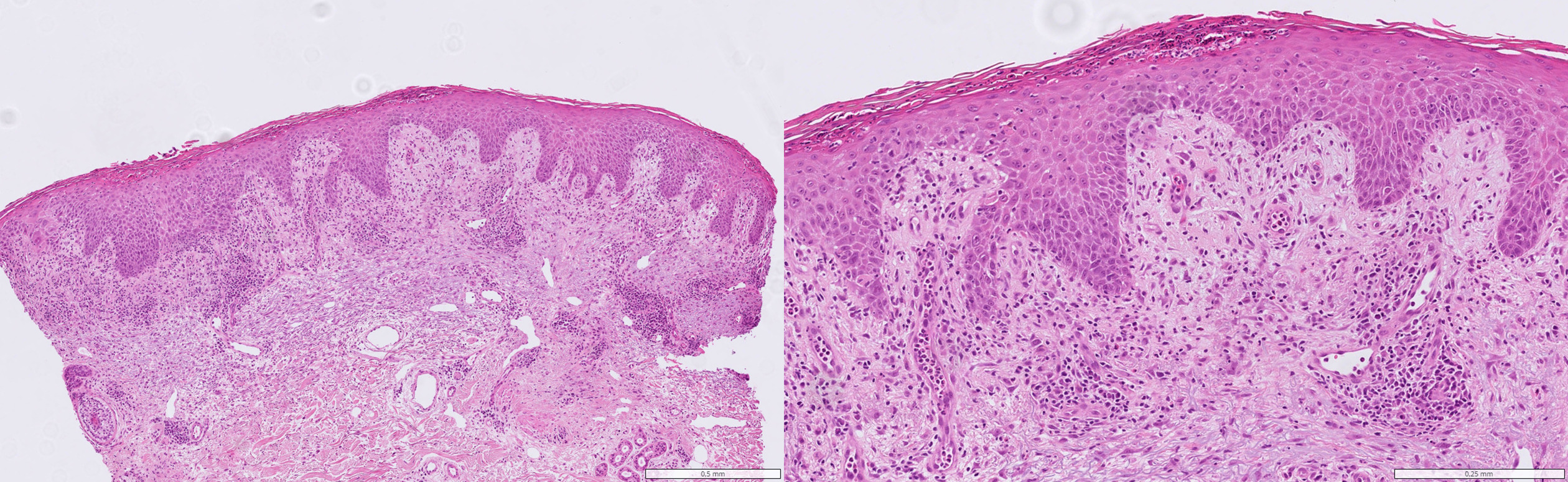
Fig. 2. Microscopic examination (htx) of the skin reveals subcorneal and intraepidermal neutrophilic pustules and associated epidermal spongiosis. A subepidermal, papillary dermal oedema is present. The superficial dermis shows an inflammatory infiltrate composed of eosinophils, plasma cells and lymphohistiocytic cells. htx, haematoxylin-eosin stain.
The initial suspicion of T-cell lymphoma could not be verified in a subsequent lymph node biopsy. As an alternative, some form of autoimmune disease was suggested in the pathology report. The final diagnosis, DRESS-syndrome, was confirmed after 7 weeks of treatment, and the patient was recommended never more to take enalapril. The steroid treatment, which had been complemented with omeprazole, trimethoprim/sulfamethoxazole, alendronate, calcium and vitamin D3, was slowly reduced. Repeated attacks of atrial fibrillation prompted several transitory treatments with rivaroxaban and atenolol. Cutaneous relapses and deranged blood chemistry lead to several adjustments of prednisolone dose. Five months after the start of prednisolone treatment, when a 4th relapse was clearly linked to reinitiation of atenolol, this was permanently terminated and prednisolone could be stopped 3 months later. All remaining drugs except alendronate, calcium and vitamin D3 were discontinued another 3 months later once the blood tests had returned to normal.
One and a half year after prednisolone treatment was finished, the patient experienced a feeling of arrhythmia. A few weeks later skin rash and night sweats with fever also returned. Again, there were obvious signs of inflammation and indications of liver damage (June 2023, Table I). Gamma-GT, which had not been elevated during the first period of the disease, was now strongly elevated. CT scan revealed multiple enlarged lymph nodes and a pulmonary infiltrate. As recurrence of the patient’s DRESS syndrome was evident, prednisolone therapy was started (40 mg daily). The effect was prompt, skin rash disappeared and inflammation markers returned to normal levels within a few days. The pulmonary infiltrate resolved. However, following several attempts with a lower dose of prednisolone, the Gamma-GT value increased again. Alendronate treatment was therefore terminated, after which all liver function tests normalized within a period of 6 months.
Patch tests with all drugs were performed 3 months after the 1st and 6 months after the 2nd termination of prednisolone. No allergy was detected. In connection with the latter test, LTT was also performed with the 2 drugs that could be analysed in the patient’s home country, ibuprofen and quinine. Both tests were negative.
In September 2025, 16 months after completing prednisolone medication, the remaining possibly responsible drugs were analysed with LTT at the IMD immunology laboratory in Berlin. The test revealed cellular sensitization to enalapril and atenolol, but not of the other drugs. Alendronate was not analysed.
DISCUSSION
Several different drugs may lead to development of DRESS syndrome in a single patient. This has been highlighted previously (6), although not with the current agents. Enalapril as well as atenolol and alendronate has been considered uncommon causes of the DRESS syndrome (7, 8, 9).
DRESS syndrome itself is an uncommon condition. The most reliable epidemiological data comes from 2 nationwide, detailed surveys in Japan 2013 and 2021 (5). The incidence of typical disease was estimated to be less than 2 per million inhabitants, and if atypical cases were included, it was between 4 and 5 per million. In Sweden, 22 patients were diagnosed with DRESS syndrome over the past 5 years (10).
The disease has a varying geographical distribution, possibly explained by the varying prevalence of the specific HLA-A and HLA-B alleles strongly associated with the development of the syndrome (8).
Cutaneous manifestations vary considerably. In the present case, it started with urticaria on the ankles, progressed to a reddened morbilliform rash on the arms, trunk, back and lower legs, and increasing swelling of the skin, then progressed to erythroderma with exfoliation involving 90% of the skin.
DRESS syndrome is often associated with the activation of a latent viral infection. This applies primarily to herpes viruses but also to cytomegalovirus and Epstein-Barr virus, as in the present case (11, 12).
It is urgent to identify the culprit drug so the patient can abstain from further exposure. It is sometimes identified from the patient history, as alendronate responsible for the late recurrence in our patient. Identification by LTT is strongly advised according to available guidelines (4, 5). The test was developed already in the 1960s and has thereafter been further refined, but there still exists a hesitance concerning this technology demanding method (13). We have followed the guidelines and have had the tests performed at an experienced laboratory (14), where the lymphocyte incubation time has been extended from 5 to 6 days as opposed to other protocols.
When there is reason to suspect that a patient has developed DRESS syndrome due to intolerance to more than one drug, it is often from chemically related agents. However, atenolol, enalapril and alendronate lack obvious chemical similarity. The current case illustrates that multiple causes of the disease should be considered, implying analysis of all medications the patient may have taken, permanently or temporarily. Our findings also emphasize the importance of performing LTT.
Acknowledgments
We appreciate the detailed analysis of dermatological manifestations performed by Dr Emelie Gunnesson, MD
REFERENCES
- Mizukawa Y, Shiohara T. Recent advances in the diagnosis and treatment of DIHS/DRESS in 2025. Allergol Int 2025; 74: 372–379. https://doi.org/10.1016/j.alit.2025.03.007
- Chen CB, Hung WK, Wang CW, Lee CC, Hung SI, Chung WH. Advances in understanding of the pathogenesis and therapeutic implications of drug reaction with eosinophilia and systemic symptoms: an updated review. Front Med 2023; 10. https://doi.org/10.3389/fmed.2023.1187937
- Wei BM, Fox LP, Kaffenberger BH, Korman AM, Micheletti RG, Mostaghimi A, et al. Drug-induced hypersensitivity syndrome/drug reaction with eosinophilia and systemic symptoms. Part II diagnosis and management. J Am Acad Dermatol 2024; 90: 911–926. https://doi.org/10.1016/j.jaad.2023.02.073
- Cabañas R, Ramírez E, Sendagorta E, Alamar R, Barranco R, Blanca-López N, et al. Spanish guidelines for diagnosis, management, treatment, and prevention of DRESS syndrome. J Investig Allergol Clin Immunol 2020; 30: 229–253. https://doi.org/10.18176/jiaci.0480
- Mizukawa Y, Hama N, Niihara H, Miyagawa F, Hashizume H, Tohyama M, et al. Guidelines for the management of drug‐induced hypersensitivity syndrome 2023. J Dermatol 2025; 52. https://doi.org/10.1111/1346-8138.17609
- Picard D, Vellar M, Janela B, Roussel A, Joly P, Musette P. Recurrence of drug-induced reactions in DRESS patients. J Eur Acad Dermatol Venereol 2015; 29: 801–804. https://doi.org/10.1111/jdv.12419
- Cabañas R, Calderón O, Ramírez E, Fiandor A, Caballero T, Heredia R, et al. Sensitivity and specificity of the lymphocyte transformation test in drug reaction with eosinophilia and systemic symptoms causality assessment. Clin Exp Allergy 2018; 48: 325–333. https://doi.org/10.1111/cea.13076
- Wei BM, Fox LP, Kaffenberger BH, Korman AM, Micheletti RG, Mostaghimi A, et al. Drug-induced hypersensitivity syndrome/drug reaction with eosinophilia and systemic symptoms. Part I. Epidemiology, pathogenesis, clinicopathological features, and prognosis. J Am Acad Dermatol 2024; 90: 885–908. https://doi.org/10.1016/j.jaad.2023.02.072
- Confavreux CB, Canoui-Poitrine F, Schott AM, Ambrosi V, Tainturier V, Chapurlat RD. Persistence at 1 year of oral antiosteoporotic drugs: a prospective study in a comprehensive health insurance database. Eur J Endocrinol 2012; 166: 735–741. https://doi.org/10.1530/EJE-11-0959
- Personal message. Swedish Medical Products Agency’s Adverse Reactions Department; 2025Dec.
- Descamps V, Valance A, Edlinger C, Fillet AM, Grossin M, Lebrun-Vignes B, et al. Association of human herpesvirus 6 infection with drug reaction with eosinophilia and systemic symptoms. Arch Dermatol 2001; 137: 301–304. https://doi.org/10.1001/archderm.137.3.301
- Zhu H, Ren V. Immunopathogenic insights on preferential human herpesvirus-6 reactivation in drug rash with eosinophilia and systemic symptoms: a scoping review. J Cutan Med Surg 2023; 27: 388–398. https://doi.org/10.1177/12034754231177590
- Sachs B, Fatangare A, Sickmann A, Glässner A. Lymphocyte transformation test: history and current approaches. J Immunol Methods 2021; 493: 113036. https://doi.org/10.1016/j.jim.2021.113036
- Chu C, Schönbrunn A, Elitok S, Kern F, Schnatbaum K, Wenschuh H, et al. T-cell proliferation assay for the detection of SARS-CoV-2-specific T-cells. Clinica Chimica Acta 2022; 532: 130–136. https://doi.org/10.1016/j.cca.2022.05.025