SHORT COMMUNICATION
Periocular Primary Cutaneous Mucinous Carcinoma in a Young Asian Male: A Rare Presentation
Cuirong XIAO1,2,3,4,5,6† , Zhixun XIAO1,3,4,5,6†, Qiuyun XU1,3,4,5,6, Jiawen CHEN1,3,4,5,6 and Bo CHENG1,3,4,5,6*
1Department of Dermatology, The First Affiliated Hospital of Fujian Medical University, Fuzhou, Fujian, China, 2Department of Dermatology, Zhangzhou Affiliated Hospital of Fujian medical University, Xiangcheng District, Zhangzhou, Fujian, China, 3Institute of Dermatology and Venereology, Fujian Medical University, Fuzhou, Fujian, China, 4Key Laboratory of Skin Cancer of Fujian higher education institutions, The Fujian Medical University, Fuzhou, Fujian, China, 5Fujian Dermatology and Venereology Research Institute, The First Affiliated Hospital, Fujian Medical University, Fuzhou, Fujian, China, and 6Fujian Provincial Clinical Research Center for Immune Skin Diseases, The First Affiliated Hospital of Fujian Medical University, Fuzhou, Fujian, China. *Email: chengbo630415@126.com
†These authors contributed equally to this work.
Citation: Acta Derm Venereol 2026; 106: adv-2026-0591. DOI: https://doi.org/10.2340/actadv.v106.adv-2026-0591.
Copyright: 2026 ©Author(s). Published by MJS Publishing, on behalf of the Society for Publication of Acta Dermato-Venereologica. This is an Open Access article distributed under the terms of the Creative Commons Attribution-NonCommercial 4.0 International License (https://creativecommons.org/licenses/by-nc/4.0/).
Submitted: Apr 7, 2026. Accepted after revision: Aug 25, 2026.
Published: Sept 16, 2026.
Competing interests and funding: The authors have no conflicts of interest to declare.
The National Natural Science Fund Youth Fund of China (No. 82573966).
The data that support the findings of this study are available from the corresponding author upon reasonable request.
This study was conducted in accordance with the Declaration of Helsinki. Written informed consent for publication of clinical details and photographs was obtained from the patient and has been uploaded as a supplementary file.
Primary cutaneous mucinous carcinoma (PCMC) is an extremely rare adnexal tumour originating from eccrine sweat glands, with an estimated incidence of 0.07 per million person-years (1). As of 2024, approximately 380 cases have been documented in the global literature, predominantly affecting elderly White individuals with a marked predilection for the periorbital region (2). Although PCMC typically exhibits indolent behaviour with distant metastasis in less than 3% of cases, local recurrence rates approach 30% in the absence of adequate margin control (2, 3). We report a rare case of periocular PCMC in a 42-year-old Asian male, highlighting the diagnostic challenges in atypical demographics and the pivotal role of margin-controlled excision.
CASE REPORT
A 42-year-old Asian male presented with a 3-year history of a slowly growing, asymptomatic left facial nodule. Physical examination identified a 1.2×1×0.8 cm firm cystic nodule with surface telangiectasia and translucent skin in the left infraorbital region, 0.5 cm below the lower eyelid margin, without eyelid involvement (Fig. 1). No lymphadenopathy or systemic symptoms were observed.
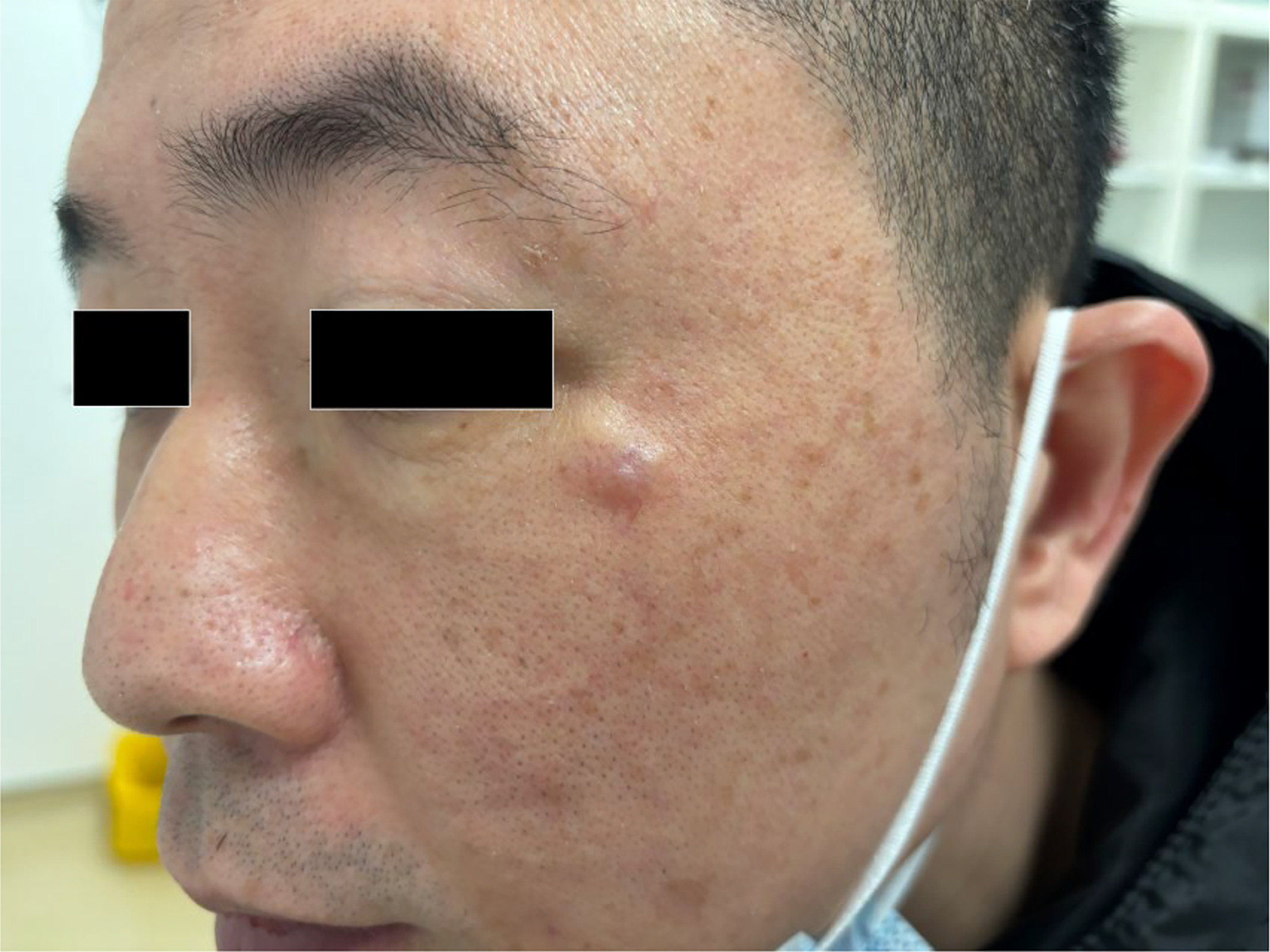
Fig. 1. Clinical and histopathological findings. A 1.2 cm firm, cystic nodule with telangiectasia on the left infraorbital region, approximately 0.5 cm below the lower eyelid margin, without involvement of the eyelid.
Pathological examination of the initially excised lesion revealed well-demarcated dermal tumor islands consisting of monomorphic basaloid cells within abundant extracellular mucin, consistent with the classic “blue islands in a mucinous sea” morphology (Fig. 2A and B). Alcian blue staining confirmed acidic mucin deposition (Fig. 2C). Immunohistochemical staining showed diffuse CK7 positivity, with focal CEA and EMA reactivity, whereas CK20, S100, GCDFP15, GATA3, ER and PR were entirely negative (Fig. 2 D–F). Systemic workup was performed to exclude occult visceral malignancy. Contrast-enhanced chest, abdominal and pelvic CT showed no primary tumor or regional lymphadenopathy. Upper gastrointestinal endoscopy and colonoscopy revealed only chronic gastritis and benign colonic polyps. Combined negative imaging, endoscopic results and targeted immunohistochemical panel excluded pulmonary, gastrointestinal and breast primary malignancies. No regional lymph node abnormalities were detected on clinical palpation or radiological assessment, and serum tumor markers (CEA, CA19-9, CA125) were all within normal ranges.
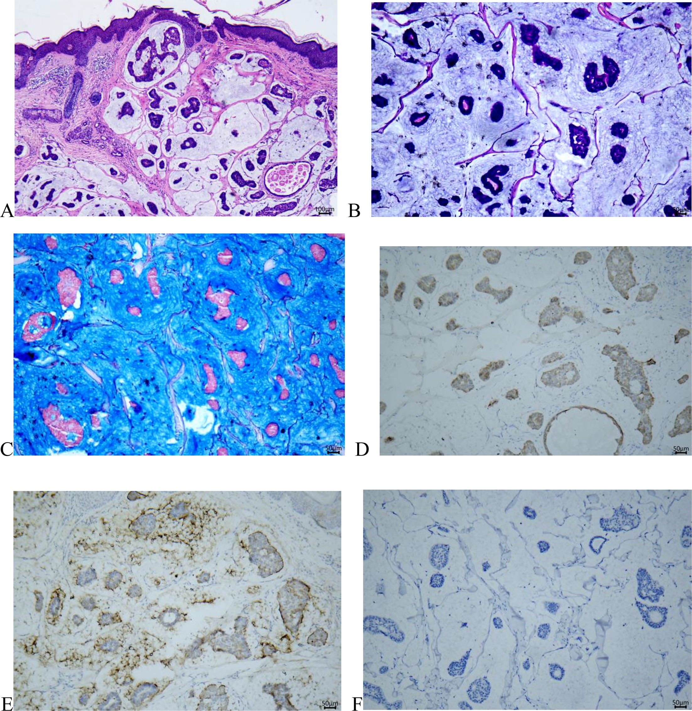
Fig. 2. Histopathological, histochemical and immunohistochemical findings of the cutaneous lesion. (A) Haematoxylin and eosin (H&E) staining (IHC, ×25). Basket-weave hyperkeratosis is visible in the epidermis; the epidermal layer is largely unremarkable, with discrete tumour islands present within the dermis. (B) H&E staining (IHC, ×100). Well-circumscribed dermal tumour islands consisting of monomorphic basaloid epithelial cells suspended within abundant extracellular mucin, demonstrating the characteristic “blue islands in a mucinous sea” morphology. (C) Alcian blue staining (IHC, ×200), verifying abundant deposition of acidic mucin in the tumoral stroma. (D) Immunohistochemistry (IHC) showing diffuse cytoplasmic CK7 positivity within tumour cells (IHC, ×200). (E) IHC demonstrating focal EMA expression in tumour cells (IHC, ×200). (F) IHC revealing negative CK20 staining in neoplastic cells (IHC, ×200).
Initial excision with 2 mm clinical margins revealed tumour involvement at the deep margin. Mohs micrographic surgery was subsequently performed with intraoperative frozen section analysis confirming complete excision. The surgical defect was closed primarily with excellent functional and cosmetic outcome. At 1 year follow-up, no local recurrence or distant metastasis was observed.
DISCUSSION
Mucinous carcinoma in the skin poses a diagnostic dilemma, as it more commonly represents metastasis from an occult visceral primary than a primary cutaneous neoplasm (4). This distinction carries profound prognostic and therapeutic implications. Our case illustrates a systematic immunohistochemical approach to this challenge.
Cutaneous mucinous carcinoma creates a critical diagnostic dilemma, as most cutaneous lesions represent metastasis from occult visceral primaries rather than primary cutaneous mucinous carcinoma (PCMC), with significant implications for clinical prognosis and treatment strategies (4). While the CK7+/CK20- immunoprofile is characteristic of PCMC, this expression pattern is also shared with mucinous carcinomas of breast and lung origin, necessitating the use of additional lineage-specific immunohistochemical markers for accurate differentiation(4). In our case, all breast-specific markers (ER, PR, GCDFP15, GATA3) and gastrointestinal-specific markers (CK20, CDX2, SATB2) yielded negative results. Combined with unremarkable systemic imaging and endoscopic findings, these results confirmed a primary cutaneous origin with no evidence of metastasis. PCMC diagnosis relies on comprehensive integration of histopathological, immunohistochemical, clinical and systemic findings, rather than reliance on a single biomarker. Although auxiliary cutaneous markers p63 and CK5/6 (expressed in 40% and 20% of PCMC cases, respectively) were not tested in this study (4, 5), this minor limitation did not compromise diagnostic accuracy due to our comprehensive diagnostic panel.
PCMC predominantly affects elderly White individuals, with a mean age of 63.5 years, while Asian patients account for only 12.7% of published cases, and young adult onset is extremely uncommon (2, 6). Our 42-year-old Asian patient is consistent with several previously reported rare young-onset cases, including a 33-year-old Chinese patient and a 29-year-old patient with misdiagnosed eyelid PCMC (7, 8). These cases expand the demographic spectrum of PCMC, indicating that this tumor should be considered in the differential diagnosis of indolent facial nodules in Asian patients regardless of age. Due to its infiltrative growth pattern and poorly defined clinical margins, PCMC exhibits a 30% local recurrence rate following conventional excision, particularly in anatomically sensitive periocular regions (3, 6). Mohs micrographic surgery (MMS) allows complete margin assessment and maximal preservation of healthy tissue, and is currently the preferred surgical modality for PCMC (3). Our patient achieved negative surgical margins and satisfactory functional and cosmetic outcomes after conversion to MMS, consistent with previous studies demonstrating the technical challenges of margin control in periocular PCMC (2).
Despite its indolent clinical behavior with a distant metastasis rate of less than 3%, PCMC requires prolonged long-term surveillance owing to the risk of late recurrence, which may occur more than 10 years after initial treatment (9). Meta-analytic data indicate that tumor size greater than 1.5 cm and truncal location predict poor prognosis, while Asian ethnicity may be associated with favorable clinical outcomes, though this association requires further large-cohort validation (6). Our patient currently follows a strict surveillance protocol with quarterly clinical examinations and annual imaging to monitor regional or distant tumor spread. In summary, this case highlights the occurrence of PCMC in young Asian patients. Standardized immunohistochemical panels effectively exclude metastatic disease, and MMS optimizes oncological safety and cosmetic outcomes for periocular PCMC. Clinicians should maintain a high index of suspicion for PCMC when evaluating persistent periocular nodules in young Asian populations.
REFERENCES
- Breiting L, Christensen L, Dahlstrøm K, Breiting V, Winther JF. Primary mucinous carcinoma of the skin: a population-based study. Int J Dermatol 2008; 47: 242–245. https://doi.org/10.1111/j.1365-4632.2008.03558.x
- Park TJ, Chung DH, Kim L, Hong MK. Primary mucinous carcinoma of the lower eyelid treated with wide excision and postoperative radiotherapy: a case report and literature review. Arch Craniofac Surg 2024; 25: 292–297. https://doi.org/10.7181/acfs.2024.00059
- Kelly Á, Anand R, Natkunarajah J, Samarasinghe V, Craythorne E. Mohs micrographic surgery for the treatment of primary cutaneous mucinous carcinoma. Clin Exp Dermatol 2023; 48: 778–780. https://doi.org/10.1093/ced/llad057
- Levy G, Finkelstein A, McNiff JM, Bolognia J, Bosenberg M, Ko CJ. Immunohistochemical techniques to compare primary vs. metastatic mucinous carcinoma of the skin. J Cutan Pathol 2010; 37: 411–415. https://doi.org/10.1111/j.1600-0560.2009.01436.x
- Qureshi HS, Salama ME, Chitale D, Bansal I, Ma CK, Raju U, et al. Primary cutaneous mucinous carcinoma: presence of myoepithelial cells as a clue to the cutaneous origin. Am J Dermatopathol 2004; 26: 353–358. https://doi.org/10.1097/00000372-200410000-00001
- Kamalpour L, Brindise RT, Nodzenski M, Bach DQ, Veledar E, Alam M. Primary cutaneous mucinous carcinoma: a systematic review and meta-analysis of outcomes after surgery. JAMA Dermatol 2014; 150: 380–384. https://doi.org/10.1001/jamadermatol.2013.6006
- Liu RF, Liu D, Wang F, Yao LF, Zhang KM. Primary cutaneous mucinous carcinoma in a young patient. Chin J Dermatol 2019; 52: 776–777.
- Alazawi SS, Katana VG. Young man with primary mucinous cell carcinoma of the eyelid: 36 month follow up. Mil Med 2024; 189: e2719–e2721. https://doi.org/10.1093/milmed/usad477
- Marra DE, Schanbacher CF, Torres A. Mohs micrographic surgery of primary cutaneous mucinous carcinoma using immunohistochemistry for margin control. Dermatol Surg 2004; 30: 799–802. https://doi.org/10.1111/j.1524-4725.2004.30223.x