SHORT COMMUNICATION
Anifrolumab in Refractory Discoid Lupus Erythematosus without Systemic Involvement: A Case Series of Five Patients
Jasmin MARSCHNER1 and Franziska SCHAUER1*
1Department of Dermatology, Medical Center-University of Freiburg, Faculty of Medicine, Hauptstraße 7, DE-79104 Freiburg, Germany. *Email: franziska.schauer@uniklinik-freiburg.de
Citation: Acta Derm Venereol 2026; 106: adv-2026-0559. DOI: https://doi.org/10.2340/actadv.v106.adv-2026-0559.
Copyright: 2026 ©Author(s). Published by MJS Publishing, on behalf of the Society for Publication of Acta Dermato-Venereologica. This is an Open Access article distributed under the terms of the Creative Commons Attribution-NonCommercial 4.0 International License (https://creativecommons.org/licenses/by-nc/4.0/).
Submitted: Apr 1, 2026. Accepted after revision: Jul 2, 2026.
Published: Sept 17, 2026.
Competing interests and funding: The data that support the findings of this study are available from the corresponding author upon reasonable request.
Written informed consent was obtained from all patients.
FS is Principle Investigator in the Lavender Study on cutaneous lupus erythematodes and treatment with anifrolumab. The Phase III study begann later than the patients discussed here.
Discoid lupus erythematosus (DLE) is a chronic form of cutaneous lupus erythematosus (CLE), characterized by scarring, erythematous plaques and pigmentation changes. CLE may occur alone or with systemic lupus erythematosus (SLE) and treatments for both often overlap.
No new FDA- or EMA-approved systemic therapies for CLE have been approved for CLE in more than 60 years. Hydroxychloroquine and corticosteroids remain the only approved systemic treatments (1). Other immunosuppressive agents – mycophenolate mofetil, methotrexate and dapsone – are widely used off-label but are often poorly tolerated or ineffective, especially in treatment-resistant disease (2).
Anifrolumab received EMA approval in 2022 for moderate-to-severe SLE (3, 4, 5). Post hoc analyses of the TULIP trials demonstrated significant improvements in skin manifestations, as measured by the Cutaneous Lupus Erythematosus Disease Area and Severity Index (CLASI) (6). Given the central role of the type I interferon pathway in lupus pathogenesis, anifrolumab is a promising candidate, though real-world evidence remains limited (1, 5).
This case series presents 5 patients with refractory CLE treated with anifrolumab and discusses clinical outcomes, safety and broader potential therapeutic implications.
CASE REPORTS
Patient: Midface DLE resistant to multiple treatments. A 47-year-old woman presented with persistent, sharply demarcated erythematous plaques on her nose and cheeks for 12 years (Fig. 1A). Multiple conventional therapies – Hydroxychloroquine, methotrexate, azathioprine, mycophenolate mofetil and laser ablation – had failed (Table SI). At the start of anifrolumab therapy, her CLASI-A score was 10 and her CLASI-D score 4. After 3 infusions, CLASI-A score dropped to 1 and CLASI-D score to 2, with significant improvement in quality of life and no adverse events (Fig. 1B). After 12 months of therapy, patient 1 requested a break from treatment but suffered a relapse as early as the fourth month, so treatment was resumed.
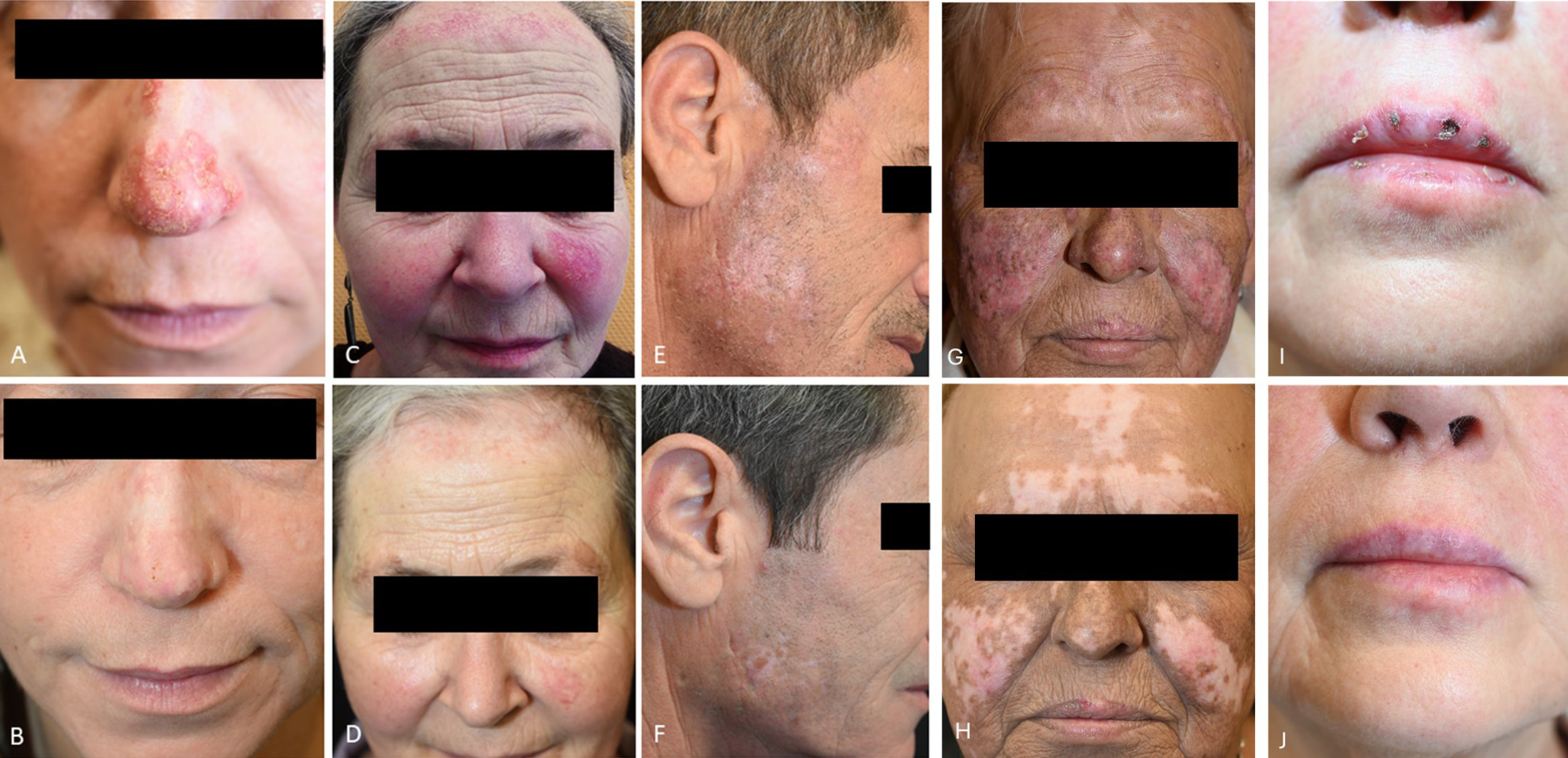
Fig. 1. (A) Patient 1: discoid, partially scarred erythematous plaques on the nose. (B) Marked improvement after 3 anifrolumab infusions. (C) Patient 2: scarring alopecia and cutaneous lupus erythematosus involving the forehead and left cheek. (D) Near skin-coloured plaques with mild atrophy after 5 infusions. (E) Patient 3: discoid plaques in the right preauricular area. (F) Hypopigmented scars without active inflammation after 3 infusions. (G) Centrofacial erythematous plaques with scarring consistent with active cutaneous lupus. (H) Residual scarring with resolution of erythema after treatment. (I) Erosive cheilitis with crusting and erythema prior to anifrolumab. (J) Complete lesion clearance with residual scarring after therapy.
Patient 2: Scarring DLE of the scalp and face. A 64-year-old woman had treatment-resistant DLE affecting her face and scalp since the 1980s, with progressive scarring and hair loss despite numerous therapies. Antimalarials, methotrexate, azathioprine, mycophenolate mofetil and corticosteroid pulse therapy were either ineffective or poorly tolerated (Table SI). At the time of anifrolumab initiation, CLASI-A score was 19, CLASI-D was 3, reflecting severe disease activity (Fig. 1C). Skin lesions improved significantly after initiation of anifrolumab. CLASI-A dropped to 5, while CLASI-D remained stable (Fig. 1D). Nonetheless, progression of alopecia on the scalp was observed. It remains uncertain whether this was attributable to disease activity, post-inflammatory remodeling or a potential adverse effect of anifrolumab (Table SI). Nonetheless, the patient reported significant improvement of quality of life.
Patient 3: Refractory facial and auricular DLE with severe pruritus. A 57-year-old man experienced persistent, intensely pruritic DLE lesions on his face, neck and ears for 10 years. Multiple systemic therapies, including hydroxychloroquine, azathioprine, methotrexate, mycophenolate, mycophenolic acid, tofacitinib and corticosteroid pulses as well as participation in a clinical trial had all failed (CLASI-A score 11, CLASI-D score 2, Fig. 1E). After 8 years of disease activity, initiation of anifrolumab led to almost complete clearance of his skin lesions (CLASI-A 0, CLASI-D 1, Fig. 1F), pruritus resolved and overall disease burden decreased substantially. The treatment was well tolerated without side-effects.
Patient 4: Early Anifrolumab use in DLE with anti-dsDNA positivity without full systemic criteria. A 61-year-old female presented with persistent centrofacial erythematous plaques and diffuse alopecia beginning in 2020, consistent with cutaneous lupus. Despite anti-dsDNA and anti-SS-A antibody positivity, she did not fulfil EULAR/ACR classification criteria for SLE. Initial therapy with hydroxychloroquine, followed by methotrexate provided limited efficacy, with ongoing disease activity and scarring alopecia. Off-label treatment with anifrolumab was initiated in April 2024 (CLASI-A 4, CLASI-D 5, Fig. 1G), resulting in a notable reduction of inflammatory activity. Residual hypopigmentation persisted (CLASI-A 0, CLASI-D 5, Fig. 1H). No adverse events occurred. Hydroxychloroquine and methotrexate were later reintroduced due to concern for systemic progression (Table SI).
Patient 5: DLE with refractory erosive and scarring labial involvement. A 58-year-old woman was diagnosed with DLE, presenting with partially scarring erythematous plaques on the face, décolleté and back, as well as circumscribed scalp alopecia. Since 2020, she had experienced recurrent erosive and crusted lip lesions with subsequent scarring. Histopathology of the lips in 2021 and 2025 confirmed chronic DLE without malignancy; herpes simplex virus infection was repeatedly excluded. Labial involvement remained refractory to hydroxychloroquine, corticosteroid pulse therapy, methotrexate, azathioprine, mycophenolate mofetil and upadacitinib. Due to therapy-resistant labial involvement, anifrolumab was initiated in October 2025 (CLASI-A 3, CLASI-D 8, Fig. 1I). During the first 2 infusions, the patient experienced chills and arthralgia, which resolved spontaneously. Subsequent infusions were well tolerated. One episode of cystitis occurred during the treatment course and was successfully treated with antibiotics. After 6 months, labial inflammation had resolved completely (CLASI-A 0, Fig. 1J), with permanent scarring remaining at previously affected sites (CLASI-D 8).
DISCUSSION
Several case reports and small case series have documented the off-label use of anifrolumab in refractory CLE and DLE, with consistently favourable outcomes (1, 5). However, most included patients with concomitant SLE or a heterogeneous mix of CLE subtypes; real-world evidence in isolated, treatment-refractory DLE without systemic involvement remains scarce. Type I IFN signalling is central to both SLE and CLE pathogenesis (7, 8). Even histologically normal skin in CLE can exhibit a strong IFN signature, indicating a primed inflammatory state (9, 10). IFN-α disrupts the epidermal barrier, induces keratinocyte apoptosis and recruits autoreactive immune cells, leading to chronic cutaneous inflammation and scarring (10, 11, 12). By attenuating these effects, anifrolumab may suppress IFN-driven skin inflammation even in the absence of systemic disease – supporting the expansion of its therapeutic use to isolated CLE (6).
Our series addresses this gap by focusing on histologically confirmed, isolated DLE. It is noteworthy that this group of patients had suffered from a long-standing condition and had not responded to several systemic therapies, including conventional immunosuppressants and JAK inhibitors; nevertheless, anifrolumab (300 mg monthly) demonstrated high efficacy within 3 months. One patient presented with severe, debilitating pruritus as a predominant symptom – a feature rarely emphasized in published DLE series – which resolved markedly. We further include a diagnostically challenging patient with severe skin hyperpigmentation associated with hydroxychloroquine and anti-dsDNA positivity without fulfilment of EULAR/ACR classification criteria for SLE, as well as a rare case of treatment-resistant erosive cheilitis responding to anifrolumab.
Anifrolumab became available in the EU as a subcutaneous formulation in December 2025, allowing for patient to self-administer treatment. Phase III data demonstrated comparable efficacy and safety to the intravenous formulation, offering a more convenient option for long-term treatment. Given the limited therapeutic options for refractory DLE, these developments may further expand the clinical applicability of Anifrolumab in everyday practice. It remains unclear how long therapy should be continued in patients with CLE, as for Pt. 1 discontinuation of therapy appears to have led to a relapse.
Several questions also remain unanswered, including predictive biomarkers of response, long-term safety and comparisons with alternative targeted therapies such as JAK inhibitors. Our findings must also be interpreted in the context of the inherent limitations of the small sample size, absence of a comparator group, potential confounding from reintroduction of concomitant systemic therapy in 1 patient. Larger prospective controlled studies are awaited to define the efficacy, safety and optimal positioning of anifrolumab in future treatment algorithms for refractory DLE.
In conclusion, anifrolumab appears to be a promising therapeutic option for patients with longstanding, treatment-resistant DLE.
REFERENCES
- Xie L, Lopes Almeida Gomes L, Stone CJ, Faden DF, Werth VP. An update on clinical trials for cutaneous lupus erythematosus. J Dermatol 2024; 51: 885–894. https://doi.org/10.1111/1346-8138.17161
- Deutsche Gesellschaft für Rheumatologie e.V., Deutsche Dermatologische Gesellschaft e.V., Gesellschaft für Pädiatrische Dermatologie. Diagnostik und Therapie des kutanen Lupus erythematodes (S2k-Leitlinie); 2020. [cited 2025 April 28]. Available from: https://www.awmf.org/uploads/tx_szleitlinien/013-060l_S2k_Kutaner_Lupus_erythematodes_2020-06-verlaengert.pdf
- Furie R, Khamashta M, Merrill JT, Werth VP, Kalunian K, Brohawn P, et al. Anifrolumab, an anti–interferon‐α receptor monoclonal antibody, in moderate-to-severe systemic lupus erythematosus. Arthritis Rheumatol 2017; 69: 376–386. https://doi.org/10.1002/art.39962
- Furie RA, Morand EF, Bruce IN, Manzi S, Kalunian KC, Vital EM, et al. Type I interferon inhibitor anifrolumab in active systemic lupus erythematosus (TULIP-1): a randomised, controlled, phase 3 trial. Lancet Rheumatol 2019; 1: e208–e219. https://doi.org/10.1016/S2665-9913(19)30076-1
- Plüß M, Piantoni S, Wincup C, Korsten P. Rapid response of refractory systemic lupus erythematosus skin manifestations to anifrolumab—a case-based review of clinical trial data suggesting a domain-based therapeutic approach. J Clin Med 2022; 11: 3449. https://doi.org/10.3390/jcm11123449
- Morand EF, Furie RA, Bruce IN, Vital EM, Dall’Era M, Maho E, et al. Efficacy of anifrolumab across organ domains in patients with moderate-to-severe systemic lupus erythematosus: a post-hoc analysis of pooled data from the TULIP-1 and TULIP-2 trials. Lancet Rheumatol 2022; 4: e282–e292. https://doi.org/10.1016/S2665-9913(21)00317-9
- Baechler EC, Batliwalla FM, Karypis G, Gaffney PM, Ortmann WA, Espe KJ, et al. Interferon-inducible gene expression signature in peripheral blood cells of patients with severe lupus. Proc Natl Acad Sci USA 2003; 100: 2610–2615. https://doi.org/10.1073/pnas.0337679100
- Braunstein I, Klein R, Okawa J, Werth VP. The interferon-regulated gene signature is elevated in subacute cutaneous lupus erythematosus and discoid lupus erythematosus and correlates with the cutaneous lupus area and severity index score. Br J Dermatol 2012; 166: 971–975. https://doi.org/10.1111/j.1365-2133.2012.10825.x
- Billi AC, Ma F, Plazyo O, Gharaee-Kermani M, Wasikowski R, Hile GA, et al. Non-lesional and lesional lupus skin share inflammatory phenotypes that drive activation of CD16+ dendritic cells. Immunology. Preprinted posted online 2021doi: 10.1101/2021.09.17.460124.
- Hile GA, Werth VP. Understanding the role of type I interferons in cutaneous lupus and dermatomyositis: toward better therapeutics. Arthritis Rheumatol 2025; 77: 1–11. https://doi.org/10.1002/art.42983
- Niewold TB. Interferon alpha as a primary pathogenic factor in human lupus. J Interferon Cytokine Res 2011; 31: 887–892. https://doi.org/10.1089/jir.2011.0071
- Rönnblom L, Leonard D. Interferon pathway in SLE: one key to unlocking the mystery of the disease. Lupus Sci Med 2019; 6: e000270. https://doi.org/10.1136/lupus-2018-000270