SHORT COMMUNICATION
Thymoma-associated Multiorgan Autoimmunity Presenting as Severe Psoriasiform Keratotic Erythroderma 35 Years after Thymoma Diagnosis
Kazuki HIGUCHI1, Mika WATANABE1* , Sho KATAYAMA1, Takamasa ITO1 and Hideyuki UJIIE1
1Department of Dermatology, Faculty of Medicine and Graduate School of Medicine, Hokkaido University, Sapporo, Japan. *Email: mika.watanabe@med.hokudai.ac.jp
Key words: Psoriasiform erythroderma; Thymoma-associated multiorgan autoimmunity.
Citation: Acta Derm Venereol 2026; 106: adv-2026-0814. DOI: https://doi.org/10.2340/actadv.v106.adv-2026-0814.
Copyright: 2026 ©Author(s). Published by MJS Publishing, on behalf of the Society for Publication of Acta Dermato-Venereologica. This is an Open Access article distributed under the terms of the Creative Commons Attribution-NonCommercial 4.0 International License (https://creativecommons.org/licenses/by-nc/4.0/).
Submitted: Jun 22, 2026. Accepted after revision: Sept 2, 2026.
Published: Sept 22, 2026.
Competing interests and funding: The authors have no conflicts of interest to declare.
The data that support the findings of this study are available from the corresponding author upon reasonable request.
Thymoma-associated multiorgan autoimmunity (TAMA) is a graft-versus-host disease (GVHD)-like syndrome that occurs in patients with thymoma and involves the skin, liver and/or gastrointestinal tract (1). Cutaneous manifestations include diffuse erythema, papulosquamous or psoriasiform eruptions and erythroderma, sometimes mimicking more common inflammatory dermatoses. A prolonged interval between thymoma diagnosis and TAMA has been reported, and a previous case described a latency of 23 years (2, 3). Presented here is a patient with severe psoriasiform keratotic erythroderma diagnosed as TAMA 35 years after thymoma diagnosis.
CASE REPORT
A 72-year-old man was referred for generalized erythema lasting more than 1 month. He had diffuse keratotic erythema, total scalp alopecia and nail dystrophy, and had initially been treated as psoriasis without improvement. His medical history was notable for myasthenia gravis at 35 years of age and thymoma at 37 years of age for which he underwent thymectomy in the same year. He had been receiving prednisolone 5 mg/day every other day and tacrolimus 2 mg/day long-term for myasthenia gravis. Physical examination showed almost generalized psoriasiform keratotic erythroderma with thick scales (Fig. 1). Laboratory tests revealed leukocytosis, hypoalbuminaemia, renal dysfunction and inflammatory activity. A skin biopsy from a keratotic erythematous lesion on the abdomen showed parakeratosis, acanthosis, interface dermatitis and scattered necrotic keratinocytes, consistent with an acute GVHD-like pattern (Fig. 2). Based on pleural thickening seen on contrast-enhanced computed tomography (Appendix S1), the patient was diagnosed with recurrent thymoma, stage IVa, which was subsequently confirmed histopathologically. Because he had no history of hematopoietic stem-cell transplantation, the combination of recur-rent thymoma and GVHD-like histology led to the diagnosis of TAMA. Topical clobetasol propio-nate was started after admission. However, his respiratory condition worsened on hospital day 18, and meropenem, trimethoprim-sulfamethoxazole, ganciclovir and high-dose prednisolone were initiated. On hospital day 21, he was diagnosed with MRSA bacteraemia and cytomegalovirus pneumonia. The keratotic eruption subsequently regressed, but his overall condition deteriorated, and he died 32 days after admission.
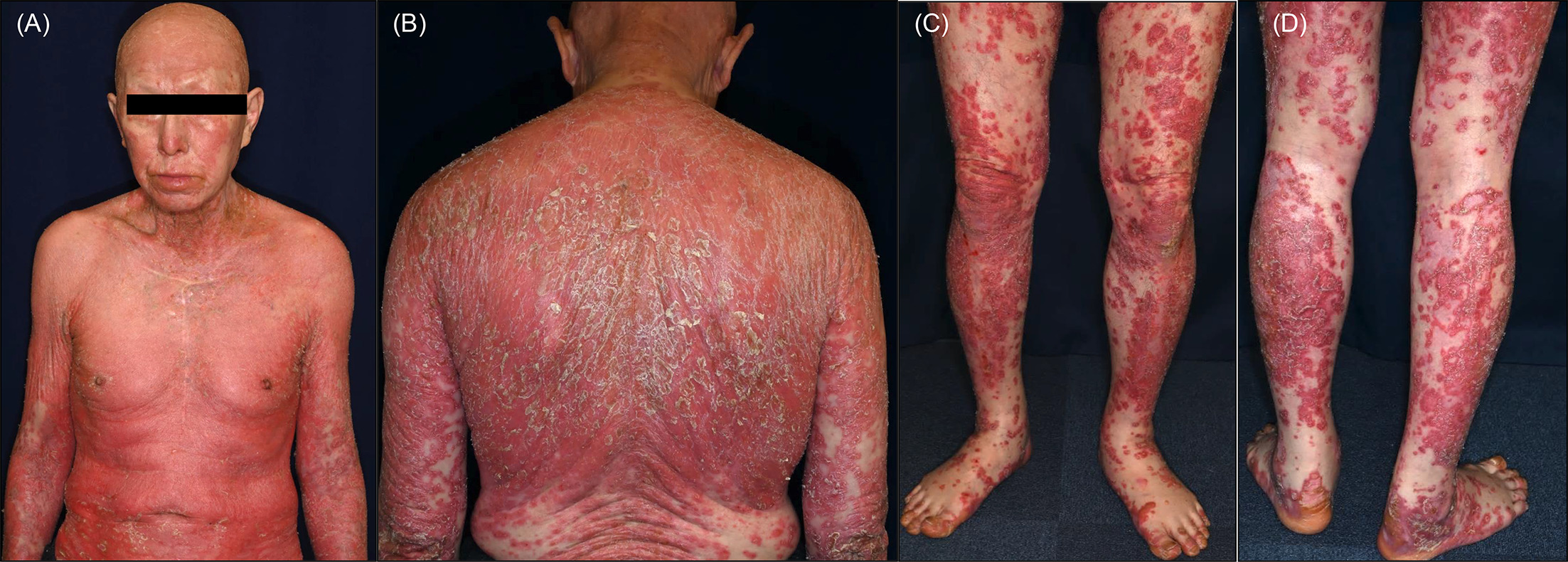
Fig. 1. Clinical features at presentation. (A) Frontal view showing generalized psoriasiform keratotic erythema with thick scales on the trunk and upper extremities. (B) Dorsal view showing confluent scaly erythema over the back. (C, D) Lower extremities showing diffuse erythematous scaly plaques with marked hyperkeratosis.
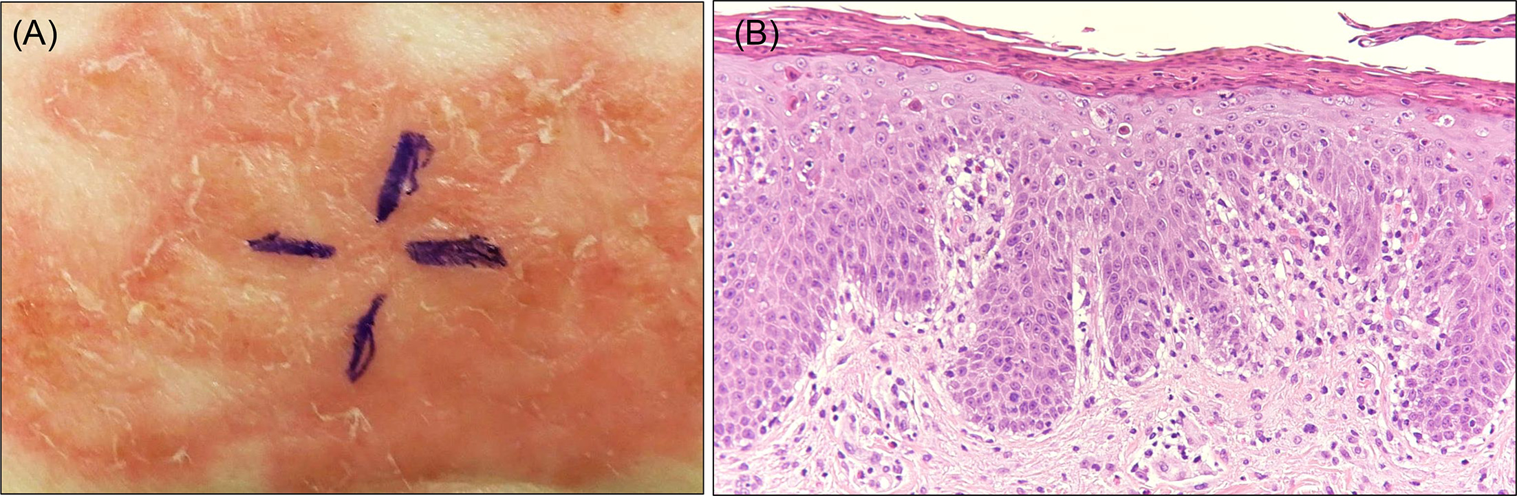
Fig. 2. Skin biopsy findings. (A) Clinical appearance of the abdominal lesion selected for biopsy. (B) Histopathological findings showing parakeratosis, acanthosis and interface changes with scattered necrotic keratinocytes, consistent with a GVHD-like pattern. Haematoxylin-eosin stain, ×200.
DISCUSSION
This case is notable for 3 reasons. First, the latency between the diagnosis of thymoma and the onset of cutaneous TAMA was 35 years. To our knowledge, the longest previously reported latency between thymoma and TAMA onset was 23 years (2, 3). Our case extends this by more than a decade, further broadening the recognized temporal window for TAMA development. Second, the patient had long-standing myasthenia gravis and chronic immunosuppressive therapy before TAMA became clinically apparent. As thymoma is associated with multiple autoimmune phenomena, this case suggests that thymoma-associated immune dysregulation may persist for decades, even under chronic immunosuppression. Third, the clinical course was rapidly fatal after the onset of extensive cutaneous involvement. Cutaneous TAMA has been proposed as a “fatal sign” (4), and a recent review reported no statistically significant difference in survival but a negative trend in outcomes among patients with skin involvement (5). Our patient died 32 days after ad-mission, supporting the view that extensive cutaneous TAMA may indicate severe systemic disease and an unfavourable prognosis.
In summary, dermatologists should consider TAMA in patients with refractory psoriasiform erythroderma, GVHD-like histology and even a remote history of thymoma.
ACKNOWLEDGEMENTS
The authors thank Dr. Yasuko Kenmotsu of Kobayashi Dermatology Clinic for referring this patient to our department and for the clinical collaboration in his care.
REFERENCES
- Wadhera A, Maverakis E, Mitsiades N, Lara PN, Fung MA, Lynch PJ. Thymoma-associated multiorgan autoimmunity: a graft-versus-host-like disease. J Am Acad Dermatol 2007; 57: 683–689. https://doi.org/10.1016/j.jaad.2007.02.027
- Shiba K, Fujita Y, Miyazawa H, Muramatsu K, Watanabe M, Nishimura M, et al. Thymoma-associated multi-organ autoimmunity: two cases and a review of the literature. J Eur Acad Dermatol Venereol 2017; 31: e324–e326. https://doi.org/10.1111/jdv.14108
- Hyobu R, Mori M, Maeda T, Fujimori K, Shimai Y, Naito M, et al. First reported case of thymoma-associated multiorgan autoimmunity induced by COVID-19. J Dermatol 2024; 51: 1674–1678. https://doi.org/10.1111/1346-8138.17519
- Muramatsu K, Fujita Y, Inokuma D, Tsukinaga I, Ito K, Nishiura Y, et al. Cutaneous Manifestations of Thymoma-associated Multi-organ Autoimmunity: A Fatal Sign. Acta Derm Venereol 2016; 96: 818–819. https://doi.org/10.2340/00015555-2367
- Carola G, Amoroso V, Cosentini D, Consoli F, Battocchio S, Di Mauro P, et al. Clinical presentation, prognosis, and treatment strategies for thymoma-associated multiorgan autoimmunity: A pooled analysis. Crit Rev Oncol Hematol 2026; 218: 105052. https://doi.org/10.1016/j.critrevonc.2025.105052