ORIGINAL REPORT
Diagnostic Accuracy of Dermoscopy for Spitzoid Tumours in Adults
Nadia VEGA1,2,3 , Sebastián PODLIPNIK1,2, Daniel MORGADO2, Natalia ESPINOSA2, Natalia CASTREJON-DE-ANTA1,4, Llucia ALÒS1,4, Josep MALVEHY1,2,5, Susana PUIG1,2,5 and Cristina CARRERA1,2,5*
1Universitat de Barcelona, Barcelona, Spain, 2Department of Dermatology, Hospital Clínic de Barcelona, IDIBAPS, Barcelona, Spain, 3Department of Dermatology, Faculty of Medicine, University of Chile, Santiago, Chile, 4Department of Pathology, Hospital Clinic de Barcelona, Barcelona, Spain, and 5Biomedical Research Networking Center on Rare Diseases (CIBERER), Instituto de Salud Carlos III, Barcelona, Spain
Corr: Cristina Carrera, Department of Dermatology, Hospital Clínic de Barcelona, Villarroel 170, 08036, Barcelona, Spain. *Email: ccarrera@clinic.cat
Key words: dermoscopy; melanoma; sensitivity and specificity; spitz nevus; spitzoid-looking.
Citation: Acta Derm Venereol 2026; 106: adv-2026-0513. DOI: https://doi.org/10.2340/actadv.v106.adv-2026-0513.
Copyright: © The Authors 2026. Published by MJS Publishing, on behalf of the Society for Publication of Acta Dermato-Venereologica. This is an Open Access article distributed under the terms of the Creative Commons Attribution-NonCommercial 4.0 International License (https://creativecommons.org/licenses/by-nc/4.0/).
Submitted: Mar 17, 2026. Accepted after revision: Aug 5, 2026.
Published: Sept 22, 2026.
Competing interests and funding: The research at the Melanoma Unit in Hospital Clinic Barcelona is partially financed by the European Commission, grant CE_HE_CANCER21_RIA. Instituto de Salud Carlos III (ISCIII), Ministerio de Ciencia, Innovación y Universidades, grants FORT23/00002_PUIG_G4, AC22/00011, PI22/01457 and PI23/0692. European Commission, grants CE_H2020-SC1_20_2s04 and CE_H2020-SC1_19_1s24. Fundació La Marató de TV3, grant 718/C/2019. La Caixa Foundation, grant LCF/PR/SP23/52950009. l'Agència de Gestió d'Ajuts Universitaris i de Recerca (AGAUR), grant 2021 SGR 01492. Part of the work was carried out at the Esther Koplowitz Center, Barcelona.
The data that support the findings of this study are available from the corresponding author upon reasonable request.
The study was conducted in accordance with the Declaration of Helsinki and approval by the Clinical Research Ethics Committee of the Clinic Hospital of Barcelona [HCB/2022/0373].
CC declares educational fees from International School of Derma SI and La Roche Posay, and support to attend congress/conferences by ISDIN, Pierre Fabre, Sanofi and Bristol Meyers Squibb. NV do not have any disclosures or conflicts of interest to report. SP declares serving as a speaker for International School of Derma SI, Olistic Research Labs SL, Top Partner - Viagens & Soluções Empresariais, S.A; Mesoestetic Sl, Industrial Farmacéutica Cantabria, S.A, Beiersdorf (Beiersdorf SA), Isdin (Isdin S.A.), You & Us, S.A, Masderm (Kervlan Lab Sl). Other declarations are educational activities for ISD, and co-founder of Athena Tech and consultant fees for Galenicum Derma SL, Bocemtium Consulting SL, Bv Healthcare Growth I, Fcr, Anima Strategic Consulting, S.l., Zurko Research Sl (Zurko Research Sl), Evidenze Health España, S.l.uDr DD do not have any disclosures or conflicts of interest to report. NE do not have any disclosures or conflicts of interest to report. NCdA do not have any disclosures or conflicts of interest to report. LA do not have any disclosures or conflicts of interest to report. JM declares serving as a speaker for Almirall, ISDIN, La Roche Posay and Sunpharma; serving on the advisory board for Almirall, ISDIN, La Roche Posay, Regeneron, Roche and Sun Pharma; research and trials for Almirall, ISDIN, La Roche Posay, and Sun Pharma; and spouse interests in Almirall, Amgen, BMS, Biofrontera, Canfield, Cantabria, Fotofinder, GSK, ISDIN, La Roche Posay, Leo, Mavig, Nevisence, Novartis, Polychem, Roche, and Sun Pharma. Other declarations are educational activities for Almirall, ISD, and co-founder of Athena Tech and stock options and consultant fees for Dermavision Solutions. SP declares serving as a speaker for Almirall, BMS, Cantabria, ISDIN, La Roche Posay, Leo Pharma, MSD, Novartis, Pfizer, Roche, Regeneron, Sanofi, and Sunpharma; serving on the advisory board for Almirall, BMS, ISDIN, La Roche Posay, Leo Pharma, Novartis, Pfizer, Regeneron, Roche, Sanofi, and Sun Pharma; research and trials for Abbvie, Almirall, Amgen, BMS, Bio-frontera, Canfield, Cantabria, Fotofinder, GSK, ISDIN, La Roche Posay, Leo Pharma, MSD, MEDA, Novartis, Polychem, Roche, and Sun Pharma; and spouse interests in Almirall, Amgen, BMS, Biofrontera, Canfield, Cantabria, Fotofinder, GSK, ISDIN, La Roche Posay, Leo, Mavig, Nevisence, Novartis, Polychem, Roche, and Sun Pharma. Other declarations are educational activities for Abbie and Lilly, ISD, and Athena Tech & Dermavision Solutions.
Spitzoid tumours (ST) represent a diagnostic challenge and the accuracy of dermoscopy in these cases remains unclear. This retrospective study evaluated dermoscopic images collected from 2011 to 2016. Two dermatologists, blinded to clinical/histopathological data, identified lesions with spitzoid dermoscopic patterns, using histopathology as the gold standard. Of 4,337 dermoscopic images, 240 (5.5%) showed spitzoid dermoscopic patterns (221 patients; median age 40; 57.9% female). Histopathology revealed 23% (55/240) ST, 62% (149/240) non-spitzoid melanocytic lesions and 15% (36/240) non-melanocytic lesions. Overall, 25.4% (61/240) were malignant, mainly melanoma/melanoma metastases (50/240). Across excised tumours, 1.5% (67/4,337) were ST. Sensitivity and specificity were 82.1% (95% CI 70.8–90.4%) and 95.7% (95% CI 95–96.3%), with an AUC of 0.88 (95% CI 0.84–0.93). Positive predictive value was 22.9% (95% CI 17.8–28.8%), and negative predictive value was 99.7% (95% CI 99.5–99.9%). Starburst pattern was more frequent in ST (p=0.007). Linear vessels (OR 5.12; 95% CI 2.08–13.1) and glomerular vessels (OR 3.03; 95% CI 1.37–6.69) correlated with malignancy. Dermoscopy demonstrated high diagnostic accuracy and negative predictive value. However, low positive predictive value highlights limited specificity in adults; since 1 in 4 lesions with a spitzoid dermoscopic pattern was malignant, any spitzoid lesion in adults warrants high clinical suspicion.
SIGNIFICANCE
Spitzoid tumours are rare and can resemble melanoma, as well as common benign lesions seen in daily practice, such as warts or pyogenic granulomas. This overlap often leads to uncertainty for both patients and clinicians. In this study, we evaluated how well dermoscopy identifies these uncommon lesions in routine practice. Among 4,337 excised tumours, only a small fraction showed a spitzoid dermoscopic pattern; however, 1 in 4 lesions was malignant. Dermoscopy showed high accuracy, helping distinguish true spitzoid tumours and features linked to malignancy. These findings highlight the importance of careful examination of lesions with spitzoid dermoscopic patterns in adults.
INTRODUCTION
Spitzoid melanocytic tumours present a diagnostic challenge for both dermatologists and pathologists due to overlapping features between benign and malignant lesions (1, 2). This diagnostic uncertainty can affect patient management, potentially leading to unnecessary treatments or, more seriously, a failure to recognize melanoma (3, 4).
Molecular classification of melanocytic tumours by the World Health Organization (WHO) defines Spitz tumours as a spectrum ranging from benign (Spitz nevus) to malignant (Spitz melanoma), including intermediate lesions (melanocytoma/atypical Spitz tumours). A proportion of these tumours can show HRAS mutations or kinase fusions (such as ALK, ROS1, NTRK1, NTRK3, MET, RET, BRAF and MAP3K8) (5, 6). The term “spitzoid” is applied to lesions that display the classic histopathological features described by Sophie Spitz, even in cases lacking molecular analysis confirming a spitzoid lineage (7, 8).
Clinical suspicion of spitzoid melanocytic tumours is difficult due to their rarity and their variable morphology (9, 10). They are predominantly seen in children and young adults, and their clinical similarity to other skin tumours leads to multiple differential diagnoses, such as pyogenic granuloma, congenital, acquired and dysplastic nevus or melanoma (2, 11). Dermoscopy has significantly improved the recognition of spitzoid tumours by identifying characteristic structures and patterns, but the diagnostic accuracy of this technique has not been well established in clinical practice (12, 13, 14).
Although current guidelines recommend excision of lesions with spitzoid dermoscopic patterns in patients aged ≥12 years to rule out melanoma (14, 15), individualized management remains controversial, highlighting the need for further research to improve clinical diagnosis and treatment strategies aligned with precision medicine.
Our study aimed to evaluate the diagnostic accuracy of dermoscopy in the detection of spitzoid melanocytic tumours in a real-world referral centre setting; to characterize the clinical and dermoscopic features of all lesions with spitzoid dermoscopic patterns; and to identify dermoscopic features associated with spitzoid histopathology and malignancy.
MATERIALS AND METHODS
We conducted a retrospective study using the database of dermoscopic images collected between 1 January 2011 and 31 December 2016, at a tertiary Spanish hospital. Only excised lesions with histopathological confirmation were included. All dermoscopic images were evaluated using a structured, multi-phase consensus approach. In the first phase, a trained dermatologist (NV) with 5 years of experience evaluated all eligible images case-by-case under standardized viewing conditions to perform the primary screening for spitzoid dermoscopic patterns. In the second phase, NV and a senior dermatologist (CC) with over 20 years of experience reviewed the selected cases together to achieve a joint consensus classification. Both evaluators remained strictly blinded to clinical and histopathological information during this process. In case of disagreement, a third senior expert (SP) reviewed the images. Crucially, SP performed this adjudication completely independently and remained blinded to the previous opinions of NV and CC, providing a blinded tie-breaking evaluation to reach the final classification. Dermoscopic images of insufficient quality and lesions lacking histopathological confirmation were excluded.
Spitzoid dermoscopic criteria were defined according to available literature (16, 17). Global patterns were classified into 7 groups: vascular (dotted vessels regularly distributed in flat lesions; glomerular, linear or hairpin vessels in raised lesions), starburst (central homogeneous area and streaks or pseudopods symmetrically distributed at the periphery), globular (brown to black globules of variable size, distributed throughout the lesion or at the periphery in multiple rows), reticular (thick, intensely pigmented, superficial black network), homogeneous (diffuse dark brown, black or bluish pigmentation in the absence of other dermoscopic structures), atypical or multicomponent (the combination of at least 2 different patterns) and nonspecific (not assignable to any of these global patterns but with local spitzoid dermoscopic features) (Fig. S1). Local dermoscopic features such as black network, negative network, shiny white lines, pseudopods, streaks, blotch, globules, regression, blue-white veil, blue-black pigmentation and vascularity were also evaluated.
Additionally, all histopathological reports containing the terms ‘Reed’, ‘Spitz’ or ‘spitzoid’ during the study period were retrieved from the pathologic archive and reviewed for correlation.
For each included lesion, the following data were recorded: patient age, sex, personal and/or family history of melanoma, Fitzpatrick skin type, anatomical location, lesion size, colour and histopathological diagnosis.
To enhance the diagnostic accuracy analysis, lesions were classified based on the concordance between dermoscopic impression and histopathological diagnosis. True positives were lesions showing a spitzoid dermoscopic pattern and diagnosed histo-pathologically as a Spitz/Reed/spitzoid nevus, atypical Spitz/spitzoid tumour or Spitz/spitzoid melanoma. False positives displayed a spitzoid dermoscopic pattern but had a different histopathological diagnosis. False negatives were those not presenting a spitzoid dermoscopic pattern but were histopathologically diagnosed as spitzoid tumour. True negatives showed no spitzoid dermoscopic pattern and were histopathologically diagnosed as non-spitzoid.
Statistical analysis
Demographic, clinical and dermoscopic variables were analysed. Categorical variables were summarized as absolute frequencies and percentages; continuous variables as mean and standard deviation (SD) or median and interquartile range (IQR), depending on their distribution. The dataset was stratified primarily into spitzoid versus non-spitzoid histopathology and secondarily into benign versus malignant tumours in general. Group comparisons were performed using Pearson’s Chi-squared or Fisher’s exact test for categorical variables and the Wilcoxon rank-sum test for continuous variables. Univariate logistic regression models were applied to calculate crude and adjusted odds ratios (OR) with 95% confidence intervals (95% CI). The diagnostic accuracy of dermoscopy for spitzoid melanocytic tumours was assessed through sensitivity, specificity, positive predictive value (PPV), negative predictive value (NPV) and the area under the curve (AUC) of the Receiver Operating Characteristic (ROC) curve with a 95% CI, using histopathology as the reference standard. The ROC curve was plotted to illustrate test performance. All statistical analyses were performed using the computing environment R version 4.5.1 (2025-06-13) and RStudio (version 2024.12.0.467). A 2-sided p-value <0.05 was considered statistically significant. Given the exploratory and descriptive nature of this study, no statistical adjustments for multiple comparisons (e.g., the Bonferroni correction) were applied. Consequently, p-values are reported as unadjusted and should be interpreted as hypothesis-generating.
The study was approved by the Clinical Research Ethics Committee of the Clínic Hospital of Barcelona (reference # HCB/2022/0373).
RESULTS
Fig. 1 shows a detailed flowchart of the selection and classification process of cases. A total of 5,499 dermoscopic images were retrieved from the database spanning 2011 to 2016. Among these, 129 cases (2.3%) were excluded due to low-quality, as well as 1,033 cases (18.7%) since they lacked histopathological confirmation. Among the remaining 4,337 dermoscopic images, 240 cases (5.5%) were classified dermoscopically as spitzoid lesions, and 55 cases (1.2% of the total) were confirmed as melanocytic spitzoid tumours by histopathology. Conversely, histopathological assessment of the 4,337 cases revealed 67 spitzoid melanocytic tumours (1.5%) and 4,270 other melanocytic and non-melanocytic diagnoses. A total of 12 lesions (0.3% of the total) classified dermoscopically as non-spitzoid were diagnosed as spitzoid tumours by histopathology (false negatives).
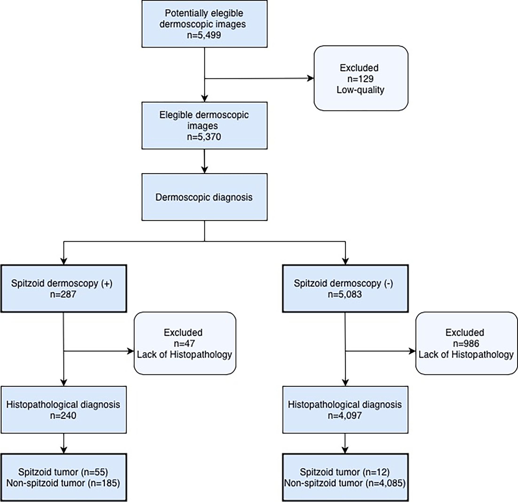
Fig. 1. Flowchart of selection and classification process of cases.
Demographic characteristics of patients with dermoscopically spitzoid lesions
Table I summarizes the demographic, clinical and dermoscopic characteristics of lesions with spitzoid dermoscopic patterns. A total of 240 dermoscopically spitzoid lesions from 221 patients were analysed. The median age of patients was 40 years (IQR, 22 years), with 57.9% females. Patients with spitzoid tumours confirmed by histopathology had a median age of 29 years (IQR, 20), compared to 45 years (IQR, 22.5 years) in cases of non-spitzoid tumours (p<0.001), with similar proportion of females between groups (67.9% vs 54.8%; p=0.091). A personal history of melanoma was more frequent in patients with non-spitzoid tumours (38.5% vs 14%; p=0.001) and family history of melanoma showed a similar trend (26.5% vs 14%; p=0.044). Conversely, patients with dermoscopically spitzoid malignant tumours had a median age of 50 years (IQR, 28.5 years) compared to 38 years (IQR, 20 years) in cases of benign lesions (p<0.001), and a personal history of melanoma was more frequent in patients with malignant tumours (46.4% vs 27.3%, p=0.009).
Table I. Demographic, clinical and dermoscopic characteristics of lesions with spitzoid dermoscopic patterns
| Overall | Spitzoid histology | Non-Spitzoid histology | P value1 | Malignant | Benign | P value1 | |
|---|---|---|---|---|---|---|---|
| Patients demographics (n=221) | |||||||
| Age, years, median (Q1, Q3) | 40 (31, 53) | 29 (18, 38) | 45 (35,57.5) | <0.001* | 50 (42, 70.5) | 38 (28, 48) | <0.001* |
| Sex, n (%) | |||||||
| Male Female |
93 (42.1%) 128 (57.9%) |
17 (32.1%) 36 (67.9%) |
76 (45.2%) 92 (54.8%) |
0.091 | 26 (46.4%) 30 (53.6%) |
67 (40.6%) 98 (59.4%) |
0.45 |
| Fitzpatrick skin type, n (%) | |||||||
| I II III IV Missing |
16 (8.8%) 70 (38.7%) 93 (51.4%) 2 (1.1%) 40 |
4 (9.5%) 13 (31%) 24 (57.1%) 1 (2.4%) 11 |
12 (8.6%) 57 (41%) 69 (49.6%) 1 (0.7%) 29 |
0.42 | 5 (10.2%) 21 (42.9%) 23 (46.9) 0 (0%) 7 |
11 (8.3%) 49 (37.1%) 70 (53%) 2 (1.5%) 33 |
0.79 |
| Personal history of melanoma, n (%) | |||||||
| No Yes Missing |
139 (67.5%) 67 (32.5%) 15 |
43 (86%) 7 (14%) 3 |
96 (61.5%) 60 (38.5%) 12 |
0.001* | 30 (53.6%) 26 (46.4%) 0 |
109 (72.7%) 41 (27.3%) 15 |
0.009 |
| Family history of melanoma, n (%) | |||||||
| No Yes Missing |
151 (76.6%) 46 (23.4%) 24 |
43 (86%) 7 (14%) 3 |
108 (73.5%) 39 (26.5%) 21 |
0.070* | 38 (74.5%) 13 (25.5%) 5 |
113 (77.4%) 33 (22.6%) 19 |
0.67 |
| Clinical and dermoscopic characteristics of lesions (n=240) | |||||||
| Size, mm, median (Q1,Q3) Missing |
5 (4, 6) 35 |
5 (3, 6) 4 |
5 (4, 6) 31 |
0.80 | 5 (4, 6) 14 |
5 (4, 6) 21 |
0.84 |
| Anatomical location, n (%) | |||||||
| Trunk Legs Arms Head/neck Acral |
109 (45.4%) 75 (31.3%) 20 (8.4%) 18 (7.5%) 18 (7.5%) |
20 (36.4%) 20 (36.4%) 6 (10.9%) 6 (10.9%) 3 (5.5%) |
89 (48.1%) 55 (29.7%) 14 (7.6%) 12 (6.5%) 15 (8.1%) |
0.39 | 21 (33.4%) 25 (41%) 7 (11.5%) 5 (8.2%) 3(3.9%) |
88 (49.2%) 50 (27.9%) 13 (7.3%) 13 (7.3%) 15 (8.4%) |
0.16 |
| Colour, n (%) | |||||||
| Brown Black Pink Blue |
125 (52.1%) 58 (24.2%) 55 (22.9%) 2 (0.8%) |
22 (40%) 27 (49.1%) 6 (10.9%) 0 (0%) |
103 (55.7%) 31 (16.8%) 49 (26.5%) 2 (1.1%) |
<0.001* | 28 (45.9%) 11 (18%) 22 (36.1%) 0 (0%) |
97 (54.2%) 47 (26.3%) 33 (18.4%) 2 (1.1%) |
0.037 |
| Symmetry, n (%) | |||||||
| No Yes |
167 (69.6%) 73 (30.4%) |
31 (56.4%) 24 (43.6%) |
136 (73.5%) 49 (26.5%) |
0.015* | 45 (73.8%) 16 (26.2%) |
122 (68.2%) 57 (31.8%) |
0.52 |
| Global dermoscopic pattern, n (%) | |||||||
| Starburst Globular Vascular Reticular Other |
78 (32.5%) 62 (25.8%) 49 (20.4%) 24 (10%) 27(11.3%) |
23 (41.8%) 10 (18.2%) 4 (7.3%) 9 (16.4%) 9 (16.4%) |
55 (29.7%) 52 (28.1%) 45 (24.3%) 15 (8.1%) 18 (9.7%) |
0.007* | 16 (26.2%) 14 (23%) 20 (32.8%) 5 (8.2%) 6 (9.8%) |
62 (34.6%) 48 (26.8%) 29 (16.2%) 19 (10.6%) 21 (11.7%) |
0.10 |
|
1Wilcoxon rank sum test; Pearson’s Chi-squared test; Fisher’s exact test. |
|||||||
Clinical and dermoscopic characteristics of lesions with spitzoid dermoscopic patterns
Among the 240 dermoscopically spitzoid lesions, the most common anatomical location was the trunk (45.4%), followed by the lower extremities (31.3%) and upper extremities (8.3%), with no significant differences between histologically spitzoid and non-spitzoid tumours or benign and malignant tumours.
Overall, the most common dermoscopic patterns were starburst (32.5%), globular (25.8%) and vascular (20.4%). The starburst and reticular patterns were significantly more frequent in histologically spitzoid tumours compared with non-spitzoid tumours (41.8% vs 29.7% and 16.4% vs 8.1%; p=0.007). No significant difference was found in dermoscopic patterns between benign and malignant tumours (p=0.10). Black colour was significantly more frequent in spitzoid tumours compared with non-spitzoid tumours (49.1% vs 16.8%; p<0.001), as was symmetry (43.6% vs 26.5%; p=0.015). Pink was a significantly more evident colour in malignant tumours than in benign lesions (36.1% vs 18.4%; p=0.037), whereas symmetry showed no significant difference (p=0.41).
Local dermoscopic features were evaluated by univariable analysis, which showed that blue-black pigmentation (OR 4.48; 95% CI, 2.31–8.75), blotch (OR 3.12; 95% CI,1.65–5.92) and streaks (OR 3.02; 95% CI, 1.60–5.72) were strongly associated with spitzoid histopathology, whereas linear vessels (OR 5.12; 95% CI, 2.08–13.1), glomerular vessels (OR 3.03; 95% CI, 1.37–6.69), milky-red areas (OR 2.84; 95% CI, 1.40–5.74) and shiny white lines (OR 2.59; 95% CI, 1.22–5.45) were associated with malignancy (Table II).
Table II. Association of dermoscopic features with definite spitzoid histopathology and malignant diagnosis
| Univariate analysis | ||||
|---|---|---|---|---|
| Spitzoid vs Non-spitzoid OR (95% CI) | p-value | Malignant vs benign OR (95% CI) | p-value | |
| Blue-black | 4.48 (2.31–8.75) | <0.001* | 0.60 (0.27–1.24) | 0.19 |
| Blotch | 3.12 (1.65–5.92) | <0.001* | 0.87 (0.43–1.66) | 0.67 |
| Streaks | 3.02 1.60–5.72 | <0.001* | 0.84 (0.42–1.61) | 0.61 |
| Pseudopods | 1.55 (0.80–2.94) | 0.18 | 0.64 (0.31–1.26) | 0.21 |
| Black network | 2.20 (0.91–5.06) | 0.069 | 0.33 (0.08–1.00) | 0.082 |
| Negative network | 0.39 (0.14–0.92) | 0.045* | 1.04 (0.50–2.08) | 0.92 |
| Dots | 0.59 (0.21–1.39) | 0.26 | 1.67 (0.77–3.47) | 0.18 |
| Globules | 0.78 (0.42–1.42) | 0.41 | 1.05 (0.58–1.88) | 0.87 |
| Regression | 0.88 (0.31–2.16) | 0.79 | 0.30 (0.07–0.90) | 0.057 |
| Blue-white veil | 1.21 (0.42–3.09) | 0.70 | 1.65 (0.63–4.02) | 0.28 |
| Ulceration | 0.29 (0.02–1.56) | 0.24 | 2.19 (0.63–7.15) | 0.19 |
| Dotted vessels | 0.45 (0.18–1.02) | 0.072 | 1.79 (0.91–3.45) | 0.088 |
| Linear vessels | 0.31 (0.05–1.11) | 0.12 | 5.12 (2.08–13.1) | <0.001* |
| Glomerular vessels | 0.21 (0.03–0.74) | 0.038 | 3.03 (1.37–6.69) | 0.006* |
| Milky-red areas | 0.78 (0.32–1.74) | 0.57 | 2.84 (1.40–5.74) | 0.004* |
| Shiny white lines | 0.66 (0.24–1.58) | 0.38 | 2.59 (1.22–5.45) | 0.012* |
Histopathological correlation of lesions with spitzoid dermoscopic patterns
Of the 240 dermoscopically spitzoid lesions, only 55 (23%) were confirmed as melanocytic spitzoid tumours by histopathology, including Spitz nevus (27/240, 11.3%), spitzoid nevus (19/240, 7.9%), atypical Spitz nevus (5/240, 2.1%) and spitzoid melanoma (4/240, 1.7%); 149 (62%) were diagnosed as common nevi (62/240, 25.8%), atypical nevi (41/240, 17.1%), conventional melanoma (37/240, 15.4%) and melanoma metastases (9/240, 3.8%). Also found were 36 (15%) non-melanocytic lesions including seborrheic keratosis (9/240, 3.8%), dermatofibromas (8/240, 3.3%) and non-melanoma skin cancer (NMSC) (11/240, 4.5%). Overall, 25.4% (61/240) of spitzoid lesions on dermoscopy were histologically diagnosed as malignant tumour (Fig. 2).
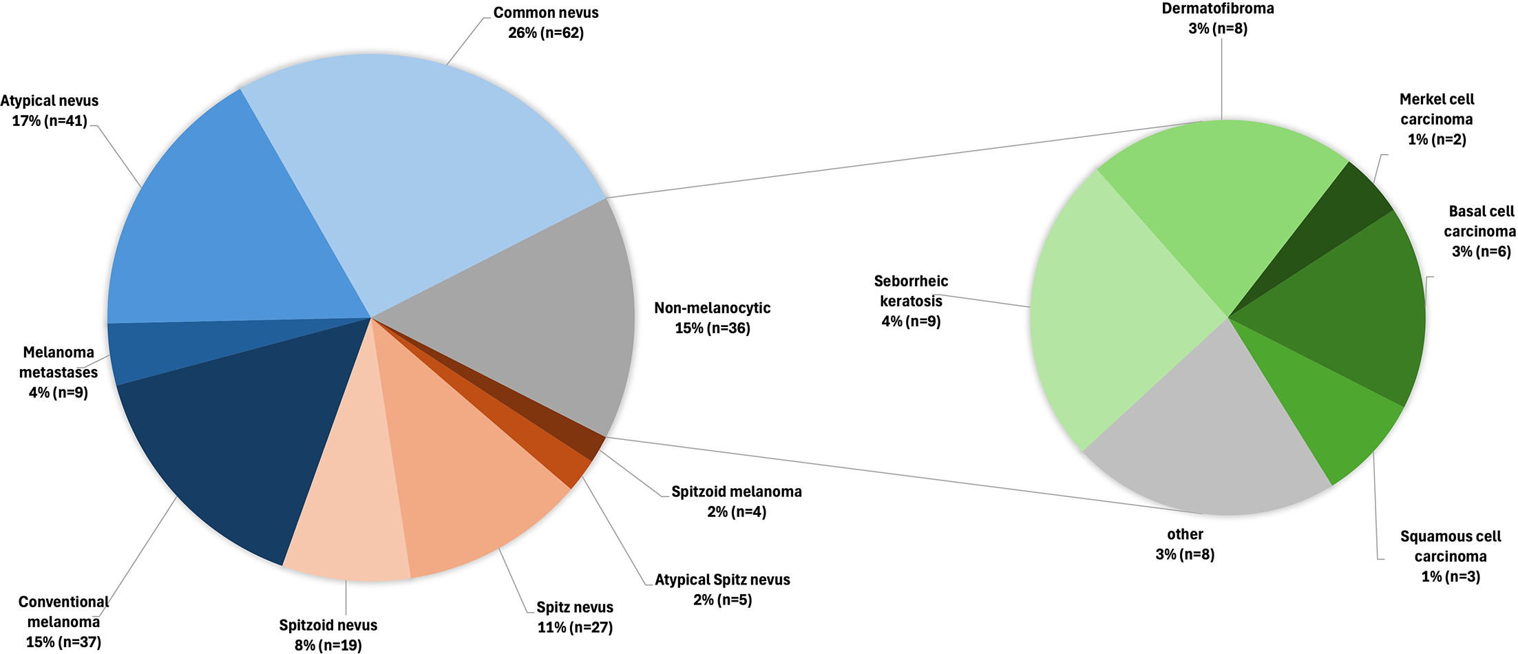
Fig. 2. Distribution of histopathological diagnoses among lesions with spitzoid dermoscopic patterns (N=240). Pie charts show the absolute number of cases. “Other” include inflammatory lesions (n=2), eccrine poroma (n=1), clear cell acanthoma (n=1), actinic keratosis (n=1), dermal vascular proliferation (n=1), acroangiodermatitis (n=1) and fibroepithelial polyp (n=1).
Sensitivity and specificity of dermoscopy
Dermoscopy showed a sensitivity of 82.1% (95% CI: 70.8%–90.4%) and a specificity of 95.7% (95% CI: 95%–96.3%) in detecting spitzoid melanocytic tumours. The PPV was 22.9% (95% CI: 17.8%–28.8%), and NPV 99.7% (95% CI: 99.5%–99.9%) (Table III). The AUC was 0.88 (95% CI: 0.84–0.93), indicating very good diagnostic accuracy (Fig. S2).
Table III. Cross tabulation of Dermoscopy results compared to Histopathology
| Histopathology | |||
|---|---|---|---|
| Dermoscopy | Spitzoid | Non-Spitzoid | Total |
| Spitzoid | 55 | 185 | 240 |
| Non-Spitzoid | 12 | 4,085 | 4,097 |
| Total | 67 | 4,270 | 4,337 |
| Sensitivity 82.1 % (95% confidence interval: 70.8%–90.4%) | |||
| Specificity 95.7 % (95% confidence interval: 95%–96.3%) | |||
| Positive predictive value 22.9 % (95% confidence interval: 17.8%–28.8%) | |||
| Negative predictive value 99.7 % (95% confidence interval: 99.5%–99.9%) | |||
DISCUSSION
The present study demonstrates that dermoscopy is highly effective in detecting spitzoid melanocytic tumours, with a sensitivity of 82.1% and a specificity of 95.7%. These results are particularly relevant considering that previous clinicopathological studies have reported that only 20% of Spitz nevi were clinically suspected, suggesting a significant risk of underdiagnosis (18, 19). These findings support the usefulness of dermoscopy both in improving the clinical recognition of these tumours and in reliably ruling them out in clinical practice. Although the PPV was low (22.9%), reflecting that only about one-quarter of dermoscopically spitzoid lesions in adults are true spitzoid tumours on histopathology, this finding carries substantial clinical weight. Since 25.4% (1 in 4) of these dermoscopically spitzoid lesions turned out to be malignant, a spitzoid dermoscopic pattern in adult patients cannot be safely monitored and should promptly lead to an excision or biopsy, reinforcing that the very high NPV (99.7%) highlights dermoscopy as a robust tool for excluding spitzoid melanocytic tumours when features are completely absent.
Our results confirm that spitzoid melanocytic tumours are infrequent, representing only 1% of all excised lesions as previously reported (9, 10). These tumours were found to appear at younger ages and were associated with a lower frequency of personal or family melanoma history compared to non-spitzoid melanocytic tumours, suggesting a distinctive molecular mechanism, as defined by the current WHO classification (5, 6). Tumours driven by gene fusions have been reported to occur more frequently in younger individuals, whereas tumours initiated by mutations tend to arise later in life, probably due to the progressive accumulation of somatic mutations over time (8, 20). In addition, the majority of spitzoid melanocytic tumours in our series were benign, and this is consistent with previous studies reporting that a low proportion of these tumours belong to an intermediate or malignant category (21, 22, 23).
In our cohort, approximately 1 in 4 lesions with spitzoid dermoscopic patterns was malignant, mainly melanoma, although NMSC were also observed. This is consistent with prior evidence showing a substantial morphological overlap between Spitz nevi and melanoma (24, 25). A retrospective study reported that the risk of a spitzoid lesion being melanoma rises markedly with age, from 2% between 12 and 20 years to 50% or higher in those over 50 years (26). Given that most of our cohort consisted of adults, maintaining a high index of suspicion is essential when evaluating spitzoid lesions in this population.
The starburst pattern was the most frequent in spitzoid tumours, aligning with established evidence (13, 17, 23). The black reticular pattern, although uncommon overall, appeared more often in spitzoid tumours and may provide limited supportive value, but should be interpreted in the clinical context (13, 27).
Vascular structures such as linear and glomerular vessels, in addition to shiny white lines, were strongly associated with malignancy, consistent with reports in atypical Spitz tumour, melanoma and NMSC (28, 29, 30). These findings support the excision of spitzoid lesions with atypical vascular structures on dermoscopy.
This study offers a novel contribution by quantifying the prevalence of spitzoid dermoscopic patterns and evaluating their clinical relevance in a real-world setting. Notably, several non-melanocytic tumours (15% of dermoscopically spitzoid lesions) can mimic spitzoid dermoscopic patterns, including irritated, clonal or regressive seborrheic keratosis (SK) (31, 32); haemosiderotic or aneurysmal dermatofibromas (33); certain basal cell carcinomas with a predominant vascular pattern, mainly on limbs (34, 35, 36); and other malignant tumours with atypical vascular patterns (37, 38) (Fig. 3). The false-negative cases lacked spitzoid features but showed atypical characteristics or changes during follow-up, raising concern and warranting excision.
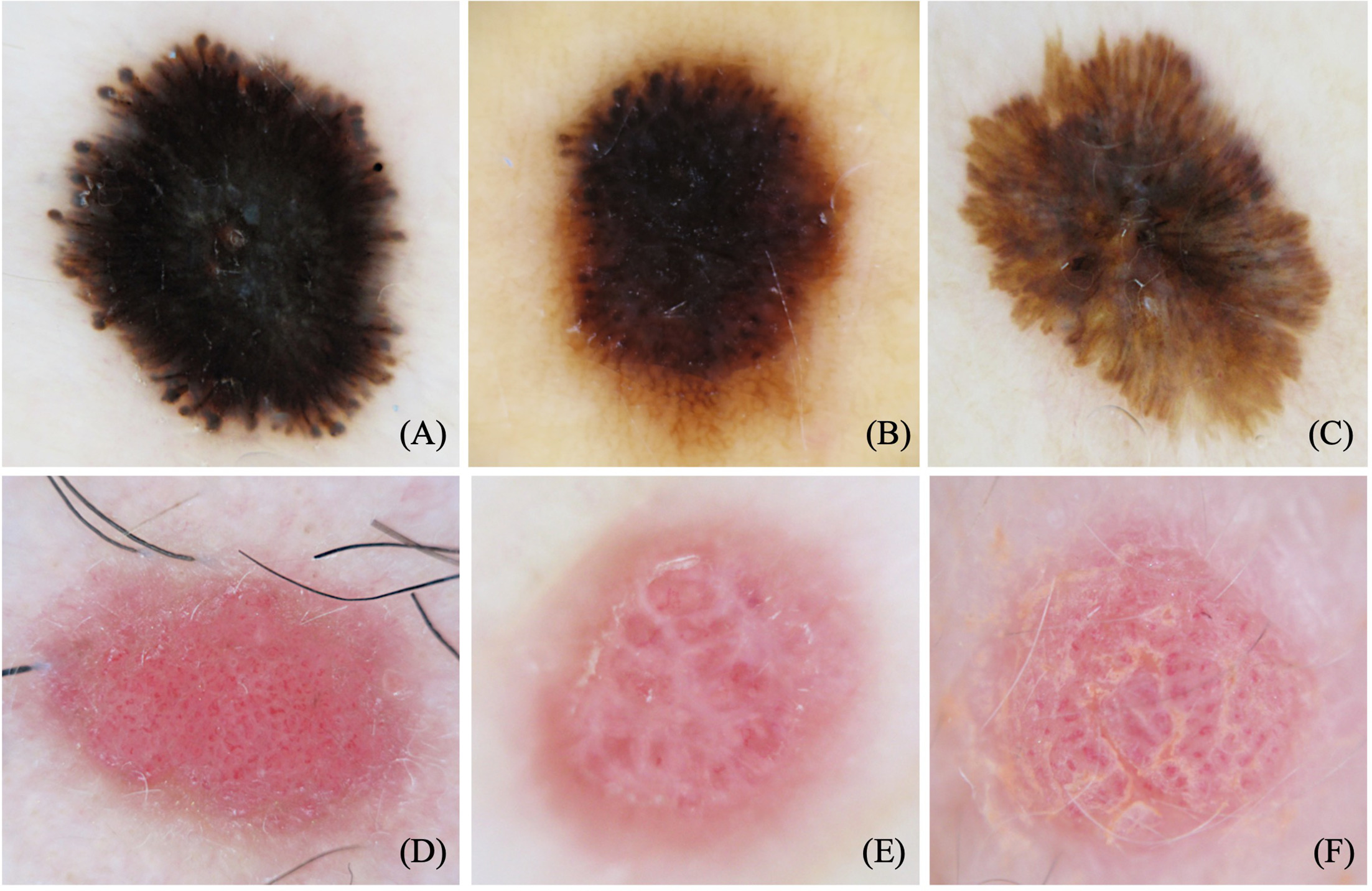
Fig. 3. Dermoscopic images of spitzoid lesions showing a starburst pattern (A–C): (A) Reed nevus; (B) Melanoma; (C) Seborrheic keratosis; and vascular pattern (D-F): (D) Spitz nevus; (E) Melanoma metastases; (F) Seborrheic keratosis.
Limitations of this study include its retrospective design; therefore, incomplete data in the medical records could have been left unaddressed. Although pediatric dermatologists refer challenging pediatric cases to our unit, most patients in this study were adults evaluated at a melanoma referral centre, which may have increased the observed prevalence of malignancy among spitzoid lesions. These factors limit the interpretation and generalizability of these results to the pediatric population and to primary care or general dermatology settings. Since only biopsied or excised lesions were included, this study may not include all spitzoid tumours seen at our institution. Conversely, histopathological diagnoses of tumours with “Spitzoid features” may not exhibit the current Spitz molecular profile and could be subject to reclassification if molecular testing were performed systematically (39). Additionally, our dermoscopic evaluation relied on a sequential screening and joint consensus-based approach rather than independent duplicate readings. This paradigm involved dermatologists with different levels of experience; while this workflow was optimized to guarantee consensus and reduce interobserver variability, initial individual screening metrics were not independently archived, precluding the calculation of formal interobserver agreement statistics. This limits the appraisal of individual test reproducibility outside a consensus panel environment. However, since diagnostic accuracy for challenging lesions like spitzoid tumours is heavily influenced by evaluator experience, the involvement of a third senior expert blinded to prior opinions for discordant cases likely maximized the reliability of our final dataset. Finally, due to the multiple statistical comparisons performed in evaluating various dermoscopic features, there is an increased risk of Type I errors (false positives); therefore, borderline significant p-values should be interpreted with caution and validated in future larger cohorts.
Further research is needed to correlate dermo-scopic features with emerging molecular classifications of Spitz tumours. Integrating molecular techniques, artificial intelligence and emerging in vivo technologies, such as reflectance confocal microscopy and line-field confocal optical coherence tomography, could potentially enhance diagnostic accuracy and provide further insight into the biological behaviour of these tumours.
Dermoscopy improves the detection of spitzoid melanocytic tumours, offering high sensitivity, specificity and NPV. In a referral centre, few excised lesions may show spitzoid features on dermoscopy and, interestingly, only a small proportion will be true spitzoid tumours. Notably, a significant percentage of dermoscopically spitzoid lesions will be malignant, primarily melanoma, while nonmelanocytic lesions may also display spitzoid features. Starburst pattern is the most frequent among true spitzoid tumours, and blue-black pigmentation, blotch and streaks are highly suggestive of spitzoid histopathology, while atypical vessels raise suspicion of malignancy.
ACKNOWLEDGEMENTS
We thank Beatriz Alejo BSc (Hons) and Abel Cano (digital imaging technician) for their substantial contributions to data collection in the Melanoma Unit of Clinic Hospital Barcelona, which made this research study possible. We thank the residents and fellows in the Dermatology department for their contribution to the image registry, and we thank all the patients, whose trust, consent and participation make the research possible.
REFERENCES
- Yeh I, Busam KJ. Spitz melanocytic tumours – A review. Histopathology 2022; 80: 122–134. https://doi.org/10.1111/his.14583
- Luo S, Sepehr A, Tsao H. Spitz nevi and other Spitzoid lesions. J Am Acad Dermatol 2011; 65: 1073–1084. https://doi.org/10.1016/j.jaad.2011.04.040
- Hillen LM, Van den Oord J, Geybels MS, Becker JC, Zur Hausen A, Winnepenninckx V. Genomic landscape of spitzoid neoplasms impacting patient management. Front Med (Lausanne) 2018; 5: 344. https://doi.org/10.3389/fmed.2018.00344
- Luo S, Sepehr A, Tsao H. Spitz nevi and other Spitzoid lesions: Part II. Natural history and management. J Am Acad Dermatol 2011; 65: 1087–1092. https://doi.org/10.1016/j.jaad.2011.06.045
- Wiesner T, Kutzner H, Cerroni L, Mihm MC, Jr Busam KJ, Murali R. Genomic aberrations in spitzoid melanocytic tumours and their implications for diagnosis, prognosis and therapy. Pathology 2016; 48: 113–131. https://doi.org/10.1016/j.pathol.2015.12.007
- Elder DE, Bastian BC, Cree IA, Massi D, Scolyer RA. The 2018 World Health Organization classification of cutaneous, mucosal, and uveal melanoma: Detailed analysis of 9 distinct subtypes defined by their evolutionary pathway. Arch Pathol Lab Med 2020; 144: 500–522. https://doi.org/10.5858/arpa.2019-0561-RA
- SPITZ S. Melanomas of childhood. Am J Pathol 1948; 24: 591–609.
- LeBoit PE. Spitz melanoma. Clin Dermatol 2025; 43: 348–355. https://doi.org/10.1016/j.clindermatol.2024.09.010
- Ko CB, Walton S, Wyatt EH, Bury HP. SPITZ NEVUS. Int J Dermatology 1993; 32: 354–357. https://doi.org/10.1111/j.1365-4362.1993.tb01472.x
- Casso EM, Grin-Jorgensen CM, Grant-Kels JM. Spitz nevi. J Am Acad Dermatol 1992; 27: 901–913. https://doi.org/10.1016/0190-9622(92)70286-o
- Sainz-Gaspar L, Sánchez-Bernal J, Noguera-Morel L, Hernández-Martín A, Colmenero I, Torrelo A. Spitz nevus and other spitzoid tumors in children —Part 1: Clinical, histopathologic, and immunohistochemical features. Actas Dermo-Sifiliográficas (English Edition) 2020; 111: 7–19. https://doi.org/10.1016/j.adengl.2019.12.006
- Steiner A, Pehamberger H, Binder M, Wolff K. Pigmented Spitz nevi: Improvement of the diagnostic accuracy by epiluminescence microscopy. J Am Acad Dermatol 1992; 27: 697–701. https://doi.org/10.1016/0190-9622(92)70240-G
- Lallas A, Apalla Z, Ioannides D, Lazaridou E, Kyrgidis A, Broganelli P, et al. Update on dermoscopy of Spitz/Reed naevi and management guidelines by the International Dermoscopy Society. Br J Dermatol 2017; 177: 645–655. https://doi.org/10.1111/bjd.15339
- Peris K, Ferrari A, Argenziano G, Soyer HP, Chimenti S. Dermoscopic classification of Spitz/Reed nevi. Clin Dermatol 2002; 20: 259–262. https://doi.org/10.1016/S0738-081X(02)00226-2
- Lallas A, Apalla Z, Papageorgiou C, Evangelou G, Ioannides D, Argenziano G. Management of flat pigmented spitz and reed nevi in children. JAMA Dermatol 2018; 154: 1353. https://doi.org/10.1001/jamadermatol.2018.3013
- Argenziano G. Spitz and Reed nevi. In: Marghoob AA, Malvehy J, Braun RP, editors. Atlas of dermoscopy. Second Edition. VolLondon: Informa Healthcare; 2012: p. 189–197. doi: 10.3109/9781841847627.022.
- Moscarella E, Lallas A, Kyrgidis A, Ferrara G, Longo C, Scalvenzi M, et al. Clinical and dermoscopic features of atypical Spitz tumors: A multicenter, retrospective, case-control study. J Am Acad Dermatol 2015; 73: 777–784. https://doi.org/10.1016/j.jaad.2015.08.018
- Requena C, Requena L, Kutzner H, Yus ES. Spitz nevus: A clinicopathological study of 349 cases. Am J Dermatopathol 2009; 31: 107–116. https://doi.org/10.1097/DAD.0b013e3181934218
- Berlingeri-Ramos AC, Morales-Burgos A, Sánchez JL, Nogales EM. Spitz nevus in a hispanic population: A clinicopathological study of 130 cases. Am J Dermatopathol 2010; 32: 267–275. https://doi.org/10.1097/DAD.0b013e3181c52b99
- Dupain C, Harttrampf AC, Urbinati G, Geoerger B, Massaad-Massade L. Relevance of fusion genes in pediatric cancers: Toward precision medicine. Mol Ther Nucleic Acids 2017; 6: 315–326. https://doi.org/10.1016/j.omtn.2017.01.005
- Patel D, Chawla R, Patel AJK. Clinical features and outcomes of paediatric Spitz-type lesions. J Plast Reconstr Aesthet Surg 2024; 97: 282–286. https://doi.org/10.1016/j.bjps.2024.07.031
- Davies OMT, Majerowski J, Segura A, Kelley SW, Sokumbi O, Humphrey SR. A sixteen‐year single‐center retrospective chart review of Spitz nevi and spitzoid neoplasms in pediatric patients. Pediatr Dermatol 2020; 37: 1073–1082. https://doi.org/10.1111/pde.14320
- Castelli R, Salvi I, Trave I, Viglizzo G, Guadagno A, Cabiddu F, et al. Clinical, dermoscopic, and histopathological features of spitzoid lesions in adult patients: Insights from a retrospective study. Dermatol Pract Concept 2025; 15: 5401. https://doi.org/10.5826/dpc.1503a5401
- Sepehr A. Long-term outcome of spitz-type melanocytic tumors. Arch Dermatol 2011; 147: 1173. https://doi.org/10.1001/archdermatol.2011.170
- Argenziano G, Scalvenzi M, Staibano S, Brunetti B, Piccolo D, Delfino M, et al. Dermatoscopic pitfalls in differentiating pigmented Spitz naevi from cutaneous melanomas. Br J Dermatol 1999; 141: 788–793. https://doi.org/10.1046/j.1365-2133.1999.03150.x
- Lallas A, Moscarella E, Longo C, Kyrgidis A, de Mestier Y, Vale G, et al. Likelihood of finding melanoma when removing a Spitzoid-looking lesion in patients aged 12 years or older. J Am Acad Dermatol 2015; 72: 47–53. https://doi.org/10.1016/j.jaad.2014.09.037
- Argenziano G, Soyer HP, Ferrara G, Piccolo D, Hofmann-Wellenhof R, Peris K, et al. Superficial black network: An additional dermoscopic clue for the diagnosis of pigmented spindle and/or epithelioid cell nevus. Dermatology 2001; 203: 333–335. https://doi.org/10.1159/000051784
- De Giorgi V, Venturi F, Silvestri F, Trane L, Savarese I, Scarfì F, et al. Atypical Spitz tumours: An epidemiological, clinical and dermoscopic multicentre study with 16 years of follow-up. Clin Exp Dermatol 2022; 47: 1464–1471. https://doi.org/10.1111/ced.15123
- Ferrara G, Cavicchini S, Corradin MT. Hypopigmented atypical Spitzoid neoplasms (atypical Spitz nevi, atypical Spitz tumors, Spitzoid melanoma): A clinicopathological update. Dermatol Pract Concept 2015; 5: 45–52. https://doi.org/10.5826/dpc.050106
- Carrera C, Scope A, Dusza SW, Argenziano G, Nazzaro G, Phan A, et al. Clinical and dermoscopic characterization of pediatric and adolescent melanomas: Multicenter study of 52 cases. J Am Acad Dermatol 2018; 78: 278–288. https://doi.org/10.1016/j.jaad.2017.09.065
- Papageorgiou V, Apalla Z, Sotiriou E, Papageorgiou C, Lazaridou E, Vakirlis S, et al. The limitations of dermoscopy: False‐positive and false‐negative tumours. Acad Dermatol Venereol 2018; 32: 879–888. https://doi.org/10.1111/jdv.14782
- Squillace L, Cappello M, Longo C, Moscarella E, Alfano R, Argenziano G. Unusual dermoscopic patterns of seborrheic keratosis. Dermatology 2016; 232: 198–202. https://doi.org/10.1159/000442439
- Zaballos P, Álvarez-Salafranca M, Llambrich À, Malvehy J, Taberner R, Medina C, et al. Dermoscopy of haemosiderotic/aneurysmal dermatofibroma: A morphological study of 110 cases. J Eur Acad Dermatol Venereol 2023; 37: 317–327. https://doi.org/10.1111/jdv.18664
- Lombardi M, Pampena R, Borsari S, Bombonato C, Benati E, Pellacani G, et al. Dermoscopic features of basal cell carcinoma on the lower limbs: A chameleon! Dermatology 2018; 233: 482–488. https://doi.org/10.1159/000487300
- Wolner ZJ, Bajaj S, Flores E, Carrera C, Navarrete-Dechent C, Dusza SW, et al. Variation in dermoscopic features of basal cell carcinoma as a function of anatomical location and pigmentation status. Br J Dermatol 2018; 178: e136–e137. https://doi.org/10.1111/bjd.15964
- Lallas A, Apalla Z, Argenziano G, Longo C, Moscarella E, Specchio F, et al. The dermatoscopic universe of basal cell carcinoma. Dermatol Pract Concept 2014; 4: 11–24. https://doi.org/10.5826/dpc.0403a02
- Zalaudek I, Kreusch J, Giacomel J, Ferrara G, Catricalà C, Argenziano G. How to diagnose nonpigmented skin tumors: A review of vascular structures seen with dermoscopy: Part I. Melanocytic skin tumors. J Am Acad Dermatol 2010; 63: 361–374;. https://doi.org/10.1016/j.jaad.2009.11.698
- Zalaudek GI, Corona R, Sera F, Cicale L, Petrillo G, et al. Vascular structures in skin tumors: A dermoscopy study. Arch Dermatol 2004; 140: 1485–1489. https://doi.org/10.1001/archderm.140.12.1485
- Moysset I, Castrejon N, Garcia-Herrera A, Castillo P, Marginet M, Teixido C, et al. Restrospective reappraisal of the prognostic classification of spitzoid melanocytic neoplasms after BRAF and NRAS mutation characterisation: A single institution experience. Histopathology 2024; 84: 1154–1166. https://doi.org/10.1111/his.15160