ORIGINAL REPORT
Physical Activity and Risk of Autoimmune Bullous Skin Diseases: A Prospective Cohort Study
Shuai JU1,2†, Guowei ZHOU1,2†, Changmin LIU3, Fangfang LI1,2, Qingyu HONG1,2, Wanyao LIU1,2, Hong LIU1,2* and Jia GUO1,2*
1Department of Dermatology, Hunan Engineering Research Center of Skin Health and Disease, Hunan Key Laboratory of Skin Cancer and Psoriasis, Xiangya Hospital, Central South University, Changsha, Hunan 410008, China, 2National Engineering Research Center of Personalized Diagnostic and Therapeutic Technology, Central South University, Changsha, Hunan 410008, China, and 3School of Foreign Languages and Literature, Shandong University, Jinan 250100, China
†These authors contributed equally to this work.
Corr: Hong Liu, Department of Dermatology, Xiangya Hospital, Central South University, No.87 Xiangya Road, Changsha, Hunan 410008, China and Jia Guo, Department of Dermatology, Xiangya Hospital, Central South University, No.87 Xiangya Road, Changsha, Hunan 410008, China. *Emails: hongliu1014@csu.edu.cn; jiaguo1128@csu.edu.cn
Keywords: physical activity; autoimmune bullous skin diseases; pemphigus; pemphigoid, bullous; body mass index.
Citation: Acta Derm Venereol 2026; 106: adv-2026-0705. DOI: https://doi.org/10.2340/actadv.v106.adv-2026-0705.
Copyright: 2026 ©Author(s). Published by MJS Publishing, on behalf of the Society for Publication of Acta Dermato-Venereologica. This is an Open Access article distributed under the terms of the Creative Commons Attribution-NonCommercial 4.0 International License (https://creativecommons.org/licenses/by-nc/4.0/).
Submitted: May 18, 2026. Accepted after revision: Aug 27, 2026.
Published: Sept 22, 2026.
Competing interests and funding: The authors have no conflicts of interest to declare.
This work was supported by the National Natural Science Foundation of China [grant numbers 82504290]; and the China Postdoctoral Science Foundation [grant numbers BX20250226, 2024M763721].
The datasets generated and/or analysed during the current study are available in the UK Biobank repository (https://www.ukbiobank.ac.uk/). Data access for this project was approved under Application Number 55257. Qualified researchers can obtain these datasets by submitting a formal application directly to the UK Biobank.
The UK Biobank received ethical approval from the North West Multi-Center Research Ethics Committee (11/NW/0382). Written informed consent was collected from all participants. The study was performed in accordance with the Helsinki Declaration of 1964, and its later amendments.
Physical activity has been linked to immune regulation in several autoimmune diseases, but its association with autoimmune bullous skin diseases (AIBD), mainly pemphigus and pemphigoid, has not been adequately evaluated. We examined the association between regular physical activity and incident AIBD in 476,057 UK Biobank participants. Multivariable-adjusted Cox models were used to estimate hazard ratios (HRs) and 95% confidence intervals (CIs) for AIBD risk according to habitual physical activity. We also assessed joint associations with polygenic risk and performed mediation analysis. During a median follow-up of 15.21 years, 272 incident AIBD cases were documented, including 61 pemphigus and 211 pemphigoid cases. Physically active participants had a 31% lower risk of AIBD than inactive participants (adjusted HR 0.69, 95% CI 0.54–0.88). Similar associations were observed for pemphigus (HR 0.53) and pemphigoid (HR 0.72). Individuals with high genetic risk who were physically inactive had a higher risk than physically active individuals with low genetic risk (HR 2.12, 95% CI 1.47–3.07). In an exploratory mediation analysis, BMI accounted for 8.86 % of the association. These findings suggest that regular physical activity is associated with a lower risk of AIBD after adjustment for genetic susceptibility and other risk factors.
SIGNIFICANCE
Autoimmune bullous skin diseases, including pemphigus and pemphigoid, are uncommon blistering disorders that can markedly impair quality of life. Evidence on modifiable lifestyle factors for these diseases is limited. In this large UK Biobank cohort, physically active participants had a lower risk of developing autoimmune bullous skin diseases, including both pemphigus and pemphigoid. The association was seen across genetic risk groups, while exploratory mediation analysis suggested a possible role for body mass index. These findings support further research on physical activity as a potentially modifiable factor in autoimmune blistering skin diseases.
INTRODUCTION
Autoimmune bullous skin diseases (AIBD) encompass a variety of organ-specific autoimmune diseases that manifest with cutaneous and/or mucosal blisters and erosions (1). Pemphigus and pemphigoid represent the 2 major groups of AIBD (2, 3), which impose a profound burden on patients’ quality of life and healthcare costs (4, 5). Consequently, there is a need to identify potentially modifiable factors associated with the occurrence of these conditions. A growing body of evidence indicates that physical activity exerts immunomodulatory effects, with regular exercise, in particular, leading to a reduction in systemic inflammatory biomarkers, and these benefits have been observed in various autoimmune diseases (6, 7, 8). However, evidence regarding the influence of physical activity on the incidence of pemphigus and pemphigoid remains limited, despite their well-characterized autoimmune pathogenesis. Therefore, data from large-scale prospective cohort studies are needed to evaluate this association and explore the underlying mechanisms.
In large cohort studies, self-reported physical activity is often summarized as metabolic equivalent-hours per week, although such estimates may be affected by recall errors in duration and intensity. We therefore used questionnaire-derived criteria based on the frequency and duration of moderate or vigorous physical activity to capture regular health-promoting activity patterns.
In the present study, we therefore aimed to investigate the association between physical activity and the risk of incident pemphigus and pemphigoid in a large-scale prospective cohort of 476,057 participants. We further explored the potential roles of genetic and inflammatory factors in this association.
MATERIALS AND METHODS
Study population
The UK Biobank is a large prospective cohort study, and it recruited over 500,000 participants between 2006 and 2010, aged 37–73 years, from 22 assessment centres throughout England, Wales and Scotland. The details of the study design and approach have been documented elsewhere (9). Briefly, a wide range of health-related data were collected through touchscreen questionnaire, physical examinations and biological specimens at baseline or follow-up assessment. Written informed consent was collected from all of the participants. UK Biobank received ethical approval from the North West Multi-Center Research Ethics Committee (11/NW/0382).
Data from a total of 502,129 participants were initially available for this study. We excluded participants with a diagnosis of pre-existing pemphigus (n=41) or pemphigoid (n=78) at baseline, incomplete data on physical activity (n=11,356) and those with a mismatch between genetic and self-reported sex (n=14,597). After these exclusions, a total of 476,057 participants were eligible for the final analytical cohort. A flow diagram of participant selection is shown in Fig. S1.
Assessment of pemphigus and pemphigoid
Pemphigus and pemphigoid cases were ascertained using linked UK Biobank health records, including primary care records, hospital inpatient records, death register records and self-reported medical conditions. Diagnoses were identified using ICD-10 codes, as detailed in Table SI. The primary outcome of this study was incident autoimmune bullous skin disease, i.e. pemphigus or pemphigoid. For the primary analysis, at least 1 qualifying diagnostic record was sufficient for case ascertainment. To further assess diagnostic validity, we additionally repeated the analysis using a stricter case definition requiring at least 2 qualifying diagnostic records (10, 11). Specialist confirmation and disease-specific treatment were not required for case ascertainment.
Assessment of Habitual Physical Activity
Assessment of habitual physical activity was based on responses to a baseline touchscreen questionnaire regarding the frequency and duration of moderate (Data-Fields 884, 894) and vigorous (Data-Fields 904, 914) activities. For the primary analysis, these data were operationalized into a summary dichotomous variable to classify participants according to regular physical activity patterns. A participant was classified as “active” if they met at least one of the following 4 criteria derived from the questionnaire: 1) engaging in moderate-intensity activity for >150 minutes/week, 2) engaging in moderate-intensity activity on >6 days/week, 3) engaging in vigorous-intensity activity for >75 minutes/week, or 4) engaging in vigorous-intensity activity on >1 day/week. These criteria were informed by established physical activity recommendations and the UK Biobank physical activity questionnaire (12). Participants not meeting any of these criteria were classified as “inactive”. Responses of “Do not know” or “Prefer not to answer” for the activity questions were treated as missing values.
Assessment of genetic susceptibility to pemphigus and pemphigoid as an effect modifier
To quantify genetic susceptibility, we constructed a polygenic risk score (PRS). The PRS was based on the GWAS of the combined “Pemphigus and pemphigoid” phenotype (PheCode 695.22) reported by Zhou et al. (GWAS Catalog: GCST90436597).(13) The PRS was constructed for our target cohort (UK Biobank) using a clumping and thresholding (C+T) method in PRSice-2. We selected 62 independent single nucleotide polymorphisms (SNPs) reaching a p-value threshold of <1 × 10⁻⁵ to build the score. The SNPs included in the PRS are listed in Table SII. For subsequent analyses, participants were categorized into 2 groups (low and high genetic risk) based on the median of the final continuous PRS.
Covariates
Baseline sociodemographic, anthropometric and lifestyle covariates were collected using a self-administered touchscreen questionnaire. These included age, sex, ethnic background, level of education, socioeconomic status, body mass index (BMI), smoking status, alcohol consumption and diet. Age was categorized into tertiles, socioeconomic status into quintiles, and BMI into nonoverweight, overweight and obesity categories. Smoking status and alcohol consumption were each grouped into 3 categories, while diet was summarized as a dichotomous variable according to adherence to healthy dietary components. Detailed data fields, coding and categorization criteria are provided in Table SIII.
Statistical analysis
Participant follow-up commenced at recruitment (2006–2010) and concluded on the date of pemphigus or pemphigoid diagnosis, death, loss to follow-up, or the study’s end date (31 May 2024), whichever occurred first. We estimated cumulative incidence using Kaplan–Meier curves stratified by physical activity status (“active” vs “inactive”) and assessed differences with the log-rank test. Ten-year cumulative risks were derived from the Kaplan-Meier estimates, and incidence rates were calculated per 100,000 person-years. To estimate hazard ratios (HRs) and 95% confidence intervals (CIs), we developed a series of multivariable-adjusted Cox proportional hazards models. Our modelling strategy involved 3 sequential adjustments to assess the influence of different covariate sets. Model 1 adjusted for a core set of sociodemographic factors: age, sex, ethnic background, socioeconomic status and level of education. Model 2 was built upon Model 1 by adding key lifestyle behaviours, including smoking status, alcohol consumption and diet. Model 3 further adjusted for BMI categories. Missing values for categorical covariates were handled by creating a separate indicator category, whereas median imputation was used for continuous variables. We verified that the proportional hazards assumption was met for all models.
We conducted subgroup analyses to evaluate whether the association between physical activity and disease risk was modified by baseline characteristics. We assessed the following variables as potential effect modifiers: age (categorized by tertiles), sex (male vs female), ethnic background (white vs nonwhite), socioeconomic status (categorized by tertiles), level of education (academic/professional degree vs other), smoking status (never, former or current), alcohol consumption (abstainers, moderate or heavy), BMI (nonoverweight, overweight or obese) and diet (healthy vs not healthy). We tested the statistical significance of interactions between physical activity and these variables by comparing Cox proportional hazards models with and without the multiplicative interaction terms, using the likelihood ratio test.
We performed several sensitivity analyses to test the robustness of our findings: (1) to reduce the potential influence of reverse causation, we excluded the first 2 and 5 years of follow-up and conducted an analysis censoring all participants at 10 years; (2) we excluded individuals with extreme BMI (<18.5 or ≥40 kg/m²) to assess the potential impact of outliers; and (3) to evaluate whether the observed association was influenced by assessment centre, we further adjusted for this variable in our models.
To assess the joint impact of physical activity and genetic risk for AIBD, we first used logistic regression to confirm that the polygenic risk score (PRS) was associated with AIBD risk in our cohort. We then compared the risk of AIBD among participants with different combinations of physical activity status (“active” vs “inactive”) and genetic risk (high vs low PRS) to those who were physically “active” and had a low genetic risk.
We further performed a mediation analysis to investigate potential pathways that might explain the association between physical activity and disease risk. Physical activity is a key regulator of body mass, and previous studies link the pathogenesis of AIBD to systemic inflammation and metabolic dysregulation (14, 15, 16, 17, 18). We therefore selected a broad panel of potential mediators. This included BMI, a range of blood cell counts and other inflammatory biomarkers, and key markers of lipid metabolism. The analysis was conducted using the “CMAverse” R package, which is based on a counterfactual framework (19). Confidence intervals for all effects were estimated using non-parametric bootstrapping (1,000 iterations) to ensure robustness. Subgroup, genetic risk and mediation analyses were considered exploratory.
All statistical analyses were performed using R (Version 4.5.1). A 2-sided p-value <0.05 was considered to indicate statistical significance for all analyses.
RESULTS
Baseline characteristics of the 476,057 participants, stratified by physical activity status, are detailed in Table I. Clear demographic and lifestyle differences emerged between the “active” and “inactive” groups. Compared to their inactive counterparts, physically active individuals were more likely to be male and have a higher level of education. They also presented with a healthier metabolic profile, evidenced by a significantly lower mean BMI (26.9 vs 27.9 kg/m2; p<0.001). Further distinctions were noted in lifestyle habits. The active group reported lower rates of current smoking (9.5% vs 11.5%) and were less likely to be abstainers from alcohol. Notably, adherence to a healthy diet was substantially higher among active participants (42.1%) than inactive participants (34.5%).
Table I. Baseline characteristics of the study participants according to physical activity status
| Characteristic | Overall N=476,057 |
Inactive N=212,974 |
Active N=263,083 |
p-valuea |
|---|---|---|---|---|
| Age, n (%) | <0.001 | |||
| Age Group 1 (age≤53) | 169,167 (35.5%) | 72,446 (34.0%) | 96,721 (36.8%) | |
| Age Group 2 (53<Age ≤ 61) | 149,341 (31.4%) | 69,981 (32.9%) | 79,360 (30.2%) | |
| Age Group 3 (age>61) | 157,549 (33.1%) | 70,547 (33.1%) | 87,002 (33.1%) | |
| Sex, n (%) | <0.001 | |||
| Female | 257,646 (54.1%) | 121,368 (57.0%) | 136,278 (51.8%) | |
| Male | 218,411 (45.9%) | 91,606 (43.0%) | 126,805 (48.2%) | |
| Ethnic background, n (%) | <0.001 | |||
| White | 450,516 (95.0%) | 201,854 (95.1%) | 248,662 (94.8%) | |
| Not White | 23,957 (5.0%) | 10,419 (4.9%) | 13,538 (5.2%) | |
| Unknown | 1,584 | 701 | 883 | |
| Body Mass Index, mean (SD) | 27.4 (4.8) | 27.9 (5.1) | 26.9 (4.4) | <0.001 |
| Unknown | 1,757 | 966 | 791 | |
| Level of education, n (%) | <0.001 | |||
| Lower degree | 244,667 (51.9%) | 112,377 (53.3%) | 132,290 (50.8%) | |
| Academic or other professional degree | 226,571 (48.1%) | 98,519 (46.7%) | 128,052 (49.2%) | |
| Unknown | 4,819 | 2,078 | 2,741 | |
| Socioeconomic status, n (%) | <0.001 | |||
| Quintile 1 (least deprived) | 97,542 (20.5%) | 42,687 (20.1%) | 54,855 (20.9%) | |
| Quintiles 2–4 | 285,990 (60.1%) | 126,893 (59.7%) | 159,097 (60.5%) | |
| Quintile 5 (most deprived) | 91,941 (19.3%) | 43,132 (20.3%) | 48,809 (18.6%) | |
| Unknown | 584 | 262 | 322 | |
| Alcohol use, n (%) | <0.001 | |||
| Abstainers | 90,789 (19.1%) | 44,994 (21.1%) | 45,795 (17.4%) | |
| Moderate | 259,161 (54.5%) | 114,284 (53.7%) | 144,877 (55.1%) | |
| Heavy | 125,734 (26.4%) | 53,505 (25.1%) | 72,229 (27.5%) | |
| Unknown | 373 | 191 | 182 | |
| Smoking, n (%) | <0.001 | |||
| Never | 259,923 (54.8%) | 115,313 (54.3%) | 144,610 (55.2%) | |
| Former smoker | 165,113 (34.8%) | 72,451 (34.1%) | 92,662 (35.3%) | |
| Current smoker | 49,359 (10.4%) | 24,429 (11.5%) | 24,930 (9.5%) | |
| Unknown | 1,662 | 781 | 881 | |
| Diet, n (%) | <0.001 | |||
| Not healthy diet | 292,008 (61.3%) | 139,569 (65.5%) | 152,439 (57.9%) | |
| Healthy diet | 184,049 (38.7%) | 73,405 (34.5%) | 110,644 (42.1%) | |
| Pemphigus, n | 61 | 37 | 24 | 0.012 |
| Pemphigoid, n | 211 | 114 | 97 | 0.007 |
|
ap-alues were calculated using the Pearson χ2 test for categorical variables and the Wilcoxon rank sum test for continuous variables. SD: standard deviation. |
||||
Over a median follow-up of 15.21 years, we documented a total of 272 incident cases of AIBD, including 61 cases of pemphigus and 211 cases of pemphigoid. The absolute incidence of AIBD was low, with incidence rates of 4.83 and 3.11 per 100,000 person-years among inactive and active participants, respectively. The corresponding 10 year cumulative risks were 0.0489% and 0.0256%. The Kaplan–Meier curves (Fig. 1) demonstrated a significantly lower cumulative incidence of AIBD in the “active” group (Fig. 1A, log-rank p<0.001). This trend was consistent for both pemphigus and pemphigoid when analysed individually (Fig. 1B and C; log-rank p=0.011 and p=0.005, respectively).
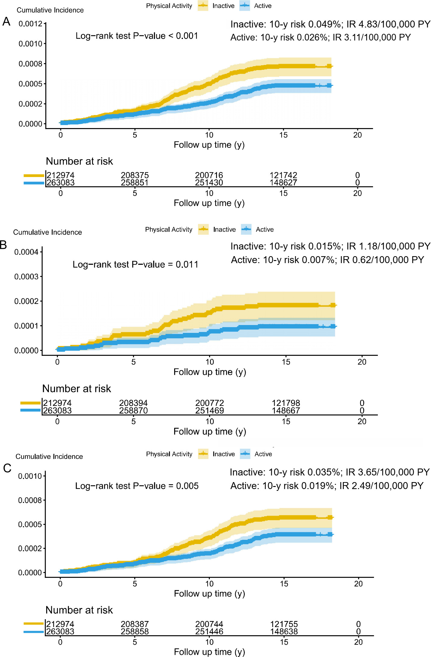
Fig. 1. Cumulative Incidence of autoimmune bullous skin diseases by physical activity level. Kaplan-Meier curves illustrating the cumulative incidence of (A) autoimmune bullous skin diseases (AIBD), (B) pemphigus, and (C) pemphigoid, stratified by physical activity status (“active” vs “Inactive”). Ten-year cumulative risks and incidence rates (IRs) per 100,000 person-years (PY) are shown for each physical activity group. Shaded areas indicate 95% confidence intervals. p-Values were calculated using the log-rank test. The tables below each plot indicate the number of participants at risk at 0, 5, 10, 15 and 20 years of follow-up.
Event numbers, person-time, and incidence rates according to physical activity status are presented in Table II, with the corresponding Cox proportional hazards model results presented in Table III. In the fully adjusted model, physical activity was associated with a lower risk of AIBD (HR, 0.69; 95% CI, 0.54–0.88), with similar associations for pemphigus (HR, 0.53; 95% CI, 0.32–0.90) and pemphigoid (HR, 0.72; 95% CI, 0.55–0.95).
Table II. Incidence of autoimmune bullous skin diseases according to physical activity status
Table III. Association between physical activity and risk of AIBD
| Disease type | Model | HR1 (95% CI) | p-value |
|---|---|---|---|
| AIBD | Model 1a | 0.66 (0.51, 0.84) | <0.001 |
| Model 2b | 0.67 (0.52, 0.85) | 0.001 | |
| Model 3c | 0.69 (0.54, 0.88) | 0.003 | |
| Pemphigus | Model 1a | 0.51 (0.30, 0.85) | 0.010 |
| Model 2b | 0.51 (0.31, 0.86) | 0.012 | |
| Model 3c | 0.53 (0.32, 0.90) | 0.018 | |
| Pemphigoid | Model 1a | 0.68 (0.52, 0.90) | 0.006 |
| Model 2b | 0.70 (0.53, 0.92) | 0.010 | |
| Model 3c | 0.72 (0.55, 0.95) | 0.020 | |
|
1Represents the risk for the ‘Active’ group compared to the ‘Inactive’ reference group. hazard ratio (HR) < 1.00 indicates a reduced risk. aModel 1 was adjusted for age, sex, ethnic background, socio-economic status, and level of education. bModel 2 was further adjusted for smoking status, alcohol use, and diet. cModel 3 was further adjusted for body mass index categories. AIBD: autoimmune bullous skin diseases; CI: confidence Interval. |
|||
We conducted several sensitivity analyses to test the robustness of our findings. For the primary outcome of AIBD, the association with lower risk remained statistically significant and the hazard ratios were not materially altered across all analyses (Table SIV). This included excluding the first 2 years of follow-up (HR, 0.69; 95% CI, 0.53–0.89), excluding the first 5 years of follow-up (HR, 0.66; 95% CI, 0.50–0.88), censoring follow-up at 10 years (HR, 0.55; 95% CI, 0.40–0.75), excluding individuals with extreme BMI (HR, 0.73; 95% CI, 0.57–0.94), and additionally adjusting for the assessment centre (HR, 0.69; 95% CI, 0.54–0.89). When analysed separately, the results for pemphigus and pemphigoid were also largely robust (Table SIV). For instance, after excluding the first 5 years of follow-up, the hazard ratios were 0.50 (95% CI, 0.26–0.95) for pemphigus and 0.68 (95% CI, 0.50–0.93) for pemphigoid. The associations generally remained significant, with the exception that the association for pemphigoid was attenuated and no longer statistically significant after excluding participants with extreme BMI (HR, 0.78; 95% CI, 0.59–1.04).
Exploratory stratified analyses were conducted to evaluate whether the association between physical activity and AIBD risk was modified by key baseline characteristics (Table IV). The association between physical activity and lower AIBD risk was generally consistent across subgroups defined by age, sex, ethnic background, level of education, socio-economic status, alcohol use, smoking status, and diet. No statistically significant interactions were observed (all p for interaction >0.05). Descriptively, lower HRs were observed in participants who were not overweight (HR, 0.54; 95% CI, 0.33–0.88) and in non-white individuals (HR, 0.30; 95% CI, 0.11–0.84). When pemphigus and pemphigoid were analysed separately, the results remained largely consistent (Table SV). For pemphigus, lower HRs were observed among individuals with a normal BMI (HR, 0.16; 95% CI, 0.03–0.75) and among those with heavy alcohol consumption (HR, 0.18; 95% CI, 0.04–0.84). For pemphigoid, a lower HR was observed among participants with obesity (HR, 0.61; 95% CI, 0.38–0.99).
Table IV. Stratified analyses for the association between physical activity and autoimmune bullous skin diseases risk
The PRS was associated with AIBD risk in both crude and adjusted models (Table SVI). We found that participants with a high genetic risk for AIBD who were also physically inactive experienced a substantially higher risk of disease (Fig. 2), even though the test for interaction between genetic risk and physical activity was not statistically significant (P for interaction=0.693, Table SVII). Compared to participants with a low genetic risk who were physically active, those with a high genetic risk and an inactive lifestyle showed a 112 % increased risk of developing AIBD (HR, 2.12; 95% CI, 1.47–3.07; Fig. 2).
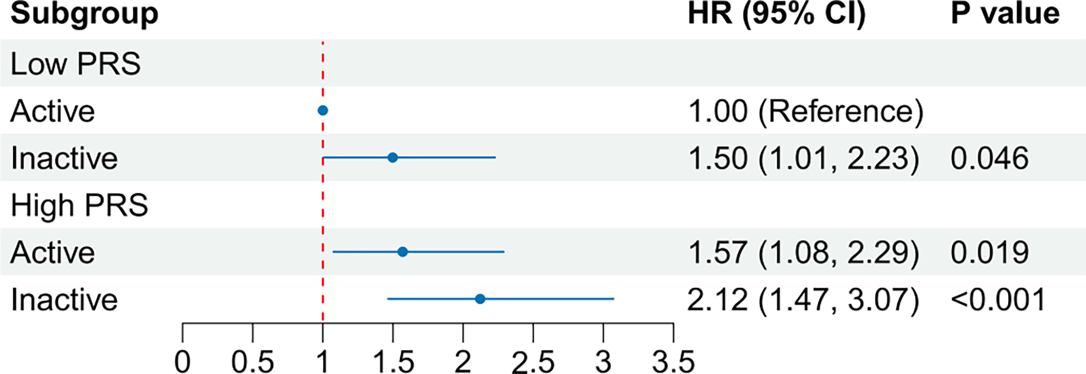
Fig. 2. Joint associations of genetic risk and physical activity with risk of autoimmune bullous skin diseases. Forest plot showing the hazard ratios (HRs) and 95 % confidence intervals (CIs) for the combined effects of polygenic risk score (PRS) and physical activity. The reference group consists of participants with a low PRS who were physically active. Models were adjusted for age, sex, ethnic background, socioeconomic status, level of education, smoking status, alcohol use, diet and body mass index.
We performed an exploratory mediation analysis to investigate potential pathways linking physical activity to AIBD risk. BMI showed a mediated proportion of 8.86% (95% CI, 2.35%–31.70%). Small indirect effects were also observed for eosinophil count (PM, 0.88%), albumin (PM, 1.17%) and alkaline phosphatase (PM, 1.09%). Lymphocyte and red blood cell counts showed indirect effects in the opposite direction (Table V).
Table V. Mediation analysis results
DISCUSSION
In this large prospective cohort study of 476,057 participants, regular physical activity was associated with a lower risk of incident AIBD. Compared with inactive participants, physically active participants had a 31% lower risk of AIBD, and this association was more pronounced for pemphigus. We also observed a joint association between physical activity and genetic risk: participants with a high polygenic risk score who were inactive had a 112% higher risk of AIBD than active participants with a low polygenic risk score. Exploratory mediation analysis suggested that BMI may account for a modest proportion of the association, with several inflammation-related biomarkers showing smaller indirect effects.
To our knowledge, this is the first large-scale prospective study to examine physical activity in relation to incident AIBD. Although evidence for the role of exercise in chronic inflammatory diseases is still evolving, our findings are biologically plausible and consistent with the established immunomodulatory effects of physical activity (6, 7, 8). Previous prospective studies have also linked regular physical activity to a lower risk of other immune-mediated conditions, including psoriasis and Crohn disease (20, 21). The observed association may be partly explained by the anti-inflammatory effects of exercise, including reduced systemic inflammation, myokine-mediated signalling and changes in immune-cell profiles, such as regulatory T cells and monocytes (6, 22, 23, 24). These mechanisms may help to temper the dysregulated immune responses involved in the pathogenesis of pemphigus and pemphigoid.
Our exploratory mediation analysis suggested a modest potential role for BMI in the observed association. Obesity is now recognized as a state of chronic low-grade inflammation driven by dysfunctional adipose tissue, including pro-inflammatory macrophage polarization and dysregulated adipokine secretion (25, 26). This finding is highly plausible, as a high BMI is now recognized as a significant risk factor for AIBD itself, particularly for pemphigoid (27). These findings suggest a possible role for adiposity in the observed association, although causal mediation cannot be established.
Beyond BMI, albumin, eosinophil count and alkaline phosphatase showed modest mediated effects, possibly reflecting changes in systemic inflammation and oxidative stress (18, 28). The opposite indirect effects observed for lymphocyte and red blood cell counts were more difficult to interpret and should be considered exploratory. Given the small effect sizes, these findings should be considered exploratory.
Strengths and limitations
The major strengths of the present study include its large sample size, prospective design, independent ascertainment of AIBD cases through linkage to comprehensive national health records, and availability of genetic data for over 470,000 participants. This design enabled investigation of the associations of physical activity and genetic risk with rare diseases such as pemphigus and pemphigoid.
Several limitations should be considered. First, physical activity was self-reported at baseline and may not reflect long-term activity patterns. Second, case ascertainment based on routinely collected diagnostic records may be subject to misclassification. In a stricter analysis requiring at least 2 qualifying diagnostic records, 63 AIBD cases remained (12 pemphigus and 51 pemphigoid). The association remained significant for overall AIBD (HR, 0.58; 95% CI, 0.35–0.98), whereas the disease-specific estimates were not statistically significant but remained inverse and directionally consistent with the primary analysis (Table SVIII). The reduced case numbers may have limited statistical precision. Clinical confirmation using immunopathology or serology was unavailable. Non-differential misclassification would likely attenuate associations, whereas differential ascertainment related to healthcare utilization or general health cannot be excluded. Third, residual confounding and healthy-user bias remain possible despite multivariable adjustment, and no negative-control outcome analysis was performed. Fourth, the low absolute incidence of AIBD and limited number of events, particularly for pemphigus, reduced the precision of subgroup and mediation analyses; grouping clinically heterogeneous subtypes may also have obscured subtype-specific associations. Finally, the healthy volunteer effect and predominance of White European ancestry in UK Biobank may limit generalizability.
Conclusion
Regular physical activity was associated with a lower risk of AIBD in this large prospective cohort. This association persisted after adjustment for established risk factors and genetic susceptibility, while exploratory mediation analyses suggested potential roles for BMI and several inflammation-related biomarkers.
ACKNOWLEDGEMENTS
This research has been conducted using the UK Biobank Resource under Application Number 55257. We are grateful to the participants of the UK Biobank for their invaluable contributions.
REFERENCES
- Saschenbrecker S, Karl I, Komorowski L, Probst C, Dähnrich C, Fechner K, et al. Serological Diagnosis of Autoimmune Bullous Skin Diseases. Front Immunol 2019; 10: 1974. https://doi.org/10.3389/fimmu.2019.01974
- Kershenovich R, Hodak E, Mimouni D. Diagnosis and classification of pemphigus and bullous pemphigoid. Autoimmun Rev 2014; 13: 477–481. https://doi.org/10.1016/j.autrev.2014.01.011
- Egami S, Yamagami J, Amagai M. Autoimmune bullous skin diseases, pemphigus and pemphigoid. J Allergy Clin Immunol 2020; 145: 1031–1047. https://doi.org/10.1016/j.jaci.2020.02.013
- Bahloul D, Dubucq H, Thomas RB, Ajith A, Boss J, Fotheringham I, et al. Burden of Disease of Bullous Pemphigoid: A Targeted Literature Review. Dermatology 2024; 240: 823–832. https://doi.org/10.1159/000540480
- Hsu D, Brieva J, Silverberg JI. Costs of Care for Hospitalization for Pemphigus in the United States. JAMA Dermatol 2016; 152: 645–654. https://doi.org/10.1001/jamadermatol.2015.5240
- Gleeson M, Bishop NC, Stensel DJ, Lindley MR, Mastana SS, Nimmo MA. The anti-inflammatory effects of exercise: mechanisms and implications for the prevention and treatment of disease. Nat Rev Immunol 2011; 11: 607–615. https://doi.org/10.1038/nri3041
- Sharif K, Watad A, Bragazzi NL, Lichtbroun M, Amital H, Shoenfeld Y. Physical activity and autoimmune diseases: Get moving and manage the disease. Autoimmun Rev 2018; 17: 53–72. https://doi.org/10.1016/j.autrev.2017.11.010
- Luo B, Xiang D, Ji X, Chen X, Li R, Zhang S, et al. The anti-inflammatory effects of exercise on autoimmune diseases: A 20-year systematic review. J Sport Health Sci 2024; 13: 353–367. https://doi.org/10.1016/j.jshs.2024.02.002
- Sudlow C, Gallacher J, Allen N, Beral V, Burton P, Danesh J, et al. UK biobank: an open access resource for identifying the causes of a wide range of complex diseases of middle and old age. PLoS Med 2015; 12: e1001779. https://doi.org/10.1371/journal.pmed.1001779
- Leisti P, Pankakoski A, Jokelainen J, Varpuluoma O, Huilaja L, Panelius J, et al. Accurate diagnosis of bullous pemphigoid requires multiple health care visits. Front Immunol 2023; 14: 1281302. https://doi.org/10.3389/fimmu.2023.1281302
- Hsu D, Brieva J, Nardone B, Silverberg JI. Validation of database search strategies for the epidemiological study of pemphigus and pemphigoid. Br J Dermatol 2016; 174: 645–648. https://doi.org/10.1111/bjd.14172
- Bull FC, Al-Ansari SS, Biddle S, Borodulin K, Buman MP, Cardon G, et al. World Health Organization 2020 guidelines on physical activity and sedentary behaviour. Br J Sports Med 2020; 54: 1451–1462. https://doi.org/10.1136/bjsports-2020-102955
- Zhou W, Nielsen JB, Fritsche LG, Dey R, Gabrielsen ME, Wolford BN, et al. Efficiently controlling for case-control imbalance and sample relatedness in large-scale genetic association studies. Nat Genet 2018; 50: 1335–1341. https://doi.org/10.1038/s41588-018-0184-y
- Sams WM Jr, Gammon WR. Mechanism of lesion production in pemphigus and pemphigoid. J Am Acad Dermatol 1982; 6: 431–452. https://doi.org/10.1016/s0190-9622(82)70036-2
- Limberg MM, Weihrauch T, Gray N, Ernst N, Hartmann K, Raap U. Eosinophils, Basophils, and Neutrophils in Bullous Pemphigoid. Biomolecules 2023; 13: 1019. https://doi.org/10.3390/biom13071019
- Gabay C, Kushner I. Acute-phase proteins and other systemic responses to inflammation. N Engl J Med 1999; 340: 448–454. https://doi.org/10.1056/NEJM199902113400607
- Arroyo V, García-Martinez R, Salvatella X. Human serum albumin, systemic inflammation, and cirrhosis. J Hepatol 2014; 61: 396–407. https://doi.org/10.1016/j.jhep.2014.04.012
- Sheinenzon A, Shehadeh M, Michelis R, Shaoul E, Ronen O. Serum albumin levels and inflammation. Int J Biol Macromol 2021; 184: 857–862. https://doi.org/10.1016/j.ijbiomac.2021.06.140
- Shi B, Choirat C, Coull BA, VanderWeele TJ, Valeri L. CMAverse: A Suite of Functions for Reproducible Causal Mediation Analyses. Epidemiology 2021; 32: e20–e22. https://doi.org/10.1097/EDE.0000000000001378
- Frankel HC, Han J, Li T, Qureshi AA. The association between physical activity and the risk of incident psoriasis. Arch Dermatol 2012; 148: 918–924. https://doi.org/10.1001/archdermatol.2012.943
- Wang Q, Xu KQ, Qin XR, Wang XY. Association between physical activity and inflammatory bowel disease risk: A meta-analysis. Dig Liver Dis 2016; 48: 1425–1431. https://doi.org/10.1016/j.dld.2016.08.129
- Pedersen BK. Anti-inflammatory effects of exercise: role in diabetes and cardiovascular disease. Eur J Clin Invest 2017; 47: 600–611. https://doi.org/10.1111/eci.12781
- Petersen AMW, Pedersen BK. The anti-inflammatory effect of exercise. J Appl Physiol 2005; 98: 1154–1162. https://doi.org/10.1152/japplphysiol.00164.2004
- Scheffer D da L, Latini A. Exercise-induced immune system response: Anti-inflammatory status on peripheral and central organs. Biochim Biophys Acta Mol Basis Dis 2020; 1866: 165823. https://doi.org/10.1016/j.bbadis.2020.165823
- Ouchi N, Parker JL, Lugus JJ, Walsh K. Adipokines in inflammation and metabolic disease. Nat Rev Immunol 2011; 11: 85–97. https://doi.org/10.1038/nri2921
- Saltiel AR, Olefsky JM. Inflammatory mechanisms linking obesity and metabolic disease. J Clin Invest 2017; 127: 1–4. https://doi.org/10.1172/JCI92035
- Zhang B, Chen X, Liu Y, Chen F, Yang N, Li L. Relationship between bullous pemphigoid and metabolic syndrome: a 12-year case-control study conducted in China. Ther Adv Chronic Dis 2022; 13: 20406223221130707. https://doi.org/10.1177/20406223221130707
- Blank M, Israeli D, Shoenfeld Y. Exercise, autoimmune diseases and T-regulatory cells. J Autoimmun 2024; 149: 103317. https://doi.org/10.1016/j.jaut.2024.103317