REVIEW ARTICLE
Summary of Best Evidence for Wound Management in Stevens–Johnson Syndrome and Toxic Epidermal Necrolysis
Shuzhen KONG1†, Haoru NIU1† , Simeng CUI2, Yuxuan KONG3, Yingnan ZHAI4, Jianqiao YE1, Yuanjie ZHANG4, Haoyang HONG4, Jiahui TIAN4, Yirui ZHOU4, Bin PENG1, Baibing MI5* and Songmei GENG1*
1Department of Dermatology, The Second Affiliated Hospital of Xi’an Jiaotong University, Xi’an, Shaanxi, China, 2Department of Neurology, The First Affiliated Hospital of Xi’an Jiaotong University, Xi’an, Shaanxi, China, 3Wuhan University Medical Center, Wuhan University, Wuhan, Hubei, China, 4Xi’an Jiaotong University Medical Center, Xi’an Jiaotong University, Xi’an, Shaanxi, China, and 5Department of Epidemiology and Biostatistics, School of Public Health, Xi’an Jiaotong University Health Science Center, Xi’an, Shaanxi, China
†These authors contributed equally to this work.
Corr: Baibing Mi, Department of Epidemiology and Biostatistics, School of Public Health, Xi’an Jiaotong University Health Science Center, Xi’an, Shaanxi, China and Songmei Geng, Department of Dermatology, The Second Affiliated Hospital of Xi’an Jiaotong University, Xi’an, Shaanxi, China. *Emails: xjtu.mi@xjtu.edu.cn; gengsongmei73@163.com
Key words: evidence-based practice; Stevens-Johnson syndrome; toxic epidermal necrolysis; wound healing; skin care.
Citation: Acta Derm Venereol 2026; 106: adv-2026-0511. DOI: https://doi.org/10.2340/actadv.v106.adv-2026-0511.
Copyright: 2026 ©Author(s). Published by MJS Publishing, on behalf of the Society for Publication of Acta Dermato-Venereologica. This is an Open Access article distributed under the terms of the Creative Commons Attribution-NonCommercial 4.0 International License (https://creativecommons.org/licenses/by-nc/4.0/).
Submitted: Mar 18, 2026. Accepted after revision: Aug 24, 2026.
Published: Sept 28, 2026.
Competing interests and funding: The authors have no conflicts of interest to declare.
This work was supported by the [National Natural Science Foundation of China] under Grant [number 82103944]. For multiple agency grants: This work was supported by [the Science and Technology Resources Open Sharing Platform of the Shaanxi Province] under Grant [number 2023-CX-PT-47].
Data sharing is not applicable to this article as no new data were created or analysed in this study.
This study did not involve human or animal subjects, therefore, no ethical approval was required.
Stevens–Johnson syndrome (SJS) and toxic epidermal necrolysis (TEN) are rare but life-threatening mucocutaneous reactions characterized by extensive epidermal detachment and systemic complications. Wound management is central to survival and long-term outcomes, yet standardized care pathways remain controversial and inconsistently implemented. This study aimed to systematically synthesize the best available evidence on wound management for patients with SJS/TEN to support clinical decision-making and optimize clinical outcomes. A comprehensive search of major international and Chinese databases and authoritative websites was conducted from January 2010 to May 2025 to collect relevant clinical decisions, guidelines, expert consensus, systematic reviews and evidence summaries. Two reviewers independently screened and evaluated the literature and then extracted and summarized evidence using the JBI grading system. A total of 14 high-quality publications met the inclusion criteria, yielding 28 pieces of best evidence across 7 domains: wound assessment, conservative management, surgical approach, dressing selection, pain management, infection prevention and surveillance, and hospital setting. Among them, 11 recommendations were graded as strong and 17 as weak. This evidence-based synthesis provides practical guidance for wound management in SJS/TEN and supports the development of standardized care protocols to promote epithelial recovery, reduce complications, and improve quality of life.
SIGNIFICANCE
Wound care in Stevens–Johnson syndrome and toxic epidermal necrolysis is difficult and essential for survival, yet clear and consistent guidance for clinicians has been lacking. This study brings together the best available research to summarize practical wound care strategies. The findings highlight simple but important practices, such as gentle skin care, appropriate dressings, effective pain control and early treatment in specialized hospitals. When applied according to specific clinical contexts, these strategies can help doctors and nurses make more consistent decisions, improve recovery, reduce complications and support better outcomes for patients with these severe skin conditions.
INTRODUCTION
Stevens-Johnson syndrome (SJS) and toxic epidermal necrolysis (TEN) are rare, life-threatening cutaneous disorders characterized by extensive keratinocyte apoptosis and epidermal detachment, predominantly mediated by type IV hypersensitivity reactions to pharmacologic agents (1, 2). Clinically, SJS/TEN manifest as progressive purpuric macules, mucosal erosions, bullous lesions and systemic manifestations (3).
SJS/TEN represent a disease continuum, differentiated by the extent of epidermal detachment: SJS involves <10% total body surface area (BSA), SJS/TEN overlap involves 10–30% BSA, and TEN involves >30% BSA.
Despite their rarity, SJS/TEN carry substantial morbidity and mortality. Overall mortality ranges from 10% to 20%, rising above 30% in TEN despite intensive treatment, particularly among elderly or immunocompromised patients (4). Beyond acute mortality, SJS/TEN are associated with long-term sequelae and considerable socioeconomic burden (2).
Given the complex, multiorgan pathophysiology, high mortality and associated healthcare costs, optimizing therapeutic strategies constitutes an urgent clinical imperative.
Current management includes immunomodulatory therapies and supportive interventions. Systemic pharmacological treatments include corticosteroids, intravenous immunoglobulin, tumor necrosis factor (TNF) inhibitors and cyclosporine (5). Supportive care, particularly wound management, has demonstrated efficacy in reducing infection risk and mortality rate (6). Wound care strategies range from conservative to surgical approaches. Conservative therapy preserves detached epidermis as a biological dressing, facilitating healing and protecting the underlying dermis. Surgical approach involves debriding detached skin and applying biosynthetic dressings, allografts or xenografts (5). Notably, conservative management accelerates re-epithelialization compared with surgical debridement (6). Optimized wound care protocols enhance cutaneous regeneration and improve long-term prognoses, underscoring their integral role.
However, standardized protocols for SJS/TEN remain undefined. Current practice varies considerably, ranging from conservative and surgical approaches to combined strategies, while high-quality evidence regarding dressing selection and application remains limited (7, 8). Consequently, clinical decision-making is largely based on expert consensus or extrapolation from burn care protocols – approaches that may be insufficient to address the extensive epidermal detachment, high susceptibility to infection and complex healing processes characteristic of SJS/TEN. This evidence gap hinders the development of standardized, evidence-based wound management strategies.
Accordingly, this study aimed to systematically synthesize and reappraise the best available evidence on wound management for SJS, TEN and SJS/TEN overlap. By consolidating high-quality evidence, this review will establish standardized wound management strategies to improve clinical outcomes, reduce complications and optimize resource utilization in the management of severe cutaneous disorders.
MATERIALS AND METHODS
Study design
This study was conducted as an evidence summary following the methodological framework established by the Joanna Briggs Institute (JBI) (9) and the reporting standards set forth by the Fudan University Center for Evidence-based Nursing (10) (registration no. ES20257947). The study aimed to identify, critically appraise and synthesize the best available evidence on wound management for patients with SJS, TEN and SJS/TEN overlap.
Problem formulation
The research question was formulated using the PIPOST framework (JBI Centre for Evidence-Based Nursing, Fudan University). The components included: Population (P): patients with SJS, TEN or SJS/TEN overlap; Intervention (I): wound management strategies; Professional (P): clinical healthcare practitioners; Outcome (O): patient outcomes, practitioner-level outcomes and system-level outcomes; Setting (S): dermatology wards and burn unit wards; Type of evidence (T): guidelines, evidence summaries, systematic reviews, expert consensus statements and randomized controlled trials (RCTs).
Literature search strategy
A comprehensive literature search was conducted using the “6S” evidence hierarchy model, prioritizing high-level evidence sources such as clinical decision-support systems, guidelines, expert consensus, systematic reviews and evidence summaries (11).
Data sources: The following databases and websites were systematically searched: UpToDate, BMJ Best Practice, JBI, Guidelines International Network (GIN), National Institute for Health and Care Excellence (NICE), American Academy of Dermatology (AAD), British Association of Dermatologists (BAD), European Academy of Dermatology and Venereology (EADV), American Journal of Clinical Dermatology (AJCD), European Burns Association (EBA), American Burns Association (ABA), JAMA Dermatology, the Cochrane Library, Embase, PubMed, Web of Science, CINAHL, CNKI, Wan Fang Database, VIP Database and Chinese Medical Journal Full-text Database.
Search terms and strategy: The search strategy combined Medical Subject Headings (MeSH) and free-text terms. The search terms related to 3 key concepts: (1) disease, including “Stevens–Johnson syndrome”, “SJS”, “toxic epidermal necrolysis”, “TEN”, “SJS/TEN overlap”, and “Lyell syndrome”; (2) wound management, including “wound care”, “wound management”, “skin care”, “skin management”, “wound healing”, “skin lesion care”, “epidermal detachment”, “erosion care”, “dressings”, and “debridement”; and (3) evidence type, including “guideline”, “clinical practice guideline”, “recommendation”, “consensus”, “expert consensus”, “systematic review”, “meta-analysis”, “evidence summary”, “best practice” and “randomized controlled trial”. The search covered publications from January 2010 to May 2025 without initial language restrictions. An example of the PubMed search strategy is provided in Fig. 1.

Fig. 1. PubMed search strategy.
Literature selection criteria
Inclusion criteria: (1) Patients diagnosed with SJS, TEN or SJS/TEN overlap; (2) interventions involved wound management strategies; (3) evidence types including guidelines, clinical practice recommendations, systematic reviews, expert consensus statements and RCTs; (4) outcomes encompassed Severity-of-Illness Score for Toxic Epidermal Necrolysis (SCORTEN) score, infection rates, epidermal detachment extent, re-epithelialization time, pain scores, hospitalization duration, mortality rates, pigmentation changes and scar incidence; (5) evidence applied to dermatology or burn unit settings; (6) publications were in Chinese or English; (7) articles were published between January 2010 and May 2025.
Exclusion criteria: (1) incomplete or duplicate references; (2) unavailable full-text articles; (3) studies failed to meet quality assessment standards; (4) outdated guidelines or consensus statements with updated versions available; (5) low-level evidence already encompassed within higher-level evidence; (6) conference abstracts, guideline interpretations, research protocols or translated guidelines.
Studies selection and data extraction process
Literature screening: Two reviewers independently screened the literature according to predefined criteria. Titles and abstracts were first assessed, followed by full-text evaluation of potentially eligible articles. Disagreements were resolved through discussion or consultation with a third reviewer.
Data extraction: Data extraction was conducted independently by the same reviewers using a standardized data extraction form. Extracted information included author or institution, publication year, source, evidence type, methodological characteristics and key evidence content.
Quality assessment methodology
Quality assessment tools: The quality of included guidelines was evaluated using the Appraisal of Guidelines for Research and Evaluation II (AGREE II) (12), which includes 23 items across 6 domains and uses a 7-point scoring system. Quality evaluation of best practice and evidence summaries involved returning to the original literature upon which the evidence was based. The quality of included systematic reviews was assessed using A Measure Tool to Assess Systematic Reviews (AMSTAR-2) (13), and expert consensus statements were evaluated using the Expert Consensus Standard (2020), developed by the JBI Centre for Evidence-based Health Care (14).
Inter-rater reliability assessment: Two reviewers conducted independent quality assessments of the retained articles. For appraisal tools yielding continuous scores, such as AGREE II, inter-rater reliability was quantified using the intraclass correlation coefficient (ICC). The following reliability standards were applied: ICC>0.9: excellent reliability; 0.9 >ICC>0.75: good reliability; 0.75 >ICC >0.5: moderate reliability; ICC<0.5: poor reliability (15). Conversely, for tools consisting primarily of categorical responses (such as AMSTAR-2 and the JBI checklist), ICC analysis was not applicable. Across all instruments, any disagreements were resolved through consensus discussion or adjudication by a third reviewer.
Evidence extraction and synthesis
Evidence extraction process: All included studies were systematically reviewed, and relevant evidence was extracted and categorized by thematic topics. When similar evidence was identified, concise and independent recommendations were retained. In cases of conflicting evidence, priority was given to evidence with higher methodological quality, higher evidence level and more recent publication.
Evidence level and recommendation level determination: Evidence grading was performed during extraction. For guideline-derived evidence, the original grading system was adopted, while evidence from systematic reviews, expert consensus statements and primary studies was graded using the JBI evidence grading system (16). If necessary, the original source documents were reviewed to determine the appropriate evidence level. Evidence levels ranged from Level 1 to Level 5 according to study design. Recommendation strength was determined using the JBI FAME framework (feasibility, appropriateness, meaningfulness and effectiveness) and classified as Grade A (strong recommendation) or Grade B (weak recommendation), taking into account both the hierarchy of evidence and implementation considerations.
RESULTS
Literature search and screening outcomes
A total of 2,342 records were retrieved from 15 of the searched databases and websites. After removing duplicates, 1,708 unique articles were screened by title and abstract, yielding 39 for full-text review. Fourteen high-quality references were ultimately included: 2 clinical decisions, 6 guidelines, 3 expert consensus statements and 3 systematic reviews (Fig. 2; Table I).
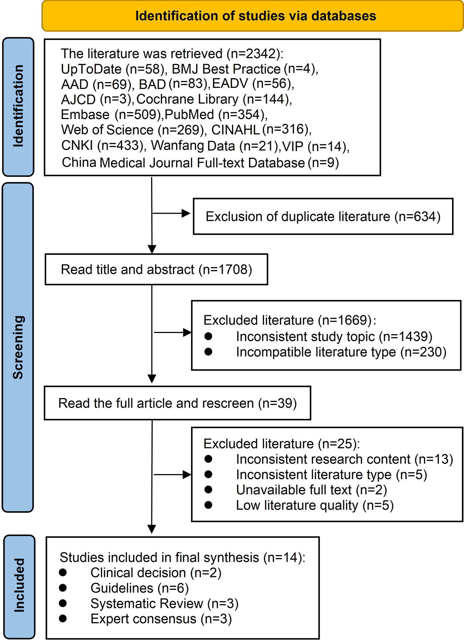
Fig. 2. Flow diagram of literature search. AAD: American Academy of Dermatology; BAD: British Association of Dermatologists; EADV: European Academy of Dermatology and Venereology; AJCD: American Journal of Clinical Dermatology.
Table I. General information of the included literature
| Included literature | Year | Literature sources | Type of evidence | Topic of the literature |
|---|---|---|---|---|
| Lee HY (31) | 2025 | UpToDate | Clinical decision | Stevens–Johnson syndrome and toxic epidermal necrolysis: Management, prognosis, and long-term sequelae |
| Kowal-Vern A (22) | 2022 | BMJ Best Practice | Clinical decision | Stevens–Johnson syndrome and toxic epidermal necrolysis – symptoms, diagnosis and treatment |
| Seminario-Vidal L et al. (24) | 2020 | CINAHL | Guideline | Society of Dermatology Hospitalists supportive care guidelines for the management of Stevens-Johnson syndrome/toxic epidermal necrolysis in adults |
| Paulmann M et al. (21) | 2024 | PubMed | Guideline | S3 guideline: diagnosis and treatment of epidermal necrolysis (Stevens-Johnson syndrome and toxic epidermal necrolysis) – part 2: supportive therapy of EN in the acute and post-acute stages |
| Creamer D et al. (17) | 2016 | Web of Science | Guideline | U.K. guidelines for the management of Stevens-Johnson syndrome/toxic epidermal necrolysis in adults 2016 |
| McPherson, T. et al. (5) | 2018 | British Journal of Dermatology | Guideline | British Association of Dermatologists' guidelines for the management of Stevens–Johnson syndrome/toxic epidermal necrolysis in children and young people, 2018 |
| Ingen-Housz-Oro S et al. (23) | 2018 | Web of Science | Guideline | Epidermal necrolysis French national diagnosis and care protocol (PNDS; protocole national de diagnostic et de soins) |
| Gupta LK et al. (25) | 2016 | PubMed | Guideline | Guidelines for the management of Stevens–Johnson syndrome/toxic epidermal necrolysis: an Indian perspective |
| Brüggen MC et al. (28) | 2021 | Embase | Expert Consensus | Supportive care in the acute phase of Stevens–Johnson syndrome and toxic epidermal necrolysis: an international, multidisciplinary Delphi-based consensus |
| White KD et al. (3) | 2018 | Embase | Expert Consensus | SJS/TEN 2017: Building Multidisciplinary Networks to Drive Science and Translation |
| Adverse Drug Reaction Research Center, Chinese Society of Dermatology, Chinese Medical Association (4). | 2021 | CMJD | Expert Consensus | Expert consensus on the diagnosis and treatment of Stevens–Johnson syndrome/toxic epidermal necrolysis |
| Castillo B et al. (19) | 2018 | PubMed | Systematic Review | Wound care for Stevens–Johnson syndrome and toxic epidermal necrolysis |
| Lee JS et al. (6) | 2024 | PubMed | Systematic Review | An individual patient data meta-analysis of wound care in patients with toxic epidermal necrolysis |
| Mahar PD et al. (18) | 2014 | Web of Science | Systematic Review | A systematic review of the management and outcome of toxic epidermal necrolysis treated in burns centres |
Quality assessment outcomes
Guideline quality: Among 6 guidelines, 2 guidelines (5, 17) achieved standardized scores ≥60% across all 6 domains, qualifying for Grade A (high rigor) classification, while the remaining 4 were rated Grade B (Table II).
Table II. Methodological quality evaluation results of the guidelines
| Included literature | Percentage of field standardization % | >60% field number(n) | >30% field number(n) | ICC | Recommendation level | |||||
|---|---|---|---|---|---|---|---|---|---|---|
| Scopes and objects | Participants | Rigor of the guidelines | Clarity of guidelines | Application of guidelines | Independence of the guide | |||||
| Creamer D et al. (17) | 91.67 | 77.78 | 93.75 | 94.44 | 64.58 | 61.11 | 6 | 6 | 0.982 | A |
| McPherson, et al.(5) | 94.40 | 75.00 | 84.38 | 86.10 | 68.75 | 95.83 | 6 | 6 | 0.962 | A |
| Seminario-Vidal L et al. (24) | 88.89 | 61.11 | 88.54 | 88.89 | 10.42 | 45.83 | 4 | 5 | 0.985 | B |
| Paulmann M et al. (21) | 88.89 | 86.11 | 88.54 | 88.89 | 54.17 | 95.83 | 5 | 6 | 0.980 | B |
| Ingen-Housz-Oro S et al. (23) | 83.33 | 58.33 | 35.42 | 88.89 | 56.25 | 87.50 | 3 | 6 | 0.943 | B |
| Gupta LK et al. (25) | 77.77 | 58.33 | 79.17 | 83.33 | 31.25 | 45.83 | 3 | 6 | 0.988 | B |
|
Note: Standardization percentage of each field = (obtained score − minimum possible score)/(maximum possible score − minimum possible score) × 100%; Recommendation level: if the standardized percentage of six fields is >60%, it is highly recommended (level A); if >3 areas have a standardized percentage > 30% and < 60% are recommended (level B); if there are ≥3 areas with a standardized percentage < 30%, it is not recommended (level C) |
||||||||||
Systematic review quality: Three systematic reviews underwent quality assessment using the AMSTAR-2 instrument. The systematic review of Lee JS et al. (6) met all criteria and was rated high quality. Mahar PD et al. (18) and Castillo B et al. (19) lacked meta-analysis and had minor methodological limitations but were classified as moderate quality due to their direct relevance to wound care and the scarcity of high-quality evidence. Table SI provides detailed quality assessments.
Expert consensus quality: Two independent evaluators assessed 3 expert consensus articles using the JBI expert opinion quality evaluation tool. Although they showed limited methodological rigor, their inclusion was justified by the scarcity of evidence in SJS/TEN wound care. Detailed quality assessments are presented in Table SI.
Clinical decision support resource quality: The UpToDate clinical decision tool and BMJ Best Practice were evaluated by reviewing their underlying primary literature. High-quality evidence identified was incorporated into the analysis.
Evidence synthesis and classification: Evidence was systematically extracted from the final included literature. The 28 evidence items were distributed as follows: Wound Assessment (2 items), Conservative Management (11 items), Surgical Approach (2 items), Dressing Selection (4 items), Pain Management (4 items), Infection Prevention and Surveillance (4 items) and Hospital Setting (1 item). Evidence grading identified 11 strong (A-level) and 17 weak (B-level) recommendations (Table III), with the remaining items requiring further evaluation or lacking sufficient evidence.
Table III. Summary of best evidence for wound care
| Evidence items | Evidence content | Level of evidence | Recommended level |
|---|---|---|---|
| Wound assessment | Conduct daily measurements of the affected skin area and denuded epidermal areas (5, 23, 25, 28). | 5b | A |
| Assessment of total body surface area (BSA) should include separate measurements of erythema and epidermal detachment using standardized tools – the Lund-Browder chart (for both adults and children) or the “rule of nines” (for adults only) for accurate body surface area estimation (5, 22, 31). | 5b | A | |
| Conservative management (anti-shear strategy) |
Careful skin handling and reducing shearing forces help to minimize epidermal peeling (5, 17, 21, 24, 31). | 5b | A |
| Limiting epidermal trauma involves avoiding sphygmomanometer cuffs, adhesive electrocardiogram (ECG) leads, adhesive dressings and identification wrist tags (5, 17). | 5b | B | |
| It is recommended to utilize silicone adhesive remover to remove adherent clothes or wound dressing (5). | 5b | B | |
| Use an air-fluidized bed or equivalent to reduce pressure and minimize shearing (23, 25, 28, 31). | 4c | B | |
| Detached epidermis should be left in situ to act as a biological dressing for the underlying dermis (4, 5, 6, 17, 21, 22, 24, 25, 31). | 3a | B | |
| Nonadherent dressings are recommended to apply to denuded, eroded or ulcerated skin areas (3, 4, 5, 6, 17, 19, 21, 22, 23, 24, 25, 28, 31). | 3c/4c | B | |
| Minimizing the frequency of dressing changes facilitates wound healing and improves patient comfort (6, 19, 22, 24, 31). | 4a | B | |
| Secondary absorptive dressings, including foam and burn dressings, are recommended for exudate management (5, 17, 22, 24). | 4c | B | |
| Large, painful or tense bullae should be aspirated or drained of fluid to relieve discomfort with careful preservation of the overlying blister roof (4, 5, 17, 21, 22, 24, 28, 31). | 5b | A | |
| Gently cleansing wounds and intact skin with warmed sterile water, saline or diluted antimicrobial ((e.g.) chlorhexidine) is recommended during dressing changes (4, 5, 17, 21, 22, 23, 24, 31). | 5b | B | |
| Applying emollients (e.g. petroleum jelly) frequently to the entire skin, including denuded areas during the acute phase, is helpful to reduce fluid loss, support barrier function, and encourage re-epithelialization (5, 21, 22, 24). | 4c | B | |
| Surgical approach | Surgical debridement should be reserved only for failed conservative management, such as clinical deterioration, worsening epidermal detachment, subepidermal pus accumulation or delayed healing (5, 17, 22, 28). | 5b | A |
| The standard surgical protocol for Stevens-Johnson syndrome/toxic epidermal necrolysis involves removal of necrotic/loose epidermis to reduce infectious risks, followed by physiological wound coverage through biosynthetic dressings or biological grafts (5, 6, 22). | 3a | A | |
| Dressing selection | Utilize petroleum jelly-impregnated non-adhesive dressings for exposed wound management (4, 25, 31). | 3d | B |
| Consider using silver-containing dressings for wound care in the treatment of SJS/TEN patients with wounds at risk of infection, which can reduce the frequency of dressing changes and improve patient comfort simultaneously (22, 24). | 4c | B | |
| For extensive treatment areas (BSA>30%), silver-containing dressings should be limited due to absorption-related systemic toxicity risks (5, 17). | 5b | A | |
| After debridement, the use of biological or (bio)synthetic dressings (e.g. porcine collagen-silicone composites) should be considered (21, 22). | 4c | B | |
| Pain management | Assess pain and medication efficacy at least once daily, and every 4 h in the acute phase or when clinically indicated ((e.g.) during wound care or dressing changes). Use validated tools (e.g.) Visual Analogue Scale, VAS) for patient-specific evaluation (5, 17, 22, 23, 24, 28, 31). | 5b | A |
| Analgesic management should combine pharmacological and non-pharmacological approaches (21). | 5b | A | |
| The choice of analgesia is dependent on the level of pain. Mild pain (VAS<4): use acetaminophen, avoid nonsteroidal anti-inflammatory drugs (NSAIDs). Moderate to severe pain (VAS≥4) or worsening pain: Prescribe regular opioids ((e.g.) morphine or fentanyl) via enteral route, infusion, or patient-controlled analgesia (PCA) as needed. For severe pain, the optimal route of administration is intravenous, which provides faster pain relief and can be titrated to meet the individual needs of the patient (17, 23, 24, 28, 31). | 4c | B | |
| Consider sedation or general analgesia for pain associated with procedures such as patient handling, repositioning and dressing changes (5, 17). | 5b | B | |
| Infection prevention and surveillance | The high risk of infection justifies strictly implementing hand hygiene, barrier nursing, and aseptic techniques in SJS/TEN care. Use chlorhexidine or suitable antiseptics for skin and hand disinfection. Apply single-use gloves, surgical masks, and avoid adhesive dressings on exposed skin. Minimize invasive devices (e.g. catheters, intravenous [IV] lines); if necessary, follow strict protocols and handle them minimally, with regular replacements (23, 25, 28, 31). | 4c | A |
| Obtain bacterial/fungal swabs from sloughy/crusted lesional skin every 48–72 h during the acute phase. Perform blood, urine and catheter cultures at admission and regularly thereafter. Collect viral swabs if HSV is suspected, and Candida spp. swabs if fungal infection is suspected (5, 17, 21, 23, 24, 25, 28, 31). | 4c | A | |
| Prophylactic systemic antibiotics are not recommended; therefore, antimicrobial therapy should only be instituted if there are clinical signs of infection (17, 18, 23, 24, 28, 31). | 5b | B | |
| If patients show clinical evidence of infection, the use of antibiotics should be guided with susceptibility pattern of bacteria cultured and local microbiology (5, 17, 21, 24, 25, 28, 31). | 5b | B | |
| Hospital setting | Patients with≥10% BSA epidermal detachment, rapid progression, or organ dysfunction should be admitted without delay to a specialist centre (e.g. burn unit, dermatology ICU, or PICU for children). Patients should be barrier-nursed in a side room with controlled humidity, ambient temperature 24–32℃, and placed on a pressure-relieving mattress (5, 17, 23, 24, 25, 28, 31). | 3c/4c | B |
Key findings: (1) Daily wound assessment with standardized evaluation using the Lund–Browder chart is recommended; (2) conservative management generally yields better outcomes than surgical interventions; (3) nonadherent dressings and gentle handling techniques are universally recommended; (4) pain management requires individualized approaches with regular assessment; (5) infection surveillance and prevention measures are essential for optimal outcomes; (6) specialized care settings significantly improve patient prognosis.
Methodological quality and evidence strength
Overall quality: The included literature demonstrated variable methodological quality, with guidelines and systematic reviews generally more rigorous than expert consensus statements. Despite limited quantity, the evidence supported recommendations across all 7 domains.
Evidence gaps: (1) Limited high-quality RCTs in wound care interventions; (2) insufficient evidence regarding optimal dressing selection protocols; (3) lack of standardized approaches for surgical intervention timing; (4) limited evidence on long-term outcomes and quality of life.
Statistical and analytical outcomes
Inter-rater reliability statistics: Excellent agreement was observed between evaluators, with ICCs of 0.943–0.988, supporting the reliability of the quality assessment.
Evidence strength distribution: 39% of recom-mendations were strong (A-level) and 61% weak (B-level), indicating a need for more high-quality research in SJS/TEN wound management.
DISCUSSION
Summary of key findings and clinical implications
This evidence synthesis compares SJS and TEN wound treatments. Our 28 evidence-based recommendations from 14 high-quality studies address 7 critical dimensions, laying the groundwork for clinical practice uniformity. The findings demonstrate that evidence-based wound management strategies can considerably improve patient outcomes and reduce healthcare burdens.
Integration with existing literature and clinical context
SJS/TEN wound management is contested due to a lack of evidence. Existing standards propose but rarely specify crucial procedures, and practice is shaped by experience or burn care protocols rather than disease-specific mechanisms (6, 20). Given these challenges, our synthesis addresses this gap by providing evidence-based guidance to support clinical decision-making and improve outcomes.
Critical analysis of evidence by domain
Wound assessment
SJS/TEN wound assessment requires dynamic tiered management: measure epidermal detachment severity by BSA percentage before developing wound treatment strategies (5, 6, 17, 21). Despite overwhelming evidence that conservative wound management is the core of wound care, for TEN patients with epidermal loss over 30% BSA who develop wound infection, show progressive disease despite conservative therapy, or present with delayed healing, surgical debridement may be considered.
In addition, early detection of wound infection is essential, as infection significantly increases mortality. Clinically manifested/suspected infection regions should undergo several sample cultures (17, 21, 22, 23). Continuous reassessment of wound progression, epidermal detachment, and local infection is essential to optimize wound protection and guide timely, individualized management decisions.
Systemic supportive care
Systemic supportive care is a critical component in the management of SJS/TEN patients. Early recognition and monitoring of systemic infection are essential: vital signs, pain levels and inflammatory markers should be monitored for sepsis. Systemic antibiotics should only treat established bacterial infections (17, 18, 23, 24).
Nutritional support is recommended, particularly in intensive care settings. Depending on the extent of oral mucosal involvement and associated pain, patients should receive oral or nasoenteric feeding (5, 17, 21, 22, 23, 25). If enteral nutrition is insufficient to meet caloric and protein requirements, or if gastrointestinal function is severely compromised, parenteral nutrition should be initiated.
Fluid management is equally important due to extensive skin barrier loss and mucosal involvement. SJS/TEN patients often have oral mucosa; hence, venous fluid replenishment is recommended. Patients need individualized fluid resuscitation to avoid hypovolaemia and hyperglycaemia. Avoid aggressive fluid resuscitation complications (5, 18, 21, 22, 24, 25, 26). Catheters are suggested for severe urogenital dysuria (5, 17, 21, 22, 24).
Conservative vs surgical wound management
Conservative wound management emphasizes gentle skin handling to minimize epidermal detachment (6, 24), reducing dressing changes to avoid unnecessary wound manipulation (6, 7, 24, 27) and using air-fluidized beds or equivalent devices to reduce pressure and friction (7, 21, 23, 24, 25, 28). As a biological dressing, detached epidermis protects the dermal layer, maintains moisture and promotes re-epithelialization (5, 6, 7, 17, 21, 23, 24, 25, 28). This treatment necessitates protecting the blister roof when aspirating or expressing blister fluid (3, 4, 5, 6, 7, 17, 24, 28).
Surgery is advised once conservative treatment fails (e.g.) clinical deterioration, epidermal detachment extension, localized sepsis or subsurface pus, delayed healing); surgical intervention is recommended (5, 6, 17, 28). However, the role of surgical debridement remains controversial; unlike burns, detached epidermis in SJS/TEN may serve as a biological dressing; therefore, conservative management is often preferred unless wound progression or infection necessitates intervention. Although conservative therapy is cheaper and more pleasant than surgical debridement, mortality is similar (6). Notably, criteria for delayed healing remain poorly defined in SJS/TEN and are therefore better interpreted as inadequate wound progression despite appropriate conservative management rather than according to a predefined duration. Future studies should standardize protocols and definitions to clarify the roles of conservative and surgical approaches.
Dressing selection and application strategies
Constant vigilance during therapeutic and nursing procedures prevents epidermal peeling. Multiple studies advise against adhesive dressings, sphygmomanometer cuffs and other adhesives to reduce iatrogenic epidermal injury (5, 17). Use silicone-based adhesive removers if needed (5). Denuded, eroded, or ulcerated skin areas should use non-adherent dressings (4, 5, 6, 17, 21, 23, 24, 25, 28). For open wounds, petroleum jelly-impregnated nonadherent dressings can be used (4, 6, 25). Minimizing skin manipulation and using nonadhesive dressings reduces scarring and hyperpigmentation, speeding wound healing (6).
Silver-containing dressings require careful risk–benefit assessment. Silver ions exhibit potent broad-spectrum antibacterial activity and are commonly recommended for wounds at high risk of bacterial colonization (29). Modern nonadherent, silver-impregnated dressings provide sustained antimicrobial effects, reduce dressing changes and improve comfort (19, 22, 24). In addition, they maintain a moist environment and may support healing by modulating matrix metalloproteinases and neovascularization (19). Thus, they may be used during wound healing after debridement (22).
However, systemic absorption of silver may occur, particularly in patients with extensive skin loss (5, 17, 21). Therefore, in cases involving large body surface area (BSA>30%), the risk–benefit profile should be carefully evaluated, with close monitoring of serum silver levels. In such patients, use should be restricted rather than contraindicated (5, 21).
Age-specific considerations are also essential. Due to increased skin permeability, a thin stratum corneum, an immature skin barrier, heightened sensitivity to external agents, and underdeveloped detoxification systems, silver-containing dressings are not recommended in infants and young children (21, 30).
Importantly, silver sulfadiazine (SSD) should be distinguished from silver-impregnated dressings. Compared with biosynthetic dressings, SSD is associated with higher infection rates and greater pain, and should be avoided in patients with suspected sulfonamide allergy.
Numerous studies propose biological or biosynthetic dressings after debridement (5, 17, 18, 21, 22, 23, 24, 31). Biological and biosynthetic dressings cover wounds and promote physiological wound healing (5, 17). They are semi-transparent and wet, allowing direct wound bed inspection. Their use also decreases dressing changes, which reduces nursing burden and patient pain, enabling earlier mobilization and functional recovery (21, 32). Note that current evidence does not show a faster healing rate than standard dressings (33).
Evidence on dressing type and healing time is limited and inconsistent (21, 23). While modern dressings may improve comfort and reduce changes, their impact on re-epithelialization remains unclear (21, 23).
According to recent studies, the total wound care technique may determine re-epithelialization time in SJS/TEN patients, but dressing type may be less important. Lee et al. confirmed that conserving the necrotic epidermis and supporting it with dressings accelerates epithelialization more than surgical debridement (6).
In summary, modern dressings offer benefits such as decreasing dressing changes, improving patient comfort and having antimicrobial qualities. Conservative dressing care may enhance re-epithelialization by maintaining a moist wound environment and enhancing healing, but further high-quality research is needed to confirm this mechanism.
Notably, porcine collagen-coated nylon scaffolds with silicone outperform traditional dressings and porcine xenografts in analgesia and healing (21). To prevent infections, gently irrigate wounds and undamaged skin with warmed sterile water, saline or an antimicrobial solution (5, 6, 17, 21, 24). Emollients should be applied to the entire skin surface during the acute phase to limit fluid loss, support barrier function, and promote re-epithelialization (3, 5, 6, 21, 24).
Surgical intervention considerations
SJS/TEN requires careful wound progression and epidermal detachment monitoring, unlike burns. Surgery is advised when conservative treatment fails (5, 17, 21, 22, 28). To limit infection risk, necrotic or loose epidermis should be removed, and physiological wound covering with biosynthetic dressings or biological grafts should be applied (5, 6, 22). Due to surgical debridement hazards such as fluid loss, infection and advanced dressings, these patients should be transferred to burn centres or adult or paediatric intensive care units [ICUs/PICUs] to improve clinical outcomes (5, 6, 17, 22, 28).
Limitations of applying burn care principles in SJS/TEN
Because both SJS/TEN and thermal burns result in extensive disruption of the epidermal barrier, leading to fluid loss, increased susceptibility to infection, and systemic inflammatory responses, transfer of patients with greater than 10% BSA epidermal loss to burn centres is appropriate (17, 24, 25). However, the 2 conditions differ fundamentally in pathophysiology. SJS/TEN is an immune-mediated cytotoxic disorder triggered by drug hypersensitivity, in which drug-specific activation of CD8+T cells and natural killer cells induces widespread keratinocyte apoptosis primarily through mediators such as granulysin, and frequently involves mucosal surfaces including the oral cavity, eyes, and genital tract (34, 35). In contrast, thermal burns result from exogenous heat exposure causing direct protein denaturation and coagulative necrosis extending from the epidermis into the dermis and deeper tissues, with comparatively greater fluid exudation (36).
Accordingly, while burn-unit infrastructure – including isolation facilities, temperature control, specialized wound care, analgesia, nutritional support, intensive monitoring, and multidisciplinary care – is highly valuable for severe SJS/TEN, management strategies such as fluid resuscitation, wound debridement, infection control and immunomodulatory therapy cannot be directly extrapolated from burn protocols, as this may lead to over-resuscitation, unnecessary tissue debridement and failure to address the underlying immune-mediated disease process.
Pain management strategies
Patients with SJS or TEN experience persistent pain from extensive mucocutaneous damage. Uncontrolled pain impairs treatment adherence, induces hypermetabolism, delays wound healing and raises post-traumatic stress risk.
Evidence 20–23 summarizes current approaches (5, 6, 17, 21, 24, 25). Regular pain assessment using validated tools such as the visual analogue scale (VAS) is essential. For mild pain (VAS<4) (3, 6, 17, 24), the use of acetaminophen is recommended, as it offers antipyretic and analgesic effects with a low risk of adverse reactions (24). Nonsteroidal anti-inflammatory drugs (NSAIDs) are advised against due to renal and gastrointestinal toxicity.
For moderate to severe pain (VAS≥4), opioids such as morphine or fentanyl are recommended (3, 6, 17, 24). Fentanyl offers a rapid onset via mucosal absorption (37). Low-dose ketamine may be considered as an alternative for opioid-tolerant or difficult-to-control pain. In addition, gabapentin or pregabalin may alleviate neuropathic pain and reduce opioid consumption (6, 24). Furthermore, sedation regimens should align with the patient’s clinical status and procedural requirements (5, 17, 21, 23).
Infection surveillance and prevention
Patients with SJS/TEN have high mortality from infection; moreover, infections have been reported in approximately 85% of SJS/TEN patients (38). Sepsis is the most common cause of death (39). Exposed dermal tissue and fibrin-rich exudative wounds promote bacterial proliferation, increasing infection risk and delaying re-epithelialization (17). Therefore, strict infection prevention measures are required, including barrier nursing, hand hygiene, use of protective equipment, careful management of invasive devices and avoidance of unnecessary invasive devices like catheterization (6, 23, 24, 25, 28).
Routine prophylactic antibiotics are not recommended, as they disrupt microbiota and promote opportunistic infections (40). Instead, vigilant surveillance of early signs is essential, and targeted antimicrobial therapy should be initiated promptly (6, 17, 23, 24, 28). During the acute phases, periodic wound cultures/susceptibility testing are essential. Viral swabs should be considered if herpes simplex virus infection is suspected (5, 6, 17, 21, 23, 24, 25, 28). Antimicrobial treatment should subsequently be modified according to the microbial diagnostic workup.
Mucosal management
Mucosal involvement is a hallmark of SJS/TEN and requires multidisciplinary management alongside cutaneous wound care. Early assessment and regular monitoring of ocular, oral and genitourinary involvement are essential during the acute phase. Key supportive measures include lubrication, pain control, infection prevention and reduction of adhesions and scarring. For significant ocular surface disease, amniotic membrane transplantation may be considered. Comprehensive mucosal care is important for minimizing long-term functional sequelae and improving patient outcomes (3, 5, 6, 17, 18, 21, 22, 23, 24, 25, 28, 32).
Hospital setting and care environment
Extensive epidermal detachment causes thermo-regulatory dysfunction, fluid loss, haemodynamic instability and risks like sepsis – the main cause of death (21). Patients with BSA involvement >10% require ICU or specialized burns/dermatology centre admission. Management includes a warm environment (24–32°C), pressure-relieving mattresses, reverse isolation, analgesic complex dressing changes and on-site bacteriology support (5, 6, 17, 18, 23, 25, 28). Early transfer improves survival and delays increase mortality (18). Children with BSA≥10% need timely admission and hygienic care.
Clinical implications and practice recommendations
This synthesis across 7 domains provides a comprehensive framework for evidence-based wound management in SJS/TEN. Key clinical implications include (1) conservative management should be the primary approach; (2) nonadherent dressings and gentle handling are universally recommended; (3) individualized pain management with regular assess-ment is essential; (4) vigilant infection surveillance and prevention measures are critical; (5) specialized care settings significantly improve outcomes; (6) early transfer to appropriate facilities improves survival rates.
Study limitations and future research priorities
Methodological limitations
This evidence synthesis, while comprehensive, exhibits several methodological limitations affecting reliability. The primary constraint is the paucity of high-quality RCTs, requiring expert consensus, case series and retrospective studies, which introduces potential bias and limits evidence strength. The reliance on lower-level evidence may reduce conclusion robustness, especially for intervention effectiveness. However, these limitations reflect the real challenges of SJS/TEN research. As rare conditions with an incidence of only 1–2 cases per million annually, SJS/TEN collectively pose major ethical and practical barriers to large RCTs, making consensus and observational studies the most feasible evidence at present. Consensus documents demonstrate high cross-regional consistency, supporting the coherence of clinical experience-based recommendations. Although the evidence hierarchy remains limited, existing findings offer practice-oriented guidance while underscoring the need for future multicentre cohorts or ethically feasible interventional trials to strengthen skin care strategies and improve patient outcomes. Furthermore, restriction to English and Chinese publications may have introduced language bias, highlighting the need for future multilingual evidence syntheses.
Comprehensive recommendations for future research priorities
Future research should prioritize multicentre comparative studies evaluating conservative vs surgical wound management, with emphasis on patient-centred outcomes such as re-epithelialization time, infection rates, pain control, quality of life, long-term functional recovery and psychological sequelae. Standardized protocols for dressing selection, dressing-change frequency, treatment duration and transition criteria are also needed to reduce practice variability and improve comparability across studies. Multicentre collaboration will be essential to improve sample size, generalizability and evidence quality, particularly for vulnerable populations such as paediatric, elderly and comorbid patients. In addition, cost-effectiveness analyses may help inform resource allocation and clinical decision-making in different healthcare settings.
This systematic evidence provides the most comprehensive evaluation of wound management interventions for SJS/TEN to date. The 28 evidence-based recommendations across 7 domains provide detailed and comprehensive guidance for clinical wound management. Implementation of these evidence-based strategies can improve patient outcomes, reduce complications and optimize resource utilization in managing these severe cutaneous disorders. The results highlight the importance of evidence-based practice in improving the wound management of SJS/TEN patients and lay a solid foundation for standardizing clinical practice.
ACKNOWLEDGEMENTS
The authors sincerely thank the research team members from the School of Public Health, Xi’an Jiaotong University, for their assistance with literature retrieval and management, and the nursing specialists at the Second Affiliated Hospital of Xi’an Jiaotong University for their critical appraisal and clinical validation of the evidence summary.
REFERENCES
- Chaby G, Maldini C, Haddad C, Lebrun-Vignes B, Hemery F, Ingen-Housz-Oro S, et al. Incidence of and mortality from epidermal necrolysis (Stevens-Johnson syndrome/toxic epidermal necrolysis) in France during 2003-16: a four-source capture-recapture estimate. Br J Dermatol 2020; 182: 618–624. https://doi.org/10.1111/bjd.18424
- Yang MS, Lee JY, Kim J, Kim GW, Kim BK, Kim JY, et al. Incidence of Stevens-Johnson syndrome and toxic epidermal necrolysis: a nationwide population-based study using National Health Insurance Database in Korea. PLoS ONE 2016; 11: e0165933. https://doi.org/10.1371/journal.pone.0165933
- White KD, Abe R, Ardern-Jones M, Beachkofsky T, Bouchard C, Carleton B, et al. SJS/TEN 2017: Building multidisciplinary networks to drive science and translation. J Allergy Clin Immunol Pract 2018; 6: 38–69. https://doi.org/10.1016/j.jaip.2017.11.023
- The Chinese Medical Association Dermatology and Venereology Branch Drug Adverse Reaction Research Center. Expert consensus on the diagnosis and treatment of Stevens-Johnson syndrome/toxic epidermal necrolysis. Chin J Dermatol 2021; 54: 376–381.
- McPherson T, Exton LS, Biswas S, Creamer D, Dziewulski P, Newell L, et al. British association of dermatologists’ guidelines for the management of Stevens-Johnson syndrome/toxic epidermal necrolysis in children and young people, 2018. Br J Dermatol 2019; 181: 37–54. https://doi.org/10.1111/bjd.17841
- Lee JS, Mallitt K, Fischer G, Saunderson RB. An individual patient data meta-analysis of wound care in patients with toxic epidermal necrolysis. Australas J Dermatol 2024; 65: 128–142. https://doi.org/10.1111/ajd.14193
- Hanson LM, Bettencourt AP. Stevens-Johnson syndrome and toxic epidermal necrolysis: A guide for nurses. AACN Adv Crit Care 2020; 31: 281–295. https://doi.org/10.4037/aacnacc2020634
- Charlton OA, Harris V, Phan K, Mewton E, Jackson C, Cooper A. Toxic Epidermal Necrolysis and Steven–Johnson Syndrome: A comprehensive review. Adv Wound Care 2020; 9: 426–439. https://doi.org/10.1089/wound.2019.0977
- JBI Manual for Evidence Synthesis. [cited 2026 February 28]. Available from: https://jbi-global-wiki.refined.site/space/jbi-global-wiki.refined.site
- Cheng L, Feng S, Hu Y. Evidence-based nursing implementation in Mainland China: A scoping review. Nurs Outlook 2017; 65: 27–35. https://doi.org/10.1016/j.outlook.2016.07.016
- DiCenso A, Bayley L, Haynes RB. ACP Journal Club. Editorial: Accessing preappraised evidence: fine-tuning the 5S model into a 6S model. Ann Intern Med 2009; 151: JC3–2. https://doi.org/10.7326/0003-4819-151-6-200909150-02002
- Brouwers MC, Kho ME, Browman GP, Burgers JS, Cluzeau F, Feder G, et al. AGREE II: advancing guideline development, reporting and evaluation in health care. Can Med Assoc J 2010; 182: E839–E842. https://doi.org/10.1503/cmaj.090449
- Zhang L, Xiao-Bing F, Chen S, Zhao ZB, Schmitz C, Weng CS. Efficacy and safety of extracorporeal shock wave therapy for acute and chronic soft tissue wounds: A systematic review and meta-analysis. Int Wound J 2018; 15: 590–599. https://doi.org/10.1111/iwj.12902
- Joanna Briggs Institute. Checklist for text and opinion—Critical Appraisal tools for use in JBI Systematic Reviews. 2020. [cited 2026 May 5]. Available from: https://jbi.global/sites/default/files/2020-08/Checklist_for_Text_and_Opinion.pdf
- Koo TK, Li MY. A Guideline of Selecting and Reporting Intraclass Correlation Coefficients for Reliability Research. J Chiropr Med 2016; 15: 155–163. https://doi.org/10.1016/j.jcm.2016.02.012
- Wang CQ, Hu Y. JBI evidence hierarchy and evidence recommendation classification system(2014).Journal of Nurses Training. J Nurses Train 2015; 30: 964–967. https://doi.org/10.16821/j.cnki.hsjx.2015.11.002
- Creamer D, Walsh SA, Dziewulski P, Exton LS, Lee HY, Dart JKG, et al. U.K. guidelines for the management of Stevens–Johnson syndrome/toxic epidermal necrolysis in adults 2016. Br J Dermatol 2016; 174: 1194–1227. https://doi.org/10.1111/bjd.14530
- Mahar PD, Wasiak J, Hii B, Cleland H, Watters DA, Gin D, et al. A systematic review of the management and outcome of toxic epidermal necrolysis treated in burns centres. Burns 2014; 40: 1245–1254. https://doi.org/10.1016/j.burns.2014.02.006
- Castillo B, Vera N, Ortega-Loayza AG, Seminario-Vidal L. Wound care for Stevens-Johnson syndrome and toxic epidermal necrolysis. J Am Acad Dermatol 2018; 79: 764–767. https://doi.org/10.1016/j.jaad.2018.03.032
- French LE. Toxic epidermal necrolysis and Stevens Johnson syndrome: our current understanding. Allergol Int 2006; 55: 9–16. https://doi.org/10.2332/allergolint.55.9
- Paulmann M, Heuer R, Annecke T, Behr B, Boch K, Boos AM, et al. S3 guideline: Diagnosis and treatment of epidermal necrolysis (Stevens‐Johnson syndrome and toxic epidermal necrolysis) – Part 2: Supportive therapy of EN in the acute and post‐acute stages. J Deutsche Derma Gesell 2024; 22: 1576–1593. https://doi.org/10.1111/ddg.15516
- Kowal-Vern A. Stevens-johnson syndrome and toxic epidermal necrolysis - symptoms, diagnosis and treatment | BMJ best practice. [cited 2025 April 17]. Available from: https://bestpractice.bmj.com/topics/en-gb
- Ingen-Housz-Oro S, Duong TA, Bensaid B, Bellon N, de Prost N, Lu D, et al. Epidermal necrolysis French national diagnosis and care protocol (PNDS; protocole national de diagnostic et de soins). Orphanet J Rare Dis 2018; 13: 56. https://doi.org/10.1186/s13023-018-0793-7
- Seminario-Vidal L, Kroshinsky D, Malachowski SJ, Sun J, Markova A, Beachkofsky TM, et al. Society of Dermatology Hospitalists supportive care guidelines for the management of Stevens-Johnson syndrome/toxic epidermal necrolysis in adults. J Am Acad Dermatol 2020; 82: 1553–1567. https://doi.org/10.1016/j.jaad.2020.02.066
- Gupta LK, Martin AM, Agarwal N, D’Souza P, Das S, Kumar R, et al. Guidelines for the management of Stevens-Johnson syndrome/toxic epidermal necrolysis: An Indian perspective. Indian J Dermatol Venereol Leprol 2016; 82: 603–625. https://doi.org/10.4103/0378-6323.191134
- Schneider JA, Cohen PR. Stevens-Johnson syndrome and toxic epidermal necrolysis: A concise review with a comprehensive summary of therapeutic interventions emphasizing supportive measures. Adv Ther 2017; 34: 1235–1244. https://doi.org/10.1007/s12325-017-0530-y
- Castillo B, Vera N, Ortega-Loayza AG, Seminario-Vidal L. Reply to: “Wound management strategies in Stevens-Johnson syndrome/toxic epidermal necrolysis: An unmet need”. J Am Acad Dermatol 2018; 79: e89. https://doi.org/10.1016/j.jaad.2018.06.017
- Brüggen MC, Le ST, Walsh S, Toussi A, de Prost N, Ranki A, et al. Supportive care in the acute phase of Stevens-Johnson syndrome and toxic epidermal necrolysis: an international, multidisciplinary Delphi-based consensus. Br J Dermatol 2021; 185: 616–626. https://doi.org/10.1111/bjd.19893
- Huang SH, Yang PS, Wu SH, Chang KP, Lin TM, Lin SD, et al. Aquacel Ag with Vaseline gauze in the management of toxic epidermal necrolysis (TEN). Burns 2010; 36: 121–126. https://doi.org/10.1016/j.burns.2009.02.018
- O’Donohoe PK, Leon R, Orr DJA, de Blacam C. Safety of silver dressings in infants; a systematic scoping review. J Burn Care Res 2025; 46: 349–360. https://doi.org/10.1093/jbcr/irae159
- Lee HY. Stevens-Johnson syndrome and toxic epidermal necrolysis: Management, prognosis, and long-term sequelae - UpToDate. [cited 2025 May 5]. Available from: https://sso.uptodate.com/contents/zh-Hans/stevens-johnson-syndrome-and-toxic-epidermal-necrolysis-management-prognosis-and-long-term-sequelae
- Boorboor P, Vogt PM, Bechara FG, Alkandari Q, Aust M, Gohritz A, et al. Toxic epidermal necrolysis: Use of Biobrane® for skin coverage reduces pain, improves mobilisation and decreases infection in elderly patients. Burns 2008; 34: 487–492. https://doi.org/10.1016/j.burns.2007.06.008
- Rogers AD, Blackport E, Cartotto R. The use of Biobrane ® for wound coverage in Stevens–Johnson Syndrome and Toxic Epidermal Necrolysis. Burns 2017; 43: 1464–1472. https://doi.org/10.1016/j.burns.2017.03.016
- Chung WH, Hung SI, Yang JY, Su SC, Huang SP, Wei CY, et al. Granulysin is a key mediator for disseminated keratinocyte death in Stevens-Johnson syndrome and toxic epidermal necrolysis. Nat Med 2008; 14: 1343–1350. https://doi.org/10.1038/nm.1884
- Noe MH, Micheletti RG. Diagnosis and management of Stevens-Johnson syndrome/toxic epidermal necrolysis. Clin Dermatol 2020; 38: 607–612. https://doi.org/10.1016/j.clindermatol.2020.06.016
- Żwierełło W, Piorun K, Skórka-Majewicz M, Maruszewska A, Antoniewski J, Gutowska I. Burns: Classification, Pathophysiology, and Treatment: A Review. Int J Mol Sci 2023; 24: 3749. https://doi.org/10.3390/ijms24043749
- Yang C, Xu XM, He GZ. Efficacy and feasibility of opioids for burn analgesia: An evidence-based qualitative review of randomized controlled trials. Burns 2018; 44: 241–248. https://doi.org/10.1016/j.burns.2017.10.012
- Lipový B, Holoubek J, Hanslianová M, Cvanová M, Klein L, Grossová I, et al. Toxic epidermal necrolysis data from the CELESTE multinational registry. Part I: Epidemiology and general microbiological characteristics. Burns 2018; 44: 1551–1560. https://doi.org/10.1016/j.burns.2018.01.019
- Kim HI, Kim SW, Park GY, Kwon EG, Kim HH, Jeong JY, et al. Causes and treatment outcomes of Stevens-Johnson syndrome and toxic epidermal necrolysis in 82 adult patients. Korean J Intern Med 2012; 27: 203–210. https://doi.org/10.3904/kjim.2012.27.2.203
- World Health Organization. Prevention and management of wound infection. 2013. [cited 2025 May 5]. Available from: https://www.who.int/publications/i/item/prevention-and-management-of-wound-infection