SHORT COMMUNICATION
A Rare Case of Minocycline-induced DIHS/DRESS with Predominant Pulmonary Involvement
Eriko OIWA1, Mayu KANAMOTO1, Hikari SAKAMOTO1, Ibuki UCHINO2 and Shunsuke TAKAHAGI1*
1Department of Dermatology, JA Hiroshima General Hospital, 1-3-3 Jigozen, Hatsukaichi, Hiroshima 738-8503, Japan, and 2Department of Respiratory Medicine, JA Hiroshima General Hospital, 1-3-3 Jigozen, Hatsukaichi, Hiroshima 738-8503, Japan. *Email: takshuns@gmail.com
Citation: Acta Derm Venereol 2026; 106: adv-2026-0829. DOI: https://doi.org/10.2340/actadv.v106.adv-2026-0829.
Copyright: 2026 ©Author(s). Published by MJS Publishing, on behalf of the Society for Publication of Acta Dermato-Venereologica. This is an Open Access article distributed under the terms of the Creative Commons Attribution-NonCommercial 4.0 International License (https://creativecommons.org/licenses/by-nc/4.0/).
Submitted: Jul 2, 2026. Accepted after revision: Sept 9, 2026.
Published: Sept 28, 2026.
Competing interests and funding: The authors have no conflicts of interest to declare.
The data that support the findings of this study are available on request from the corresponding author.
Drug-induced hypersensitivity syndrome (DIHS), also known as drug reaction with eosinophilia and systemic symptoms (DRESS), is a severe reaction that typically develops several weeks after drug exposure and is characterized by fever, widespread rashes, haematologic abnormalities and organ dysfunction (1). Although the liver is the most commonly affected organ, pulmonary involvement may also occur and can be clinically significant, presenting with diverse manifestations, such as interstitial pneumonia, eosinophilic pneumonia and acute respiratory distress syndrome (2). When pulmonary involvement predominates in the absence of liver dysfunction, DIHS/DRESS may be difficult to distinguish from infectious pneumonia or other lung diseases, delaying diagnosis. Here, we report a case of minocycline-induced DIHS/DRESS with predominant pulmonary involvement, highlighting the need to consider DIHS/DRESS even in the absence of liver dysfunction.
CASE REPORT
A 77-year-old Japanese woman with scrub typhus received minocycline for 10 days, after which her symptoms resolved. Thirteen days after completing medication, she presented to our department with fever and a generalized rash, mainly on the trunk (Day 1) (Fig. 1a). A skin biopsy specimen from the erythematous lesion of the abdomen showed lymphocytic infiltration of the epidermis with subtle individual keratinocyte necrosis and perivascular infiltration of lymphocytes and eosinophils in the superficial dermis (Fig. 1b). On Day 2, the patient’s condition deteriorated, and she developed respiratory failure. Laboratory tests revealed leukocytosis (19,290/μL), eosinophilia (1,582/μL), elevated C-reactive protein (CRP) level (23.5 mg/dL) and markedly elevated thymus and activation-regulated chemokine (TARC) level (28,628 pg/mL). Atypical lymphocytes were detected, accounting for 9.5% of leukocytes. No liver or kidney dysfunction was observed. Human herpesvirus 6 DNA and cytomegalovirus (CMV) pp65 antigen were not detected; Epstein–Barr virus serology was consistent with prior infection. Chest computed tomography showed extensive ground-glass opacities, infiltrates and interlobular septal thickening, mainly in the bilateral upper lobes (Fig. 2a). Cervical, mediastinal and hilar lymphadenopathy was also observed.
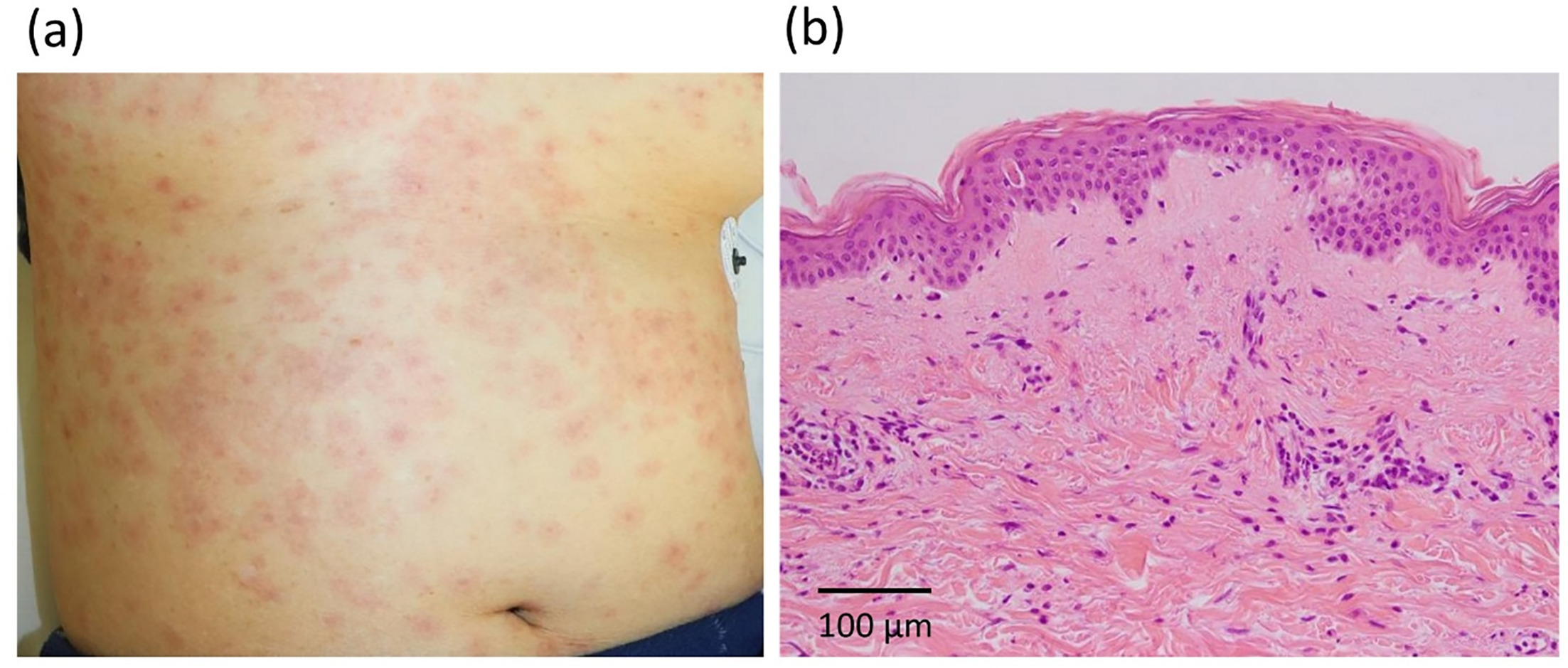
Fig. 1. (a) Clinical presentation showing edematous erythema scattered on the trunk. (b) Histopathological findings of the erythematous lesion of the abdomen (hematoxylin and eosin staining). Lymphocytes infiltrate in the epidermis, along with subtle individual keratinocyte necrosis. Perivascular infiltration of lymphocytes and eosinophils is observed in the superficial dermis.
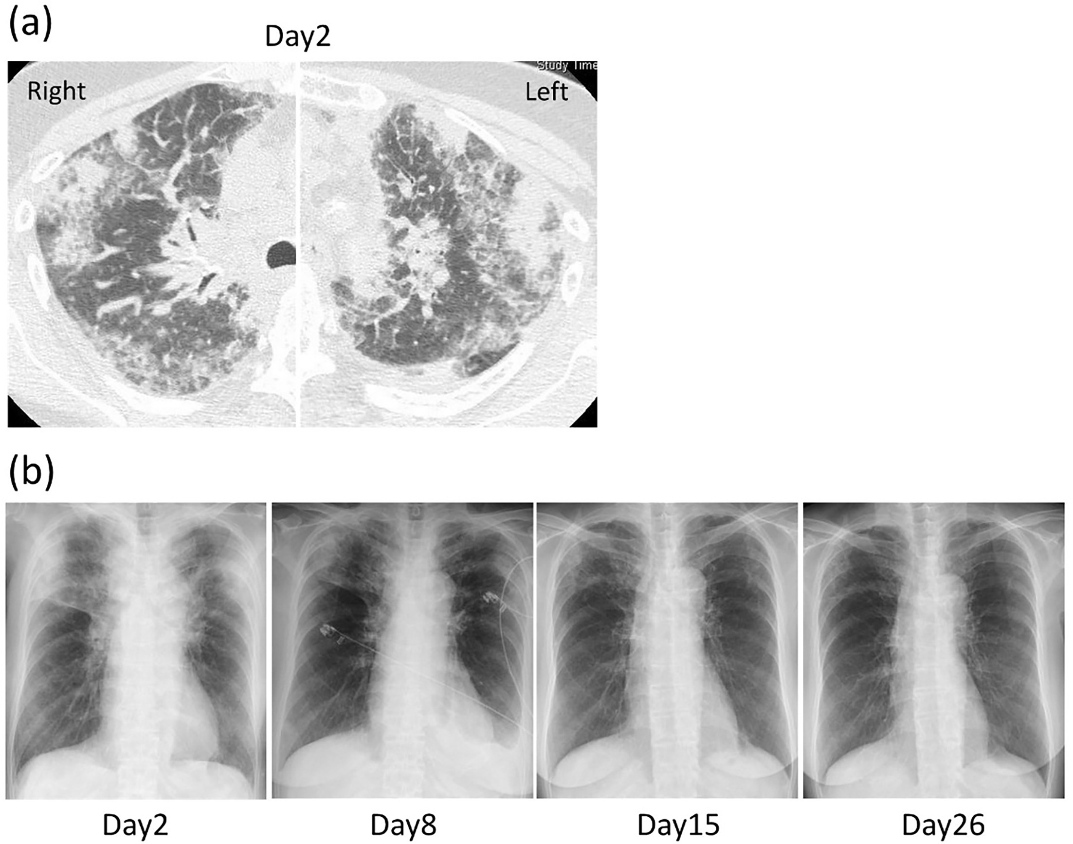
Fig. 2. (a) Chest computed tomography findings on Day 2. Extensive ground-glass opacities, infiltrates and interlobular septal thickening are observed mainly in the bilateral upper lobes. (b) Changes in chest radiographic findings during the clinical course.
The patient was diagnosed with minocycline-induced DIHS/DRESS based on the Japanese diagnostic criteria (3) and the RegiSCAR scoring system (4). Prednisolone at 1 mg/kg/day was started on Day 2 because of severe pulmonary involvement with respiratory failure. Ampicillin/sulbactam was administered temporarily in the setting of marked inflammation and respiratory failure because infectious pneumonia could not be excluded. The rash, respiratory status and chest opa-cities gradually improved (Fig. 2b), accompanied by decreases in the leukocyte count, eosinophil count and levels of CRP and TARC. Multiple episodes of thrombocytopenia and positive CMV antigenemia occurred during steroid tapering; therefore, prednisolone was tapered with caution. Prednisolone was discontinued 7 months after treatment initiation, with no recurrence. The results of a drug-induced lymphocyte stimulation test for minocycline were negative.
DISCUSSION
The present case was diagnosed as atypical DIHS according to the Japanese criteria and definite DRESS according to a RegiSCAR score of 6 (3, 4). The diagnosis was supported by a delayed onset after minocycline exposure, fever, a generalized rash, leukocytosis, eosinophilia, atypical lymphocytosis, lymphadenopathy and markedly elevated TARC level (3, 5). Notably, no liver or kidney dysfunction was observed, and the pulmonary lesions were the only evidence of visceral organ involvement. Episodes of thrombocytopenia and positive CMV antigenemia during steroid tapering may suggest immunological abnormalities associated with DIHS/DRESS (3).
Pulmonary involvement in DIHS/DRESS is relatively uncommon compared with liver or kidney involvement (2). In a systematic review, 21 of 22 cases of DRESS with pulmonary involvement showed additional extrapulmonary organ involvement, indicating that isolated pulmonary involvement is rare (2). A case of DIHS previously reported by Naniwa et al. showed predominant pulmonary involvement in the absence of other organ dysfunction and was related to an increase in anti-paramyxovirus antibody titres (6). Pulmonary lesions associated with DIHS/DRESS may mimic other pulmonary disorders, especially infectious pneumonia, as DIHS/DRESS can present with fever, elevated inflammatory markers and ground-glass opacities or infiltrates. Indeed, 45% of DRESS cases with pulmonary involvement were initially misdiagnosed as bacterial pneumonia and treated with empirical antibiotics (2). In the present case in the absence of extrapulmonary organ involvement, although infection could not be completely excluded at disease onset, the clinical course, eosinophilia, appearance of atypical lymphocytes, lymphadenopathy and marked TARC elevation (3, 5) supported pulmonary involvement as a manifestation of DIHS/DRESS. The prompt improvement after corticosteroid therapy also supported this interpretation. Recognition of DIHS/DRESS presenting with predominant pulmonary lesions in the absence of liver dysfunction is important because delayed diagnosis may delay corticosteroid therapy and lead to unnecessary antibiotic use.
In summary, rare cases of DIHS/DRESS may present with isolated or predominant pulmonary lesions, which can be difficult to distinguish from other pulmonary disorders, leading to delayed diagnosis. When delayed- onset fever, generalized rashes, eosinophilia, atypical lymphocytosis and pulmonary lesions occur after drug exposure, DIHS/DRESS should be included in the differential diagnosis, even in the absence of liver dysfunction.
ACKNOWLEDGEMENTS
The patient gave consent for her photographs and medical information to be published in print and online and with the understanding that this information may be publicly available.
REFERENCES
- Hama N, Abe R, Gibson A, Phillips EJ. Drug-Induced Hypersensitivity Syndrome (DIHS)/Drug Reaction With Eosinophilia and Systemic Symptoms (DRESS): Clinical Features and Pathogenesis. J Allergy Clin Immunol Pract 2022; 10: 1155–1167. https://doi.org/10.1016/j.jaip.2022.02.004
- Taweesedt PT, Nordstrom CW, Stoeckel J, Dumic I. Pulmonary Manifestations of Drug Reaction with Eosinophilia and Systemic Symptoms (DRESS) Syndrome: A Systematic Review. Biomed Res Int 2019; 2019: 1–10. https://doi.org/10.1155/2019/7863815
- Mizukawa Y, Hama N, Niihara H, Miyagawa F, Hashizume H, Tohyama M, et al. Guidelines for the management of drug-induced hypersensitivity syndrome 2023. J Dermatol 2025; 52: e189–e209. https://doi.org/10.1111/1346-8138.17609
- Calle AM, Aguirre N, Ardila JC, Cardona Villa R. DRESS syndrome: A literature review and treatment algorithm. World Allergy Organ J 2023; 16: 100673. https://doi.org/10.1016/j.waojou.2022.100673
- Ogawa K, Morito H, Hasegawa A, Miyagawa F, Kobayashi N, Watanabe H, et al. Elevated serum thymus and activation-regulated chemokine (TARC/CCL17) relates to reactivation of human herpesvirus 6 in drug reaction with eosinophilia and systemic symptoms (DRESS)/drug-induced hypersensitivity syndrome (DIHS). Br J Dermatol 2014; 171: 425–427. https://doi.org/10.1111/bjd.12948
- Naniwa T, Maeda S, Sawada H, Watanabe Y, Osawa T, Hayami Y, et al. Drug-induced hypersensitivity syndrome associated with a marked increase in anti-paramyxovirus antibody titers in a scleroderma patient. Allergol Int 2007; 56: 303–308. https://doi.org/10.2332/allergolint.C-06-49