SHORT COMMUNICATION
Linear Atrophoderma of Moulin with Telangiectatic Erythema: An Atypical Presentation
Shiwen GUO1 , Yanjing CHEN1 and Lin WANG1*
1Department of Dermatology and Venereology, West China Hospital, Sichuan University, Chengdu, China. *Email: lkzwl@126.com
Citation: Acta Derm Venereol 2026; 106: adv-2026-0951. DOI: https://doi.org/10.2340/actadv.v106.adv-2026-0951.
Copyright: 2026 ©Author(s). Published by MJS Publishing, on behalf of the Society for Publication of Acta Dermato-Venereologica. This is an Open Access article distributed under the terms of the Creative Commons Attribution-NonCommercial 4.0 International License (https://creativecommons.org/licenses/by-nc/4.0/).
Submitted: Aug 16, 2026. Accepted after revision: Sept 9, 2026.
Published: Sept 28, 2026.
Competing interests and funding: The authors have no conflicts of interest to declare.
The data that support the findings of this study are available from the corresponding author upon reasonable request.
Linear atrophoderma of Moulin (LAM) is a rare acquired dermatosis first described by Moulin et al. in 1992 (1). It typically presents as unilateral, asymptomatic, mildly atrophic, nonsclerotic hyper-pigmented patches following Blaschko's lines, pre-dominantly involving the trunk and extremities, whereas facial involvement is uncommon (2, 3). Herein, we report an atypical case involving the left side of the face and the right lower extremity, characterized by prominent telangiectatic erythema within the atrophic lesions. To our knowledge, only 3 patients with this telangiectatic presentation have previously been reported (4, 5).
CASE REPORT
An 18-year-old woman presented with a 10-year history of asymptomatic hyperpigmented patches and prominent telangiectatic erythema involving the right lower extremity and the left side of the face. There were no family history of similar lesions and no pre-ceding inflammatory phase or subsequent induration or sclerosis. Physical examination revealed linear, mildly atrophic hyperpigmented patches with prominent telangiectatic erythema following Blaschko’s lines on the left side of the face and the right lower extremity (Fig. 1A-D). Laboratory investigations, including a complete blood count, serum chemistry panel and autoimmune serologic testing, were unremarkable. Histopathological examination showed epidermal hyperkeratosis and acanthosis, mild dermal thinning and numerous dilated capillaries in the papillary dermis, accompanied by a sparse superficial perivascular lymphocytic infiltration. Dermal collagen showed no thickening or hyalinization (Fig. 1E-F). Based on the clinical presentation and histopathological examination, a diagnosis of LAM was established. No treatment was initiated, and the lesions remained stable during 5 months of follow-up.
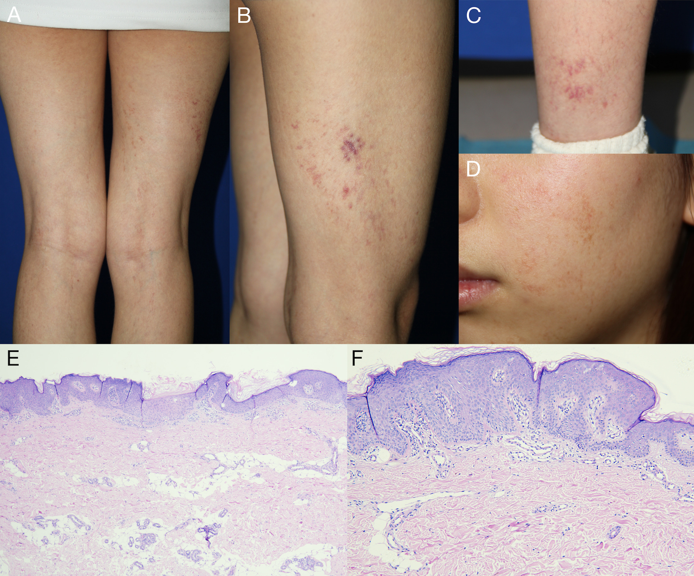
Fig. 1. Clinical and histopathological features of linear atrophoderma of moulin with prominent telangiectatic erythema. (A, B, C) Hyperpigmented atrophic patches with prominent telangiectatic erythema on the right lower extremity and (D) the left side of the face, distributed along Blaschko’s lines. (E) Histopathological examination showing epidermal hyperkeratosis, acanthosis and mild dermal thinning (haematoxylin and eosin [H&E] staining, ×40). (F) Dilated and increased capillaries within the papillary dermis and a sparse perivascular lymphocytic infiltration in the superficial dermis (H&E staining, ×100).
DISCUSSION
LAM typically presents during childhood or adolescence as unilateral, asymptomatic, hyper-pigmented atrophic lesions following Blaschko’s lines, most commonly on the trunk and extremities, whereas facial and cervical involvement is uncommon (3). In our patient, lesions involved the left side of the face and the right lower extremity, representing anatomically separate sites on opposite sides of the body, and were accompanied by prominent telangiectatic erythema, which is atypical for LAM. Including our patient, only 4 cases of LAM with clinically prominent telangiectasia have been reported (Table I). Although histopathological findings are generally nonspecific, dilation of papillary dermal capillaries may account for the clinically observed telangiectatic erythema. Utikal et al. proposed that prominent telangiectatic erythema within atrophic lesions may represent a distinct variant of LAM (4). Our case further supports the existence of this uncommon telangiectatic phenotype. The aetiology and pathogenesis of LAM are still unclear. A postzygotic mutation involving a predisposing gene, followed by loss of the corresponding wild-type allele, has been proposed, although molecular confirmation is lacking (6). Another hypothesis pointed out that LAM was an autoimmune or connective tissue disorder, forming a disease spectrum alongside atrophoderma of Pasini and Pierini and linear morphea. However, the exact underlying mechanism remains to be further elucidated.
Table I. Summary of reported cases of linear atrophoderma of Moulin with telangiectatic erythema
| Reference (first author) |
Sex | Age at onset (years) |
Location | Clinical features | Histopathological features | Treatment | Outcome |
|---|---|---|---|---|---|---|---|
| Miteva L (5) | Female | 16 | Face, right buttock and right extremities | Unilateral Blaschko-linear hypopigmented atrophic lesions with telangiectatic macules | Psoriasiform epidermal hyperplasia, papillary dermal edema, dilated/hyalinized vessels, sparse lymphoplasmacytic infiltrate, focal collagen hyalinization and reduced elastic fibres | NR | Stable |
| Utikal (4) | Male | 23 | Trunk, buttocks and extremities | Blaschko-linear telangiectatic erythematous to brownish atrophic macules and patches; no preceding inflammatory lesions | Normal epidermis with slight dermal edema and sparse perivascular lymphohistiocytic infiltrate; DIF: negative | Penicillin V+shower PUVA | Partial improvement |
| Female | 2 | Left lower extremity and left buttock | Blaschko-linear erythematous telangiectatic atrophic macules; no preceding inflammatory lesions | Normal epidermis with slight dermal edema and minimal perivascular mononuclear infiltrate; DIF: negative | Self-tanning cream | Homogenization of skin pigmentation | |
| Our case | Female | 8 | Left side of the face and right lower extremity | Hyperpigmented atrophic patches with prominent telangiectatic erythema; no induration or inflammatory signs | Hyperkeratosis, acanthosis, mild dermal atrophy, dilated and increased capillaries in the papillary dermis and sparse superficial perivascular lymphocytic infiltrate | No treatment | Stable |
|
DIF: Direct immunofluorescence; NR: not reported. |
|||||||
The principal differential diagnoses were atropho-derma of Pasini and Pierini (APP) and linear morphea. APP generally presents as sharply demarcated, depressed pigmented patches and does not characteristically follow Blaschko’s lines. Its histopathological findings are subtle and may include dermal thinning and mild collagen alterations (7). Linear morphea may also follow Blaschko’s lines but more commonly progresses from an inflammatory phase to induration and sclerosis, with thickened, homogenized collagen bundles and entrapment or loss of adnexal structures (8). In our patient, the absence of preceding inflammation or subsequent induration and sclerosis, together with the lack of collagen homogenization or adnexal alteration, favoured LAM. Other Blaschko-linear pigmentary disorders and telangiectatic dermatoses, including focal dermal hypoplasia, angioma serpiginosum, unilateral nevoid telangiectasia and cutis marmorata telangiectatica congenita, were considered less likely because of their congenital or early onset, distinct morphology or associated extracutaneous abnormalities.
Currently, no standard therapeutic regimen exists for LAM. Previous therapeutic attempts, including ultraviolet therapy, topical corticosteroids, high-dose penicillin and heparin, have generally proven ineffective (9). Observation is reasonable for stable, asymptomatic lesions when treatment for cosmetic concerns is not requested.
REFERENCES
- Moulin G, Hill MP, Guillaud V, Barrut D, Chevallier J, Thomas L. Bandes pigmentées atrophiques acquises suivant les lignes de Blaschko. Ann Dermatol Venereol 1992; 119: 729–736.
- Darung I, Rudra O, Samanta A, Agarwal M, Ghosh A. Linear Atrophoderma of Moulin over Face: An Exceedingly Rare Entity. Indian J Dermatol 2017; 62: 214–215. https://doi.org/10.4103/ijd.IJD_469_16
- Villani AP, Amini-Adlé M, Wagschal D, Balme B, Thomas L. Linear atrophoderma of moulin: report of 4 cases and 20th anniversary case review. Dermatology 2013; 227: 5–9. https://doi.org/10.1159/000347110
- Utikal J, Keil D, Klemke CD, Bayerl C, Goerdt S. Predominant telangiectatic erythema in linear atrophoderma of Moulin: novel variant or separate entity? Dermatology 2003; 207: 310–315. https://doi.org/10.1159/000073096
- Miteva L, Obreshkova E. An unusual manifestation of linear atrophoderma of Moulin. Acta Derm Venereol 2002; 82: 479–480. https://doi.org/10.1080/000155502762064737
- Danarti R, Bittar M, Happle R, König A. Linear atrophoderma of Moulin: postulation of mosaicism for a predisposing gene. J Am Acad Dermatol 2003; 49: 492–498. https://doi.org/10.1067/s0190-9622(03)00895-8
- Saleh Z, Abbas O, Dahdah MJ, Kibbi AG, Zaynoun S, Ghosn S. Atrophoderma of Pasini and Pierini: a clinical and histopathological study. J Cutan Pathol 2008; 35: 1108–1114. https://doi.org/10.1111/j.1600-0560.2008.00986.x
- Papara C, De Luca DA, Bieber K, Vorobyev A, Ludwig RJ. Morphea: The 2023 update. Front Med (Lausanne) 2023; 10: 1108623. https://doi.org/10.3389/fmed.2023.1108623
- Zahedi Niaki O, Sissons W, Nguyen VH, Zargham R, Jafarian F. Linear atrophoderma of Moulin: an underrecognized entity. Pediatr Rheumatol Online J 2015; 13: 39. https://doi.org/10.1186/s12969-015-0036-6