QUIZ SECTION
Three Progressive Upper Trunk Nodules: A Quiz
Sophie REISER1* and Kai-Philipp LINSE1
1Department of Dermatology, University Hospital Heidelberg, Im Neuenheimer Feld 440, 69120 Heidelberg, Germany. *Email: sophie.reiser@med.uni-heidelberg.de
Citation: Acta Derm Venereol 2026; 106: adv-2025-0161. DOI: https://doi.org/10.2340/actadv.v106.adv-2025-0161.
Copyright: 2026 ©Author(s). Published by MJS Publishing, on behalf of the Society for Publication of Acta Dermato-Venereologica. This is an Open Access article distributed under the terms of the Creative Commons Attribution-NonCommercial 4.0 International License (https://creativecommons.org/licenses/by-nc/4.0/).
Submitted: Nov 5, 2025. Accepted after revision:
Published: Sept 28, 2026.
Competing interests and funding:
A 32-year-old Caucasian man was referred to our dermatology department presenting with three ill-defined, violaceous to erythematous nodules that had been present for several months. The lesions were located along the left posterior axillary line, the left lumbar paravertebral region, and the right upper shoulder, each measuring approximately 2×1 cm (Fig. 1). The nodules had exhibited gradual enlargement over the preceding weeks. The patient’s dermatological and medical history was unremarkable. His occupational history was notable for employment as a gardener and retail nursery specialist. He reported being in a stable, long-term relationship. External histopathological examination of prior biopsies had raised suspicion of dermatofibrosarcoma protuberans in the left posterior axilla and right upper shoulder, and pyogenic granuloma in the left paravertebral region. Subsequent histopathological evaluation of the three excision specimens demonstrated exophytic vascular neoplasms collared by a hyperplastic epidermal fringe. The proliferating endothelial and stromal cells exhibited a dimorphic pattern, displaying both spindled and epithelioid cytomorphological features. Extravasated erythrocytes were dissecting between the vascular slits, accompanied by scattered mitotic figures and a prominent perivascular lymphoplasmacytic infiltrate. Immunohistochemical analysis demonstrated strong, diffuse immunoreactivity for D2-40 (podoplanin), nuclear ERG (ETS-related gene), and CD31 (Fig. 2).
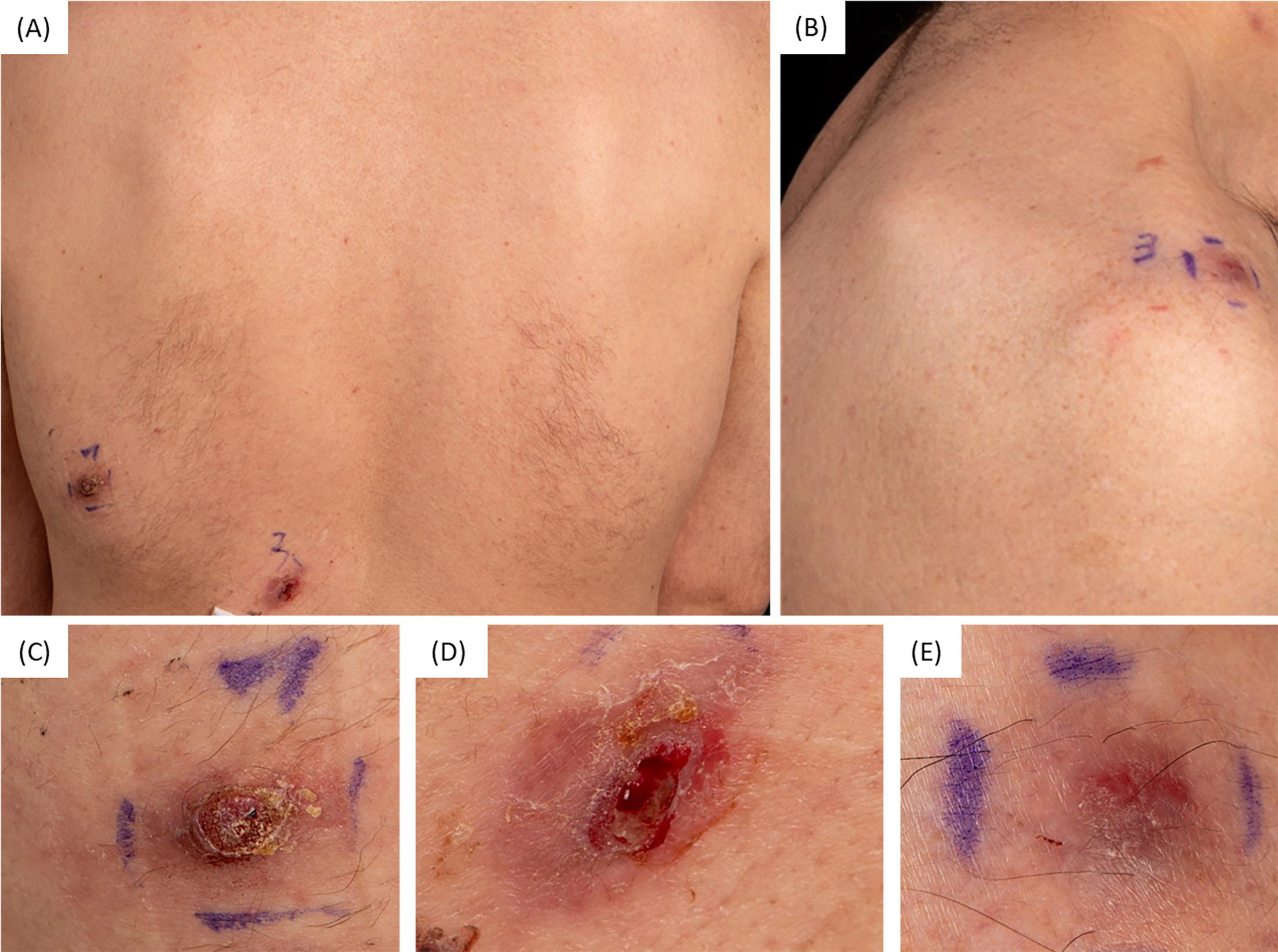
Fig. 1. (A) Overview of two erythematous to violaceous nodules on the left back and (B) one solitary nodule on the right shoulder. (C, D) Close-up views of the respective nodules on the back and (E) detailed view of the nodule on the right shoulder.
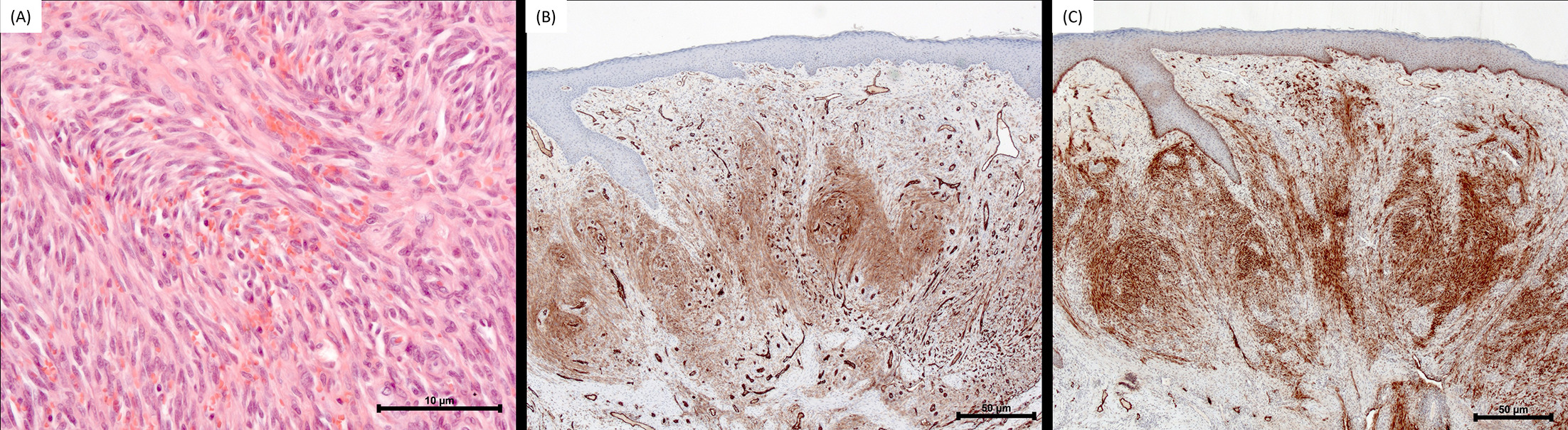
Fig. 2. Histopathological and immunohistochemical findings. (A) Hematoxylin and eosin staining demonstrating a proliferation of spindle-shaped and epithelioid endothelial cells forming slit-like vascular spaces, accompanied by extravasated erythrocytes and scattered mitotic figures (original magnification ×200). (B) Immunohistochemical staining showing strong endothelial membranous positivity for CD31 (original magnification ×25). (C) Diffuse reactivity for the lymphatic endothelial marker D2-40 (original magnification ×25).
What is your diagnosis?
1: Dermatofibrosarcoma protuberans
2: Angiosarcoma
3: Kaposi sarcoma
4: Pyogenic granuloma
See next page for answer.
ANSWERS TO QUIZ
Three Progressive Upper Trunk Nodules: A Commentary
Diagnosis: Kaposi sarcoma
Additionally, immunohistochemical staining for human herpesvirus 8 (HHV-8) latent nuclear antigen-1 demonstrated strong and diffuse nuclear positivity, confirming the diagnosis of Kaposi sarcoma (KS). Upon detailed history taking, the patient reported a history of sex with men (MSM).
Further laboratory investigations revealed a reactive human immunodeficiency virus (HIV) rapid screening test, with an HIV immunoblot confirming diagnostic reactivity against specific HIV-1 viral proteins. Quantitative HIV-1 RNA PCR revealed a viral load of 1,550,000 copies/mL and a CD4⁺ T-cell count of 168 cells/μL (reference range: 493–1,522 cells/μL), indicating advanced immunodeficiency. Further screening for syphilis revealed a reactive Treponema pallidum particle agglutination (TPPA) assay and a Rapid Plasma Reagin (RPR) titer of 1:128 (reference <1:2), confirming active syphilis. Cutaneous KS was therefore the index presentation of the underlying HIV infection, which subsequently prompted the diagnosis of concurrent active syphilis. The diagnosis of AIDS was made, with KS identified as an AIDS-defining illness. For the management of HIV-associated KS, antiretroviral therapy was initiated as the primary treatment to restore immune function. The patient was started on a fixed-dose combination of bictegravir, tenofovir alafenamide, and emtricitabine (Biktarvy), a potent antiretroviral regimen. At 1-month follow-up, a rapid virological response was observed, with the HIV-1 RNA viral load declining to 125 copies/mL. Staging examinations revealed no evidence of mucosal or visceral involvement, and no new cutaneous lesions or signs of progression were noted.
For syphilis treatment, the patient was prescribed Benzathine penicillin G (Tardocillin 2.4 million units intramuscularly) according to current clinical guidelines. Follow-up serologic testing was conducted to assess the therapeutic response. In addition, screening for sexually transmitted diseases was initiated for contact persons.
The differential diagnosis included multifocal dermatofibrosarcoma protuberans (DFSP), pyogenic granuloma, and angiosarcoma.
DFSP is a superficial, locally aggressive cutaneous tumour of low malignant potential, characterised by uniform spindle-shaped neoplastic cells with storiform growth pattern (1). Unlike KS, extravasated erythrocytes are not a primary feature of DFSP and typically present as a solitary lesion. The benign vascular lesion pyogenic granuloma often shows a well-defined lobular growth pattern with numerous endothelium-lined vascular spaces (2). Further proliferation of fibroblasts and budding endothelial cells is characteristic (2).
Unlike KS, extravasated erythrocytes are not a characteristic feature of DFSP, which typically presents as a solitary lesion. Although angiosarcoma can show positive staining for ERG and CD31, it is distinguished from KS by pronounced cytologic atypia, endothelial multilayering, and brisk mitotic activity. Angiosarcoma is a rare malignant mesenchymal tumour and is often more infiltrative and aggressive, with areas of necrosis (3).
None of these three differential diagnoses is associated with HHV-8, which represents a key diagnostic hallmark of KS (4)1, 2, 3, 4. Additional immunohistochemical markers, including D2-40, CD31, and ERG, are employed in the diagnostic workup of KS (5, 6, 7). CD31 is an immunohistochemical endothelial cell marker confirming the vascular nature of KS, supporting the characteristic endothelial cell proliferation seen in KS (6). Also, ERG, a transcription factor expressed in endothelial cells, strongly indicates vascular origin with nuclear staining in endothelial cells (7). ERG is involved in regulating vascular development and angiogenesis. It is indicative of endothelial differentiation and vascular endothelial lineage (7). D2-40, a marker for lymphatic endothelium, is often used in the evaluation of vascular tumours (5). D2-40 positivity suggests a lymphatic component in KS, which is a common feature.KS lesions are typically characterized by extravasated erythrocytes and a plasma cell-rich infiltrate around vascular channels and spindle cells, also distinguishing KS from other vascular tumours.
Histopathological re-evaluation was requested after initial external biopsies had suggested DFSP in the left posterior axilla and right upper shoulder, as well as pyogenic granuloma in the left paravertebral region. However, further histopathological investigation, coupled with immunohistochemical analysis, led to the final diagnosis of HIV-associated KS, highlighting the importance of comprehensive evaluation in cases with atypical presentations.
In addition to HIV-associated KS, other subtypes include classic, endemic and iatrogenic KS (8).
As exemplified in our case, KS typically presents as nodules, though it can also manifest as plaques or patches, ranging in colour from red to purple or brownish (8). The extravasation of red blood cells contributes significantly to the clinical appearance of the lesions (9). In addition to cutaneous involvement, KS can affect other sites such as the lymphatic system, mucosal surfaces and visceral organs. Visceral KS may affect organs such as the lungs, liver and gastrointestinal tract, leading to more severe complications (8).
This case underscores the necessity of a thorough diagnostic approach when unusual vascular lesions are observed and highlights the clinical value of an extensive social history, even in the presence of an otherwise unremarkable medical background. Additionally, any inconsistencies should prompt a thorough examination of the histology to ensure accurate diagnosis and effective treatment planning. HIV-associated KS is often the first clinical sign of an undiagnosed HIV infection. In such cases, rigorous screening for concurrent sexually transmitted infections is essential. Early initiation of antiretroviral therapy remains the cornerstone of treatment for HIV-associated KS and can prevent the progression of both HIV and KS.
REFERENCES
- Larbcharoensub N, Kayankarnnavee J, Sanpaphant S, Kiranantawat K, Wirojtananugoon C, Sirikulchayanonta V. Clinicopathological features of dermatofibrosarcoma protuberans. Oncol Lett 2016; 11: 661–667. https://doi.org/10.3892/ol.2015.3966
- Marla V, Shrestha A, Goel K, Shrestha S. The histopathological spectrum of pyogenic granuloma: a case series. Case Rep Dent 2016; 2016: 1323798. https://doi.org/10.1155/2016/1323798
- Ichiki T, Yamada Y, Ito T, Nakahara T, Nakashima Y, Nakamura M, et al. Histological and immunohistochemical prognostic factors of primary angiosarcoma. Virchows Arch 2023; 483: 59–69. https://doi.org/10.1007/s00428-023-03572-z
- Lebbé C. Humanes herpesvirus 8 (HHV-8) and Kaposi sarcoma. Hautarzt 2008; 59: 18–25. https://doi.org/10.1007/s00105-007-1445-3
- Kahn HJ, Bailey D, Marks A. Monoclonal antibody D2-40, a new marker of lymphatic endothelium, reacts with Kaposi’s sarcoma and a subset of angiosarcomas. Mod Pathol 2002; 15: 434–440. https://doi.org/10.1038/modpathol.3880543
- Russell Jones R, Orchard G, Zelger B, Wilson Jones E. Immunostaining for CD31 and CD34 in Kaposi sarcoma. J Clin Pathol 1995; 48: 1011–1016. https://doi.org/10.1136/jcp.48.11.1011
- Miettinen M, Wang ZF, Paetau A, Tan SH, Dobi A, Srivastava S, et al. ERG transcription factor as an immunohistochemical marker for vascular endothelial tumors and prostatic carcinoma. Am J Surg Pathol 2011; 35: 432–441. https://doi.org/10.1097/PAS.0b013e318206b67b
- Vangipuram R, Tyring SK. Epidemiology of Kaposi sarcoma: review and description of the nonepidemic variant. Int J Dermatol 2019; 58: 538–542. https://doi.org/10.1111/ijd.14080
- Bishop BN, Lynch DT. Kaposi Sarcoma. In: StatPearls. Treasure Island (FL): StatPearls Publishing; 2023 Jun 5.