QUIZ SECTION
Multiple Eruptive Facial Tumours in a 72-year-old Man Receiving Pembrolizumab: A Quiz
Charlotte DESJONQUERES1* , Baptiste SOURTY2 and Hervé MAILLARD1
1Dermatology department, General Hospital Le Mans, Le Mans, France, and 2Pathology department, General Hospital Le Mans, Le Mans, France. *Email: charlotte.desjonqueres@chu-angers.fr
Citation: Acta Derm Venereol 2026; 106: adv-2026-0500. DOI: https://doi.org/10.2340/actadv.v106.adv-2026-0500.
Copyright: 2026 ©Author(s). Published by MJS Publishing, on behalf of the Society for Publication of Acta Dermato-Venereologica. This is an Open Access article distributed under the terms of the Creative Commons Attribution-NonCommercial 4.0 International License (https://creativecommons.org/licenses/by-nc/4.0/).
Submitted: Mar 15, 2026. Accepted after revision:
Published: Sept 30, 2026.
Competing interests and funding:
A 72-year-old man with a history of urothelial cancer treated with pembrolizumab since 2023 developed a bullous eruption in June 2025. Skin biopsy showed a subepidermal blister, and direct immunofluorescence demonstrated linear deposition of IgG and C3 along the basement membrane zone. ELISA testing was positive for anti-BP180 antibodies, confirming the diagnosis of bullous pemphigoid induced by anti–PD-1 therapy.
Because topical corticosteroids were insufficient, methotrexate was initiated, allowing continuation of pembrolizumab therapy. Remission of the bullous pemphigoid was achieved within 6 weeks.
In October 2025, the patient returned with eruptive tumours on the face that had appeared approximately 1 month earlier. He had previously developed a similar nodule on the right hand that had been surgically excised several weeks earlier. Histologic examination of that lesion was consistent with a pyogenic granuloma.
Physical examination revealed multiple tumours of different ages. The more recent lesions appeared as erythematous nodules with central ulceration, whereas older lesions presented as erythematous lobulated tumours with slight keratinization (Fig. 1). Two skin biopsies were performed from 2 different facial lesions, and representative histopathologic features are shown in Fig. 2.
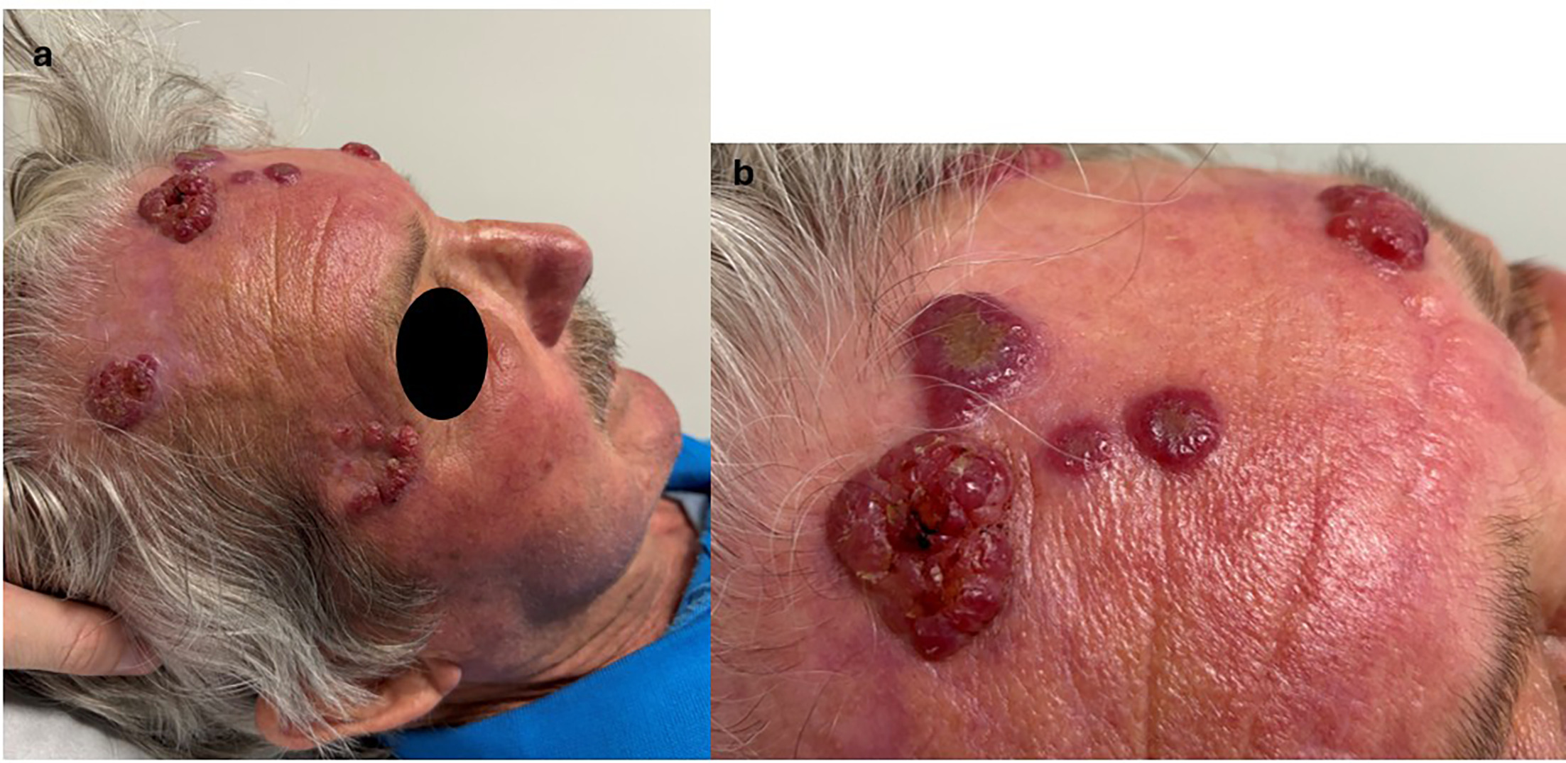
Fig. 1. (a,b) Eruptive erythematous tumours with necrotic centre.
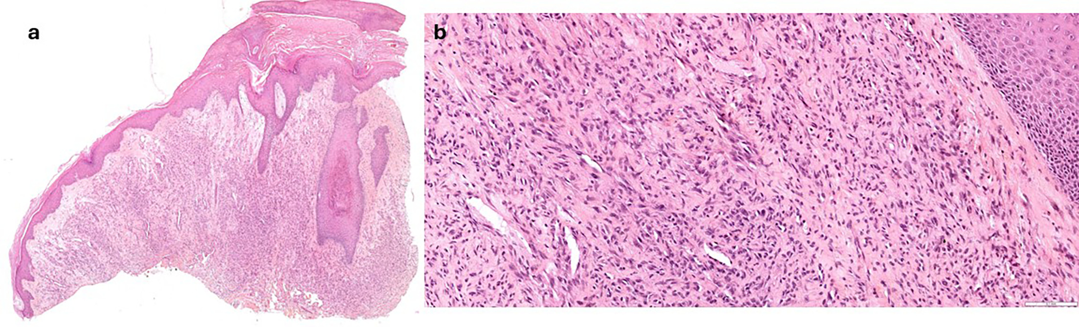
Fig. 2. (a) Low-power view showing a vascular lobulated proliferation beneath a nonulcerated epidermis (HES x 25). (b) At higher magnification, numerous capillary-sized vessels, lined by only slightly plump endothelial cells, with some mitoses, within a loose stroma containing scattered mononucleated cells (HES x 200). HES: Hematoxylin-Eosin-Saffron.
What is your diagnosis?
1: Bacillary angiomatosis.
2: Cutaneous metastasis of urothelial cancer.
3: Eruptive capillary haemangiomas.
4: Neutrophilic dermatosis (Sweet syndrome).
See next page for answer.
ANSWERS TO QUIZ
Multiple Eruptive Facial Tumours in a 72-year-old Man Receiving Pembrolizumab: A Commentary
Diagnosis: Eruptive capillary haemangiomas.
Histologic examination of both biopsies revealed a dermal vascular proliferation composed of numerous small vessels arranged in lobules within an edematous stroma with sparse inflammatory infiltrate, consistent with features of pyogenic granuloma. Immunohistochemistry for human herpesvirus 8 was negative.
Given these findings, the main differential diagnosis considered was bacillary angiomatosis, particularly as the patient was immunosuppressed due to methotrexate therapy. Additional biopsies were therefore obtained for microbiological investigations, and treatment with doxycycline (200 mg/day) was initiated. PCR for Bartonella henselae on the skin biopsy and Bartonella serology were both negative.
Although the histopathologic findings were not entirely specific, endothelial cells were less prominent than typically described in bacillary angiomatosis, and no significant neutrophilic infiltrate was observed.
Based on the clinical history and histologic findings, a diagnosis of eruptive pyogenic granulomas associated with treatment with pembrolizumab was made. Fifteen days after once-daily application of an ultrapotent topical corticosteroid (clobetasol propionate cream), the lesions completely resolved (Fig. 3).
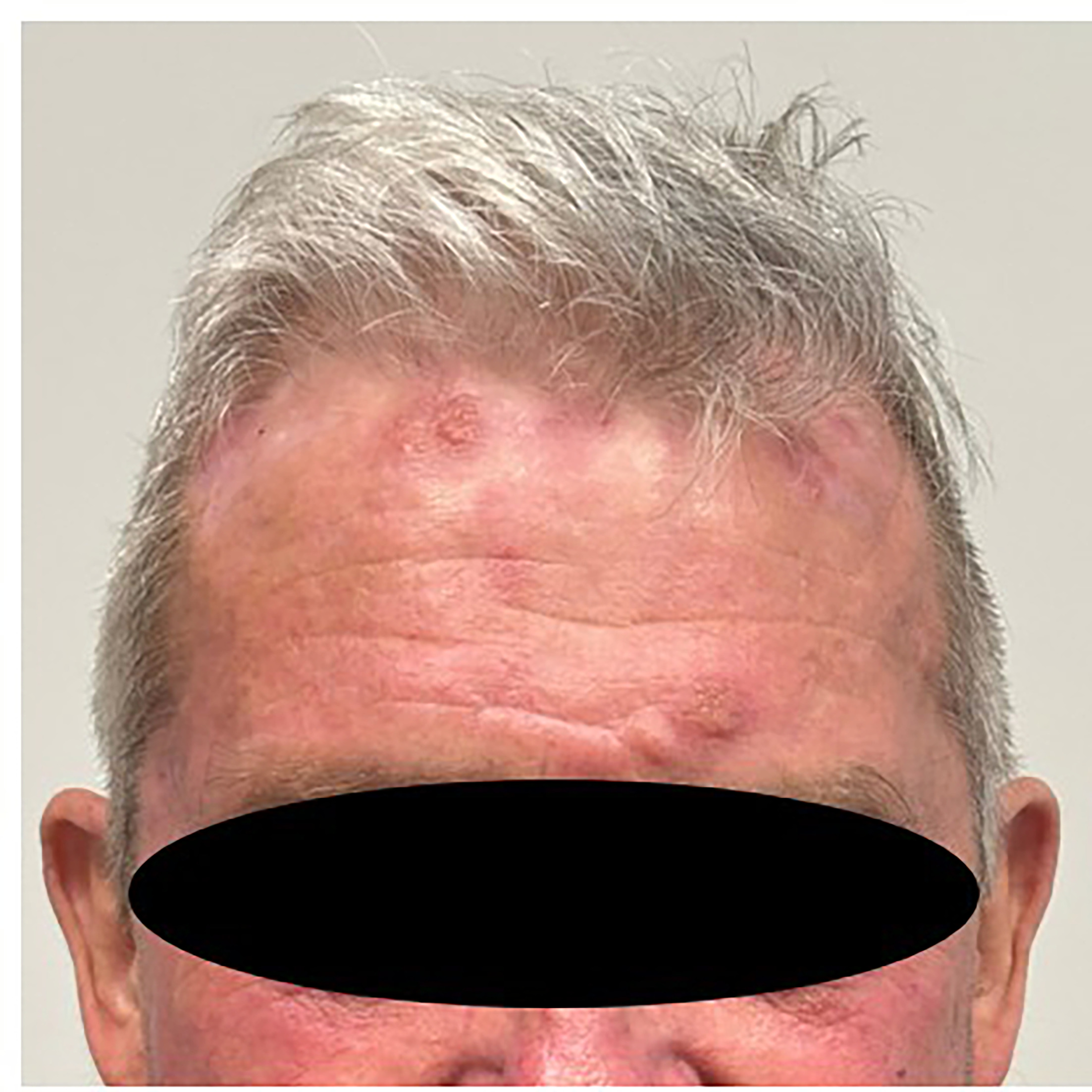
Fig. 3. Involution of eruption after the application of ultrapotent topical steroids.
Eruptive pyogenic granulomas are rare and have mainly been reported following trauma, including burns or surgical procedures. Only a few reports have described similar lesions occurring during immunotherapy. Chen et al. (1) reported 84 cases of reactive capillary hemangiomas among 98 patients treated with camrelizumab in a Chinese phase I clinical trial.( On average, the eruption peaked 2.5 months after treatment initiation. Most cases were grade one adverse events. Lesions preferentially involved the face, neck, trunk and extremities, with occasional mucosal involvement. They typically regressed spontaneously after reaching their maximum size, although new lesions could develop during ongoing treatment, generally of smaller size. Histologically, these lesions consisted of small capillaries in the papillary dermis or endothelial cell proliferation. To our knowledge, this adverse event has not been reported with commonly used anti–PD-1 agents such as pembrolizumab or nivolumab. One possible explanation is an off-target interaction of SHR-1210 with VEGFR-2. Reactive cutaneous capillary proliferations have been described as the most frequent adverse event associated with camrelizumab (2).
Pyogenic granulomas are more commonly associated with treatment with BRAF inhibitors, through paradoxical activation of the MAPK pathway (3). Indeed, mutations affecting this pathway, including BRAF V600 or NRAS mutations, have been identified in some pyogenic granulomas (4). In our patient, targeted high-throughput sequencing did not identify mutations in these genes.
In our case, the hypothesis of eruptive pyogenic granulomas arising on previously bullous areas was unlikely, as the first lesion appeared on the hand, where no previous bullous lesion had been documented. We therefore conclude that this case represents eruptive capillary hemangiomas associated with pembrolizumab therapy.
REFERENCES
- Chen X, Ma L, Wang X, Mo H, Wu D, Lan B, et al. Reactive capillary hemangiomas: a novel dermatologic toxicity following anti-PD-1 treatment with SHR-1210. Cancer Biol Med 2019; 16: 173–181. https://doi.org/10.20892/j.issn.2095-3941.2018.0172
- Lin Y, Lin Y, Zhong X, Chen Q, Tang S, Chen J. A case report and literature review on reactive cutaneous capillary endothelial proliferation induced by camrelizumab in a nasopharyngeal carcinoma patient. Front Oncol 2023; 13: 1280208. https://doi.org/10.3389/fonc.2023.1280208
- Sammut SJ, Tomson N, Corrie P. Pyogenic granuloma as a cutaneous adverse effect of vemurafenib. N Engl J Med 2014; 371: 1265–1267. https://doi.org/10.1056/NEJMc1407683
- Groesser L, Peterhof E, Evert M, Landthaler M, Berneburg M, Hafner C. BRAF and RAS mutations in sporadic and secondary pyogenic granuloma. J Invest Dermatol 2016; 136: 481–486. https://doi.org/10.1038/JID.2015.376